
1286
Int. J. Morphol.,
37(4):1286-1293, 2019.
Profile and Reference Values for Body Fat and Skeletal
Muscle Mass Percent at Females, Aged from 18.0 to 69.9,
Measured by Multichannel Segmental Bioimpedance Method:
Serbian Population Study
Perfil y Valores de Referencia del Porcentaje de Grasa Corporal y Masa Muscular en
Mujeres, con Edades Comprendidas entre 18,0 y 69,9 Años, Medido por el Método de
Bioimpedancia Segmentaria Multicanal: Estudio en Población Serbia
Rakic Sladjana
1
; Dopsaj Milivoj
1,5
; Djordjevic-Nikic Marina
1
; Vasiljevic Nadja
2
;
Dopsaj Violeta
3
; Maksimovic Milos
2
; Tomanic, S. Milena
2
& Miljus Dragan
4
RAKIC, S.; DOPSAJ, M.; DJORDJEVIC-NIKIC, M.; VASILJEVIC, N.; DOPSAJ, V.; MAKSIMOVIC, M.; TOMANIC, S. M.
& MILJUS, D. Profile and reference values for body fat and skeletal muscle mass percent at females, aged from 18.0 to 69.9, measured
by multichannel segmental bioimpedance method: Serbian population study. Int. J. Morphol., 37(4):1286-1293, 2019.
SUMMARY: Profile and standards for the diagnostics of percent of body fat and muscles were defined on a sample of 1924
women from the Republic of Serbia, aged 18.0 to 69.9, where the body structure of subjects was measured by applying multichannel
segmental bioimpedance. Total sample was divided into six age groups, for the purpose of the definition of standard with regards to age.
When it comes to body fat percentage results have shown that the average value of the total sample was 28.51±9.26 %, and between the
range of 23.81 and 39.94 % for age groups 18.0-19.9 yr and 60.0-69.9 yr, respectively. Regression analysis results have shown that the
constant of body fat percentage increase by trend of 3.417 % per decade, and that 25.1 % of mutual variance trend was explained by the
model, with prediction error of 4.55 %. With regards to the percentage of skeletal muscles in the body, the results have shown that the
average value of the total sample was 39.30±5.25 %, and within the range of 42.25 to 32.58 % for age groups 18.0-19.9 yr and 60.0-69.9
yr, respectively. Regression analysis results have shown that the constant of the skeletal muscles decrease by tend of -2.016 % per decade
and that the model explained 23.8 % of mutual variance trend with prediction error of 8.08 %.
KEY WORDS: Females; Body Composition Profile; Bioimpedance; Body Fat; Skeletal Muscle Mass.
INTRODUCTION
It is a global fact that the modern way of life is
characterized by sedentariness or insufficient level of
physical activity (Haskell et al., 2007; Owen et al., 2010;
Maksimovic et al., 2016). Being very useful on the one hand,
technical achievements of civilization negatively impact life
quality on the other hand, which relates to the health aspect
in particular, where non-infectious diseases are concerned.
Unfortunately, the business aspect and business sector to
which the modern man belongs, condition increasing
physical passivity, because thanks to workplace
modernization all the work is being done in a sitting position.
On the other side, even though the modern age man has an
ever increasing amount of free time, the population has
become increasingly physically inactive, the main reasons
being computer use, the Internet, mobile phones and
watching television (Hallal et al., 2012). Additionally, via
the phenomenon of the cause-effect connection, lack of
physical activity has as a consequence led to decreased
motivation for practicing it (Sallis et al., 2015).
Unfortunately, overabundant and irregular diet, in addition
to physical activity that is inconsistent and of insufficient
scope, are increasingly becoming dominant habits and a
comprising part of modern man’s life (Menotti et al., 2014;
Maksimovic et al.).
1
Faculty of Sport and Physical Education, University of Belgrade, Belgrade, Serbia.
2
Institute of Hygiene and Medical Ecology, Faculty of Medicine, University of Belgrade, Belgrade, Serbia.
3
Pharmaceutical Faculty, University of Belgrade, Belgrade, Serbia.
4
Institute of Public Health of Serbia “Dr Milan Jovanovic Batut”, Belgrade, Serbia.
5
SUSU, South Ural State University, Institute of Sport, Tourism and Service, Chelyabinsk, Russia.
1287
According to data from many scientific studies the
phenomenon of hypokinesis, i.e. insufficient physical
activity, has become the global public health problem in the
21st century (Hallal et al.), while epidemiological studies
have shown that three factors, such as: bad diet, stress
connected to the modern way of life and hypokinesis, are
the main cause of increase in the prevalence of non-infectious
diseases (Haskell et al.; Owen et al.; Menoti et al.).
A system for controlling the status of body
composition, through continued monitoring of the given state
of the organism, is increasingly becoming a part of the very
important health mechanism with the goal of tracking the
condition and ascertaining the trends of change in the given
conditions among the general populace (Gába & Pridalová,
2014; Ihász et al., 2015) or some specific population, such
as athletes, regardless of sex, age or type of sport (Dopsaj et
al., 2017; Bankovic et al., 2018). Also, defining the
characteristics of the model for the body composition status
condition among people represents an expert and scientific
attitude of decision making on whether a certain tissue
component is insufficiently, normally or predominantly
represented in the body (Bankovic et al.; Saraykin et al.,
2018), that is, if there exists an effect of an applied treatment
and how big it is, regardless whether it is diet correction or
physical activity treatment (Rocha et al., 2018). This is even
more pronounced with the advent of new technologies for
the measurement of body components, based on the principle
of bioimpedance, and especially new technology of
multichannel multisegment bioimpedance as being very
medically precise and easily available measurement
technology for all body composition characteristics (Ling
et al., 2011; Saraykin et al.).
From the perspective of anatomy, physiology and
biology the female organism is very specific. Cyclical
secretion of sex hormones during the reproductive period,
pregnancy, childbirth, maternity, menopause, professional
obligations and daily activities all have a great effect on body
status (Thompson et al., 2004; Kukic et al., 2019).
On the other hand, along with a considerable number
of published researches that tested body composition of
women of various ages by applying the method of segmental
multichannel bioimpedance (Gába & Pridalová; Ihász et al.;
Kukic et al., 2019) there are still no researches that define
the standards and normatives for evaluating the status of
two very important, if not the most important, body status
characteristics, and those are: percentage of body fat (PBF)
and percentage of skeletal muscles in the body (PSMM).
The goal of this paper is to define standards and
normatives for evaluation of body status among women of
various ages according to two variables: percentage of body
fat (PBF) and percentage of skeletal muscles in the body
(PSMM), measured by the method of segmental
multichannel bioimpedance. Acquired results will serve as
a qualitative and quantitative criterion for evaluation of given
variables among general population of healthy women of
various ages, with regards to the medical, health and
scientific aspect.
MATERIAL AND METHOD
This research belongs to the Transversal Survey
Study, whereas regarding meauserement it belongs to the
method of laboratory research with direct measurement. The
sample of respondents was gathered via a randomized
method with a combined approach to selecting the
respondents (measurement announcements were given
through the media, personal acquaintances and systematic
testing of different companies).
Subjects Sample. The research was conducted on a sample
of 1924 women (Ages = 35.5±10.8 yr, BH = 168.3±7.2 cm,
BM=68.1±14.5 kg, BMI = 24.08±5.2 km•m-2; 47.6 % of
respondents were from the urban, 34.8 % from suburban
and 17.6 % from rural areas; also, 46.3 % were from the
central, 33.9 % from the southern, and 19.8 % from the
northern part of Republic of Serbia). At the time of the
measurement all respondents were without acute clinical
health problems. Measurements for the purpose of this study
were conducted in the period between 2015 and 2018 using
the same apparatus (InBody720) and applying the same
measurement technology in two institutions: University of
Belgrade’s Faculty of Sport and Physical Education’s and
Faculty of Medicine’s Institute for Hygiene and Medical
Ecology in Belgrade. During the research period both
measurement instruments were regularly maintenanced and
calibrated according to the manufacturer’s instructions.
All respondents have been informed as to the purpose
of the research and have willingly consented to it. The
research was realized in accordance with the roles of
Declaration of Helsinki: Recommendations Guiding
Physicians in Biomedical Research Involving Human
Subjects (Christie, 2000).
Testing. Body composition was measured using the
standardized method of multichannel bioelectrical
impedance analysis (BIA). BIA machine InBody 720
(Biospace, Co., Ltd), with tetra-polar 8-point tactile electrode
system sends bioelectrical currents of different frequencies,
each of which estimates the amount of the corresponding
RAKIC, S.; DOPSAJ, M.; DJORDJEVIC-NIKIC, M.; VASILJEVIC, N.; DOPSAJ, V.; MAKSIMOVIC, M.; TOMANIC, S. M. & MILJUS, D. Profile and reference values for body fat and
skeletal muscle mass percent at females, aged from 18.0 to 69.9, measured by multichannel segmental bioimpedance method: Serbian population study. Int. J. Morphol., 37(4):1286-1293, 2019.

1288
tissue by the electrical resistance that certain tissue provides.
InBody 720 was shown measurement reliability with ICC =
0.97, and comparing to DXA method correlate with r = 0.93
overall and segmental body composition in female (Ling et
al.; Esco et al., 2015).
Variables. Only two variables were chosen for the purpose
of this research, these being standardly used in defining the
body status, and they represent relative values of the total
mass of fat tissue and total mass of skeletal muscle tissue in
relation to voluminosity dependence. All variables were
analyzed with regards to the following six age groups: 18.0-
19.9 yr, 20.0-29.9 yr, 30.0-39.9 yr, 40.0-49.9 yr, 50.0-59.9
yr, and 60.-69.9 yr, and the given variables were:
1. PBF – percentage of body fat mass, calculated as: relation
between body fat (BF) and body mass (BM) in kg,
expressed in %;
2. PSMM – percentage of skeletal muscle mass, calculated
as: relation between skeletal muscle mass (SMM) and
body mass (BM) in kg, expressed in %.
Statistics. After the measurement all the results were
transferred to Microsoft Excel database, where logical data
verification was performed. After that they were subjected
to basic descriptive analysis to calculate mean value
(MEAN), standard deviation (SD), variation coefficient (cV
%), minimum and maximum values (Min and Max), as well
as 95 % confidence intervals of variables. For the purpose
of defining measurement precision, as result validation of
this multicentric study, standard measurement error (SEM)
was calculated in relative values (in %). Criteria values of
distribution classes were defined at the level of 2.5, 5.0, 10.0,
25.0, 50.0, 75.0, 90.0, 95.0 and 97.5 percentiles, while
procedure according to Zactsiorsky (Zatsiorsky, 1982) was
used in relation to the athletic metrological criterion of
qualitative definition of seven part normative (Zatsiorsky).
Differences between analyzed age groups were established
by applying a single variant variance analysis ANOVA,
whereas Bonferoni post-hoc criterion was used for
establishing the differences between individual pair groups.
Trend analysis of observed phenomena in the function of
age groups was defined with the help of linear regression
analysis. The Statistical Package for Social Sciences (IBM,
SPSS Statistics 20) was used for all statistical analyses, while
the significance level was set at 95 % level of confidence at
p < 0.05 (Hair et al., 1998).
RESULTS
Basic descriptive results are shown in Table I.
Results showed that PBF among respondents is in
the range of 23.81 to 39.94 % for age groups 18.0-19.9 yr
and 60.0-69.9 yr, respectively, with result variation at the
level of 32.48 %, and standard measurement error of 1.71
%. For the variable PSMM it was established that values are
in the range of 42.25 to 32.58 % for age groups 18.0-19.9 yr
and 60.0-69.9 %, respectively, with result variation at the
level of 13.36 %, and standard measurement error of only
0.82 %.
* 18-19.9 vs 20-29.9, 30-39.9, 40-49.9, 50-59.9, 60-69.9, p ≤ 0.05; £ 20-29.9 vs 30-39.9, 40-49.9, 50-59.9, 60-69.9, p ≤ 0.05;¤ 30-39.9 vs 40-49.9,
50-59.9, 60-69.9, p ≤ 0.05; ¥ 40-49.9 vs 50-59.9, 60-69.9, p ≤ 0.05; # 50-59.9 vs 60-69.9, p ≤ 0.05.
Table I. Basic descriptive results of examined variables for the function of age groups
RAKIC, S.; DOPSAJ, M.; DJORDJEVIC-NIKIC, M.; VASILJEVIC, N.; DOPSAJ, V.; MAKSIMOVIC, M.; TOMANIC, S. M. & MILJUS, D. Profile and reference values for body fat and
skeletal muscle mass percent at females, aged from 18.0 to 69.9, measured by multichannel segmental bioimpedance method: Serbian population study. Int. J. Morphol., 37(4):1286-1293, 2019.
PBF
(
%
)
95 % Conf. Int.
Mean ( %) SD (%) cV ( %) SEM (%) Min (%) Max (%)
Low. Upper.
All 28.51 9.26 32.48 1.71 5.82 55.75 27.54 29.48
18.0-19.9
y
r 23.81 6.77 28.43 2.42 9.98 52.47 22.68 24.94
20.0-29.9
y
r 24.77 7.40 29.87 1.26 5.82 55.28 24.16 25.38
30.0-39.9
y
r 28.14*
,£
9.25 32.87 1.31 7.04 55.75 27.42 28.87
40.0-49.9
y
r 32.42*
,£,_
8.27 25.51 1.41 11.92 55.18 31.53 33.32
50.0-59.9
y
r 36.33*
,£,_,¥
7.90 21.75 1.64 13.58 51.82 35.16 37.49
60.0-69.9
y
r 39.94*
,£,_,¥,#
7.91 19.80 2.23 19.94 55.15 38.20 41.69
PSMM
(
%
)
All 39.30 5.25 13.36 0.82 19.35 55.10 38.67 39.93
18.0-19.9
y
r 42.25 3.83 9.07 0.77 27.11 50.74 41.62 42.89
20.0-29.9
y
r 41.37 4.30 10.39 0.42 24.80 55.10 41.03 41.72
30.0-39.9
y
r 39.47*
,£
5.18 13.12 0.53 19.35 51.51 39.07 39.88
40.0-49.9
y
r 37.17*
,£,_
4.58 12.32 0.69 25.18 51.84 36.66 37.67
50.0-59.9
y
r 34.73*
,£,_,¥
4.35 12.53 0.96 23.94 47.17 34.08 35.39
60.0-69.9
y
r 32.58*
,£,_,¥,#
4.19 12.86 1.54 24.94 49.84 31.60 33.56

1289
ANOVA results with differences between the
examined age groups for the function of individual varia-
bles are shown in Table II.
It was established that there are statistically significant
differences between groups in relation to both examined
variables at the level of p = 0.000, where the variable PBF
carries 24.6 %, and variable PSMM carries 25.6 % of
established differences between age groups, with the power
analysis at the level of 100 %, respectively (Table II).
Results of percentile distribution of all variables for
the function of examined age groups as well as qualitative-
normative values of examined variables were presented
within Tables III and IV.
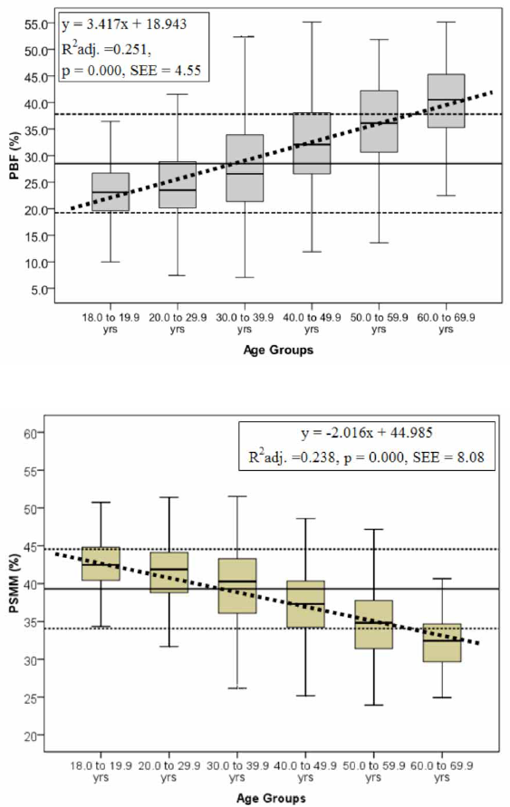
Trends of change of analysed variables for the
function of age groups are shown in Figures 1 and 2, with
defined prediction models.
Variables Percentile
PBF ( %) 2.5 5.0 10.0 25.0 50.0 75.0 90.0 95.0 97.5
All Sample 13.96 15.77 17.84 21.57 27.04 34.50 42.27 46.10 49.03
18.0-19.9 yr 13.62 14.96 16.58 19.58 23.08 26.72 31.07 35.82 45.89
20.0-29.9 yr 12.95 14.57 16.49 20.12 23.51 28.88 34.38 38.15 42.63
30.0-39.9 yr 13.74 15.06 17.81 21.34 26.56 33.89 41.46 46.15 50.34
40.0-49.9 yr 17.68 19.20 22.14 26.51 32.07 38.18 43.71 46.29 48.29
50.0-59.9 yr 22.22 23.32 26.34 30.60 36.13 42.34 47.17 49.97 50.81
60.0-69.9 yr 17.45 24.80 29.67 35.21 40.51 45.41 48.99 50.93 54.87
PSMM ( %) 2.5 5.0 10.0 25.0 50.0 75.0 90.0 95.0 97.5
All Sample 27.77 29.57 31.69 35.95 40.00 43.11 45.44 47.08 48.07
18.0-19.9 yr 30.43 35.36 37.67 40.38 42.48 44.81 46.50 47.66 48.58
20.0-29.9 yr 31.80 33.91 35.86 38.79 41.86 44.14 46.23 47.78 48.55
30.0-39.9 yr 27.39 29.83 32.24 36.08 40.27 43.27 45.17 47.30 47.96
40.0-49.9 yr 28.32 29.72 30.91 34.21 37.29 40.33 43.09 44.52 45.95
50.0-59.9 yr 26.84 27.54 28.90 31.39 34.82 37.83 39.80 41.70 43.06
60.0-69.9
y
r 25.07 26.99 27.36 29.64 32.43 34.79 38.07 39.59 42.71
Tests of Between-Subjects Effects
Source Type III Sum of Squares df Mean Square F Sig. Partial Eta
2
Observed Power
b
PBF 40398.958 5 8079.79 124.78 0.000 0.246 1.000
PSMM 13520.827 5 2704.17 131.51 0.000 0.256 1.000
Table IV. Qualitative-normative values of examined variables.
Table III. Results of percentile distribution of all variables for the function of examined age groups.
Table II. ANOVA results – differences between groups with regards to examined variables (PBF and PSMM).
RAKIC, S.; DOPSAJ, M.; DJORDJEVIC-NIKIC, M.; VASILJEVIC, N.; DOPSAJ, V.; MAKSIMOVIC, M.; TOMANIC, S. M. & MILJUS, D. Profile and reference values for body fat and
skeletal muscle mass percent at females, aged from 18.0 to 69.9, measured by multichannel segmental bioimpedance method: Serbian population study. Int. J. Morphol., 37(4):1286-1293, 2019.
18.0-19.9
y
r 20.0-29.9
y
r 30.0-39.9
y
r 40.0-49.9
y
r 50.0-59.9
y
r 60.0-69.9
y
r
PBF
(
%
)
Superior ≤ 10.2 ≤ 10.0 9.6 ≤ 15.8 ≤ 20.4 ≤ 24.1 ≤
Excellent 10.3-17.0 10.1-17.4 9.7-18.8 15.9-24.1 20.5-28.4 24.2-32.0
Above Av
g
. 17.1-20.4 17.5-21.1 18.9-23.5 24.2-28.2 28.5-32.3 32.1-35.9
Avera
g
e 20.5-27.2 21.2-28.5 23.6-32.7 28.3-36. 6 32.4-40.2 36.0-43.8
Under Avg. 27.3-30.5 28.6-32.2 32.8-37.3 36.7-40.6 40.3-44.2 43.9-47.8
Bad 30.6-37.3 32.3-39.6 37.4-46.6 40.70-48.9 44.3-52.1 47.9-55.6
Ver
y
Bad 37.4 ≥ 39.7 ≥ 46.7 ≥ 49.0 ≥ 52.2 ≥ 55.9 ≥
PSMM
(
%
)
Su
p
erior ≥ 49.9 ≥ 49.9 49.8 ≥ 46.3 ≥ 43.4 ≥ 41.0 ≥
Excellent 49.8-46.1 49.8-45.7 49.7-44.6 46.2-41.8 43.3-39.1 40.9-36.8
Above Av
g
. 46.0-44.2 45.6-43.5 44.5-42.1 41.7-39.5 39.0-36.9 36.7-34.8
Avera
g
e 44.1-40.4 43.4-39.2 42.0-36.9 39.4-34.9 36.8-32.6 34.7-30.5
Under Av
g
. 40.3-38.4 39.1-37.1 36.8-34.3 34.8-32.6 32.5-30.4 30.4-28.4
Bad 38.3-34.6 37.0-32.7 34.2-29.1 32.5-28.1 30.3-26.1 28.3-24.2
Very Bad ≤ 34.5 ≤ 32.6 29.0 ≤ 28.0 ≤ 26.0 ≤ 24.1 ≤
1290
DISCUSSION
Besides basic morphologic variables, that is, variables providing
information about absolute measurements of body composition, index
(derived) variables that give data on the relative presence of a certain
element in the body have a great practical and medically diagnostic
value (De Rosa et al., 2015; Maksimovic et al., 2016). One of the most
valid index variables are partialized in relation to the extent of body
voluminosity by which body status is evaluated, along with variables
by which fat percentage and skeletal muscle percentage are defined
(PBF and PSMM, respectively) (Ling et al.; Gába & Pridalová; Bankovic
et al.).
According to data published in the most
recent researches it has been shown that, year
after year, the level of PBF value is on the rise
among all the age categories of the population
in the area of Serbia (Boricic et al., 2014). In the
period between two researches, from 2006 and
2013, a significant degree of increase of obese
population was established (from 17.3 % to 21.2
%), especially in the age category of 45-84
(Boricic et al.). According to the results of this
research it was established (Table I) that the ave-
rage PBF value is at the level of 28.51±9.26 %
of body fat, from 23.81 % for the youngest (18.0-
19.9 yr of age) to 39.94 % for the oldest sample
(60.0-69.9 yr).
With regards to age groups, a high
statistically significant difference was found
between almost everyone (Table II, ANOVA F =
124.775, p = 0.000), in a way that apart from the
two youngest ones differing from each other
(18.0-19.9 yr and 20.0-29.9 yr) all the other
groups differ among themselves crosswise (Table
I). In other words, the percentage of body fat
among respondents was not different from 18.0
all until the age of 29.9, whereas after that age,
i.e. from 30.0 yr of age onwards, it increased in
a statistically significant way, decade after decade
all up to 69.9
yr of age. Regression analysis results
have shown that the constant of body fat
percentage increase amounted to 3.417 % per life
decade, and that the defined regression model
explains 25.1 % of mutual trend variance (Fig. 1).
However, only a 1⁄4 of the examined phenomenon
was explained by the defined equation model (PBF
= 3.417 • age group + 18.943). With the remaining
3⁄4 of the sample the PBF value change occ
urs
differently from the defined trend of 4.55 %, i.e.
either at a higher or a lower level. This only points
to the complexity of the examined phenomenon,
as well as to multidimensionality of factors that
influence body status in the sense of change in
the body fat in females during their life, and
especially after 30.0 yr of life. It was previously
found that different social, life and health factors,
such as raising a family, maternity status,
hormone status, professional-work environment
and obligations, level of physical activity, diet
habits etc. significantly influence variability fac-
tor of body composition changes among women
(Thompson et al.; Menotti et al.; Gába &
Pridalová; Sarayakin et al., 2018; Kukic et al.,
2019).
Fig. 1. PBF change trend in relation to examined age groups.
Fig. 2. PSMM change trend in relation to examined age groups
RAKIC, S.; DOPSAJ, M.; DJORDJEVIC-NIKIC, M.; VASILJEVIC, N.; DOPSAJ, V.; MAKSIMOVIC, M.; TOMANIC, S. M. & MILJUS, D. Profile and reference values for body fat and
skeletal muscle mass percent at females, aged from 18.0 to 69.9, measured by multichannel segmental bioimpedance method: Serbian population study. Int. J. Morphol., 37(4):1286-1293, 2019.
1291
In a research conducted using the same measurement
method and the same instrument, on a sample of women
from Czech Republic, an average PBF value at the level of
29.1±8.9 % was measured. Range of results in relation to
age groups was from 23.5±5.9 % for the group aged 18-29
yr, 26.3±7.2 % for the group aged 30-39 yr, 29.3±7.4 % for
the group aged 40-49 yr, 34.7±7.7 % for the group aged 50-
59 yr and 36.0±6.5 % for the group aged 60-69 yr (Gába &
Pridalová). Generally speaking, even though our results are
greater on average than the Czech by about 3.0 %, it can be
claimed that the results of both studies are still very similar
when it comes to PBF values. This only serves as proof of
the external scientific validity of our results in relation to
women from the Eastern European region. The established
difference in PBF of about 3.0 % among Serbian women
can most probably be ascribed to the fact that respondents
in this study were women 3.6 cm taller (168.3 vs 164.7 cm,
difference of 2.14 %) and 2.6 kg heavier (68.1 vs 65.5 kg,
difference 3.82 %), i.e. they were 2.98 % physically larger
than the women from the Czech research.
The next two researches performed using the same
measurement methodology and the application of the same
instrument, where some respondents were from the same
European region, i.e. Hungary (Ihász et al.), and the others
from the region of Arabian Peninsula, i.e. from the UAE,
Abu Dhabi to be precise (Kukic et al., 2019), showed that
the average PBF value among Hungarian women was at the
level of 34.32 %, and among those from UAE 40.09 %. When
compared to Serbian women, respondents from Hungary had
16.9 % more body fat on average, whereas the UAE
respondents had 28.9 % more, respectively.
These differences, i.e. the greater amount of body fat
in the sample of
women from Hungary, are a probably
consequence of numerous factors, with different diet habits
being one of the most probable. Namely, according to the data
from a recently published epidemiological study (Grasgruber
et al., 2016), it was found that differences in diet significantly
influence prevalence, and also cause the rise in incidence of
non-infectious diseases in the context of citizens from different
European countries. It is stressed in the results that a more
significant connection among women than among men was
established with regards to total fat and animal protein intake
and other indicators of fat and protein intake, where it was
found that the level of total fat consumption among Hungarian
w
omen amounts to over 130 g/day, while it is at the level of
118 and 110 g/day among women from Czech Republic and
Serbia, respectively (Grasgruber et al.). When it comes to
the UAE women, aside from those already mentioned,
differences can also be ascribed to ethnicity in the sense of
social and cultural heritage, as well as to geographical and
climate environment (Kukic et al.).
In relation to results of skeletal muscle percentage in
the body (Table I, PSMM) it was established that the avera-
ge value is at the level of 39.30±5.25 %, and that it is within
range of 42.25 % for the youngest sample (18.0-19.9 yr of
age) to 32.58 % for the oldest sample (60.0-69.9 yr). In the
research conducted using the same measurement method and
instrument, on the sample of Hungarian women (Ihász et
al.) and UAE women (Kukic et al., 2019) an average PSMM
val
ue at the level of 35.79 % was measured, i.e. 32.73 %,
which is an 8.93 %, i.e. 20.06 % smaller percentage of skeletal
muscles in the body when compared to the results of this study,
which proves the validity of the variable in relation to
international researches of the skeletal muscle amount status
among women of different nationality and race.
A high statistically significant difference was found
between almost everyone (Table II, ANOVA F = 131.506, p =
0.000), and apart from the two youngest groups differing from
each other (18.0-19.9 and 20.0-29.9) all the other older age
groups differ crosswise (Table I). As in the case of PBF,
percentage of muscle presence in the body was similar with
respondents who are 18.0 all up to 29.9 yr of age, whereas it
decreased in a statistically significant way
after that age, i.e.
after 30.0 yr of life, decade after decade, all until 69.9 yr of life.
Regression analysis results showed that the constant
of decrease in percentage of skeletal muscle in the body
amounted to -2.016 % per life decade, and also that 23.8 %
of mutual variance trend was explained by the defined
regression model (Fig. 2). As in the case of PBF, only about
1⁄4 of the examined phenomenon was explained by the
defined equation model (PSMM = -2.016 • age group +
44.985), with evaluation error of 8.08 %, while with the
remaining 3⁄4 of the sample change in the value of PSMM
occurs differently from the defined trend. As in the case of
this variable, this only points to the complexity and
multidimensionality of the examined phenomenon, in a way
that a greater number of factors influence body status in the
sense of change in the amount of skeletal muscle in the
organism among women during their life, and especially after
30.0 yr of life. Generally speaking, this variable is directly
dependent on the total body mass and the amount of SMM
(since it belongs to it) which was previously established as
gradually declining, especially after the 30
th
year. Previous
results have shown that body mass declines at the rate of
between 3 % and 8 % for each life decade after the year 30,
while the degree of muscle mass loss is increased to 5 and
up to 10 % or about 0.4 kg a year per every life decade after
the 50
th
year (Janssen et al., 2000).
When the age effect on the variable PSMM is
concerned, it was found that between the 18 and 40 year it is
not connected to the absolute muscle mass in the body,
RAKIC, S.; DOPSAJ, M.; DJORDJEVIC-NIKIC, M.; VASILJEVIC, N.; DOPSAJ, V.; MAKSIMOVIC, M.; TOMANIC, S. M. & MILJUS, D. Profile and reference values for body fat and
skeletal muscle mass percent at females, aged from 18.0 to 69.9, measured by multichannel segmental bioimpedance method: Serbian population study. Int. J. Morphol., 37(4):1286-1293, 2019.

1292
because the increase of total body mass up to the 40th year
of age occurs based on the mechanism of fat tissue increase,
regardless of sex. Only after year 50 there occurs the
beginning of absolute decrease of skeletal muscle mass, i.e.
a real decline of PSMM, as such, where the decrease in SMM
approximated 1.9 and 1.1 kg/decade in the men and women,
respectively (Janssen et al.).
Based on the results of this research it can be stated
that the average value of body fat percentage among Serbian
women is at the level of 28.51±9.26 % and also that presence
of fat tissue in the body in examined women is increased by
a trend of 3.417 % per decade. A statistically significant
increase in the percentage of body fat starts after the year
30.0, i.e. from the third decade of life. When it comes to
presence of skeletal muscles in the body, it was found that
the average value was 39.30±5.25 %, and that the presence
of skeletal muscle tissue in the body of examined women is
decreasing by a trend of -2.016 % per decade. A statistically
significant decrease in the percentage of skeletal muscles in
the body starts after the year 30.0, i.e. from the third decade
of life on. Results of this study are in agreement with the
findings of previous researches in relation to change of given
variables depending on age, while the defined standards can
for now be used from a practical aspect as well, on a national
level, and also comparatively with regards to scientific needs
of international researches.
ACKNOWLEDGEMENTS
The paper is a part of the national project III47015,
funded by the Ministry of Education, Science and
Technological Development of the Republic of Serbia -
Scientific Project 2011-2019.
RAKIC, S.; DOPSAJ, M.; DJORDJEVIC-NIKIC, M.;
VASILJEVIC, N.; DOPSAJ, V.; MAKSIMOVIC, M.;
TOMANIC, S. M. & MILJUS, D. Perfil y valores de referencia
del porcentaje de grasa corporal y masa muscular en mujeres, con
edades comprendidas entre 18,0 y 69,9 años, medido por el méto-
do de bioimpedancia segmentaria multicanal: Estudio en pobla-
ción Serbia. Int. J. Morphol., 37(4):1286-1293, 2019.
RESUMEN: El perfil y estándares para el diagnóstico del
porcentaje de grasa corporal y masa muscular fueron definidos en
una muestra de 1924 mujeres de la República de Serbia, con eda-
des comprendidas entre 18,0 y 69,9 años, donde la composición
corporal de los sujetos fue medida por bioimpedancia segmentaria
multicanal. La muestra fue dividida en seis grupos, con el propósi-
to de definir los estándares respecto a la edad. Respecto al porcen-
taje de grasa corporal los resultados han mostrado que el valor
promedio de la muestra fue de 28,51±9,26 %, y entre los rangos de
23,81 y 39,94 para los grupos de edad de 18,0-19,9 años y 60,0-
69,9 años, respectivamente. Los resultados del análisis de regre-
sión mostraron que la constante del porcentaje de grasa corporal
aumentó 3,417 % por década, y que un 25,1 % de la varianza fue
explicada por el modelo, con un error de predicción de 4,55 %.
Con respecto al porcentaje de masa muscular, los resultados han
mostrado que el valor promedio de la muestra fue de 39,30±5,25
%, y entre los rangos de 42,24 y 32,58 para los grupos de edad de
18,0-19,9 años y 60,0-69,9 años, respectivamente. Los resultados
del análisis de regresión han mostrado que la constante de masa
muscular decreció -2,016 % por década y que el modelo explicó
23,8 % de la varianza con un error de predicción de 8,08 %.
PALABRAS CLAVE: Mujeres; Perfil de composición
corporal; Bioimpedancia; Grasa corporal; Masa muscular
esquelética.
REFERENCES
Bankovic, V.; Dopsaj, M.; Terzic, Z. & Nesic, G. Descriptive body
composition profile in female olympic volleyball medalists defined
using multichannel bioimpedance measurement: Rio 2016 team case
study. Int. J. Morphol., 36(2):699-708, 2018.
Boricic, K.; Vasic, M.; Grozdanov, J.; Rakic, J.G.; Sulovic-Zivkovic, M.;
Knezevic-Jacovic, N.; Jovanovic, V.; Kilibarda, B.; Knezevic, T.; Krstic,
M.; et al., Rezultati istrazivanja zdravlja stanovnistva Srbije: 2013.
godina. Beograd, Sluzbeni glasnik, 2014.
Christie, B. Doctors revise Declaration of Helsinki. B. M. J., 321(7266):913,
2000.
De Rosa, E.; Santarpia, L.; Marra, M.; Sammarco, R.; Amato, V.; Onufrio,
M.; De Simone, G.; Contaldo, F. & Pasanisi, F. Preliminary evaluation
of the prevalence of sarcopenia in obese patients from Southern Italy.
Nutrition, 31(1):79-83, 2015.
Dopsaj, M.; Markovic, M.; Kasum, G.; Jovanovic, S.; Koropanovski, N.;
Vukovic, M. & Mudric, M. Discrimination of different body structure
indexes of elite athletes in combat sports measured by multi frequency
bioimpedance method. Int. J. Morphol., 35(1):199-207, 2017.
Esco, M. R.; Snarr, R. L.; Leatherwood, M. D.; Chamberlain, N. A.;
Redding, M. L.; Flatt, A. A.; Moon, J. R. & Williford, H. N. Comparison
of total and segmental body composition using DXA and multifrequency
bioimpedance in collegiate female athletes. J. Strength Cond. Res.,
29(4):918-25, 2015.
Gába, A. & Pridalová, M. Age-related changes in body composition in a
sample of Czech women aged 18-89 yr: a cross-sectional study. Eur. J.
Nutr., 53(1):167-76, 2014.
Grasgruber, P.; Sebera, M.; Hrazdira, E.; Hrebickova, S. & Cacek, J. Food
consumption and the actual statistics of cardiovascular diseases: an
epidemiological comparison of 42 European countries. Food Nutr. Res.,
60:31694, 2016.
Hair, J.; Anderson, R.; Tatham, R. & Black, W. Multivariate Data Analysis.
5
th
ed. New Jersey, Prentice-Hall Inc., 1998.
Hallal, P. C.; Andersen, L. B.; Bull, F. C.; Guthold, R.; Haskell, W.; Ekelund,
U. & Lancet Physical Activity Series Working Group. Global physical
activity levels: surveillance progress, pitfalls, and prospects. Lancet,
380(9838):247-57, 2012.
Haskell, W. L.; Lee, I. M.; Pate, R. R.; Powell, K. E.; Blair, S. N.; Franklin,
B. A.; Macera, C. A.; Heath, G. W.; Thompson, P. D. & Bauman, A.
Physical activity and public health: updated recommendation for adults
from the American College of Sports Medicine and the American Heart
Association. Med. Sci. Sports Exerc., 39(8):1423-34, 2007.
RAKIC, S.; DOPSAJ, M.; DJORDJEVIC-NIKIC, M.; VASILJEVIC, N.; DOPSAJ, V.; MAKSIMOVIC, M.; TOMANIC, S. M. & MILJUS, D. Profile and reference values for body fat and
skeletal muscle mass percent at females, aged from 18.0 to 69.9, measured by multichannel segmental bioimpedance method: Serbian population study. Int. J. Morphol., 37(4):1286-1293, 2019.
1293
Ihász, F.; Finn, K. J.; Lepes, J.; Halasi, S. & Szabó, P. Body composition
comparisons by age groups in Hungarian adults. Int. J. Morphol.,
33(3):850-4, 2015.
Janssen, I.; Heymsfield, S. B.; Wang, Z. M. & Ross, R. Skeletal muscle
mass and distribution in 468 men and women aged 18-88 yr. J. Appl.
Physiol. (1985), 89(1):81-8, 2000.
Kukic, F.; Scekic, A.; Koropanovski, N.; Cvorovic, A.; Dawes, J. J. &
Dopsaj, M. Age-related body composition differences in female police
officers. Int. J. Morphol., 37(1):302-7, 2019.
Ling, C. H.; de Craen, A. J.; Slagboom, P. E.; Gunn, D. A.; Stokkel, M. P.;
Westendorp, R. G. & Maier, A. B. Accuracy of direct segmental multi-
frequency bioimpedance analysis in the assessment of total body and
segmental body composition in middle-aged adult population. Clin.
Nutr., 30(5):610-5, 2011.
Maksimovic, M. Z.; Gudelj Rakic, J. M.; Vlajinac, H. D.; Vasiljevic, N. D.
& Marinkovic, J. M. Relationship between health behaviour and body
mass index in the Serbian adult population: data from National Health
Survey 2013. Int. J. Public Health, 61(1):57-68, 2016.
Menotti, A.; Puddu, P. E.; Lanti, M.; Maiani, G.; Catasta, G. & Fidanza, A.
A. Lifestyle habits and mortality from all and specific causes of death:
40-year follow-up in the Italian Rural Areas of the Seven Countries
Study. J. Nutr. Health Aging, 18(3):314-21, 2014.
Owen, N.; Sparling, P. B.; Healy, G. N.; Dunstan, D. W. & Matthews, C. E.
Sedentary behavior: emerging evidence for a new health risk. Mayo
Clin. Proc., 85(12):1138-41, 2010.
Rocha, C. A. Q. C.; Guimarães, A. C.; Maia, B. D. C.; Santos, C. A.; Moreira,
M. H. R.; Dantas, E. H. M.; Ochoa-Martínez, P. Y.; Hall-López, J. A. &
Alarcón-Meza, E. I. Effects of a 20-week concurrent training program
on bone metabolism in elderly women. Int. J. Morphol., 36(2):655-60,
2018.
Sallis, R.; Franklin, B.; Joy, L.; Ross, R.; Sabgir, D. & Stone, J. Strategies
for promoting physical activity in clinical practice. Prog. Cardiovasc.
Dis., 57(4):375-86, 2015.
Saraykin D.; Khusnutdinova A.; Pavlova V.; Kamskova Y. & Yushkov B.
Adaptation of professional athletes to various physical loads by means
of body composition changes. Hum. Sport Med., 18(3):47-59, 2018.
Thompson, D. L.; Rakow, J. & Perdue, S. M. Relationship between
accumulated walking and body composition in middle-aged women.
Med. Sci. Sports Exerc., 36(5):911-4, 2004.
Zatsiorsky, V. Sports Metrology. Moscow, Physical Education and Sports,
1982.
Corresponding author:
Sladjana Rakic
Faculty of Sport and Physical Education
University of Belgrade
Blagoja Parovica 156
Belgrade
SERBIA
Email: [email protected]
Received: 09-02-2019
Accepted: 23-05-2019
RAKIC, S.; DOPSAJ, M.; DJORDJEVIC-NIKIC, M.; VASILJEVIC, N.; DOPSAJ, V.; MAKSIMOVIC, M.; TOMANIC, S. M. & MILJUS, D. Profile and reference values for body fat and
skeletal muscle mass percent at females, aged from 18.0 to 69.9, measured by multichannel segmental bioimpedance method: Serbian population study. Int. J. Morphol., 37(4):1286-1293, 2019.
