
DOE-HDBK-1130-2007
December 2007
DOE HANDBOOK
Radiological Worker Training
U.S. Department of Energy AREA TRNG
Washington, D.C. 20585
DISTRIBUTION STATEMENT A. Approved for public release; distribution is unlimited.
TS
NOT MEASUREMENT
SENSITIVE
DOE-HDBK-1130-2007
ii
This document has been reproduced directly from the best available copy.
Available to DOE and DOE contractors from ES&H Technical Information Services, U.S.
Department of Energy, (800) 473-4375, fax: (301) 903-9823.
Available to the public from the U.S. Department of Commerce, Technology Administration,
National Technical Information Service, Springfield, VA 22161; (703) 605-6000.

DOE-HDBK-1130-2007
iii
Foreword
This Handbook describes an implementation process for core training as recommended in chapter 14 to
Implementation Guide G441.1-1B , Radiation Protection Programs for Use with Title 10, Code of
Federal Regulations, Part 835, Occupational Radiation Protection, and as outlined in the DOE standard,
Radiological Control (RCS). The Handbook is meant to assist those individuals within the Department of
Energy, Managing and Operating contractors, and Managing and Integrating contractors identified as
having responsibility for implementing core training recommended by the RCS. This training is intended
for radiological workers to assist in meeting their job-specific training requirements of 10 CFR 835.
While this Handbook addresses many requirements of 10 CFR 835 Subpart J, it must be supplemented
with facility/site-specific information to achieve full compliance.
This Handbook contains recommended training materials consistent with other DOE radiological training
materials. The training material consists of the following documents:
Program Management Guide
- This document contains detailed information on how to use the
Handbook material.
Instructor’s Guide
- This document contains a lesson plan for instructor use, including notation of
key points for inclusion of facility/site-specific information. Instructor’s notes are added
parenthetically through the text in Italic.
Student’s Guide
- This document contains student handout material and also should be augmented
by facility/site-specific information.
This Handbook was produced in Word format. Copies of this Handbook may be obtained from the DOE
Radiation Safety Training Home Page Internet site
(http://www.hss.energy.gov/radiation/RST/rstmater.htm) or the DOE Technical Standards Program
Internet site (http://www.hss.energy.gov/NuclearSafety/techstds/standard/standard.html).
Documents downloaded from the DOE Radiation Safety Training Home Page Internet site may be
manipulated using the software noted above.
DOE-HDBK-1130-2007
iv
This page intentionally left blank.

DOE-HDBK-1130-2007
Part 1 of 3
Radiological Worker Training
Program Management Guide
Coordinated and Conducted
for the
Office of Health, Safety and Security
U.S. Department of Energy
DOE-HDBK-1130-2007
ii
This page intentionally left blank.
DOE-HDBK-1130-2007
iii
Table of Contents
Page
Introduction.................................................................................................................................................1
Purpose and Scope..................................................................................................................................1
Compliance with 10 CFR 835-Subpart J................................................................................................ 1
Goal of Training Program ......................................................................................................................2
Organizational Relationships and Reporting Structure .......................................................................... 2
Training Program Descriptions.................................................................................................................2
Overview of Training Program ..............................................................................................................2
Description of Programs.........................................................................................................................3
Radiological Fundamentals....................................................................................................................3
Biological Effects...................................................................................................................................4
Radiation Dose Limits............................................................................................................................ 4
ALARA Program ...................................................................................................................................4
Personnel Monitoring Programs.............................................................................................................4
Radiological Access Controls and Postings ........................................................................................... 5
Radiological Emergencies...................................................................................................................... 5
Practical Factors for RW I......................................................................................................................5
High /Very High Radiation Area Training............................................................................................. 6
Practical Factors for High Radiation Areas............................................................................................6
Radiological Worker II...........................................................................................................................7
Radioactive Contamination Control....................................................................................................... 8
Practical Factors for RW II ....................................................................................................................8
Specialized Radiological Worker Training ............................................................................................9
Refresher Training.................................................................................................................................. 9
Proficiency Requirements ....................................................................................................................10
Retraining.............................................................................................................................................11
Instructor Training and Qualifications .................................................................................................12
Training Program Material Development.............................................................................................. 13
Training Material Presentation.............................................................................................................13
Training Certificates.............................................................................................................................13
Training Aids........................................................................................................................................14
Training Program Standards and Policies ............................................................................................14
Training Examinations ......................................................................................................................... 14
Lectures, Seminars, Training Exercises, etc......................................................................................... 16
Delinquent Training/Failure Procedures and Policies..........................................................................16
Exceptions and Waivers.......................................................................................................................17
Administration .......................................................................................................................................... 17
Training Records.................................................................................................................................. 17
Training Program Development/Change Requests .............................................................................. 17
Audits (internal and external)...............................................................................................................17
Evaluating Training Program Effectiveness.........................................................................................18
References.................................................................................................................................................. 20
DOE-HDBK-1130-2007
iv
This page intentionally left blank.
DOE-HDBK-1130-2007
Radiological Worker Training Program Management Guide
1
Introduction
Purpose and Scope
This guide describes the DOE Radiological Worker I and II (RW I and II) training
programs. It includes standards and policies as well as recommendations for material
development and program administration. It is intended for use by the DOE and DOE
contractors for the development of facility/site-specific radiological worker training.
Compliance with 10 CFR 835-Subpart J
The DOE core training materials for RW Training reflect the requirements identified in
10 CFR 835-Subpart J, “Radiation Safety Training” and recommendations identified in
chapter 14, Radiation Safety Training, of DOE Implementation Guide G441.1-1B,
Radiation Protection Programs for Use with Title 10, Code of Federal Regulations, Part
835, Occupational Radiation Protection Radiation Safety Training, and in the DOE
standard, Radiological Control. When implemented in its entirety and supplemented as
noted with appropriate facility/site-specific information, this Handbook will generally
meet the requirements of 10 CFR 835-Subpart J for radiological worker training.
However, it is incumbent on management of each facility/site to review the content of
this course against the radiological hazards present to ensure that the training content is
appropriate to each individual’s prior training, anticipated and actual assignments, and
degree of exposure to potential radiological hazards.
Training described in this Handbook does not eliminate the need for additional training
for facility/site-specific hazards. Notations throughout the program documents indicate
the need for facility/site-specific information. If the noted section is not applicable to the
DOE-HDBK-1130-2007
Radiological Worker Training Program Management Guide
2
facility/site, no information is required to be presented. The site Radiological Control
Manager or designee should concur in facility/site-generated radiological training
material.
Goal of Training Program
The goal of the core training program is to provide a high level of knowledge and skills
in radiological fundamentals for the radiological worker at all DOE facilities.
Organizational Relationships and Reporting Structure
DOE Office of Worker Safety and Health Policy (DOE HS-11) is responsible for
approving and maintaining the training materials associated with the RW I and II training
programs.
The establishment of a comprehensive and effective contractor site radiological control
training program is the responsibility of line management and their subordinates. The
training function may be performed by a separate training organization, but the
responsibility for quality and effectiveness rests with line management.
Training Program Descriptions
Overview of Training Program
Radiological Worker I Training is intended for radiological workers whose job
assignments require unescorted access to Radiological Buffer Areas, Radiation Areas, or
Radioactive Materials Areas. The RW I program consists of the core academic material
plus the appropriate practical factors evaluation and lessons learned.
The High/Very High Radiation (HR/VHR) Area module may be added to the
DOE-HDBK-1130-2007
Radiological Worker Training Program Management Guide
3
Radiological Worker I course to give personnel unescorted entry into High Radiation
Areas where contamination is not present.
Radiological Worker II Training is intended for radiological workers whose job
assignments involve unescorted entry to High Radiation Areas, Contamination Areas,
High Contamination Areas and Airborne Radioactivity Areas. Further, workers who
have potential contact with hot particles or use of gloveboxes with high contamination
levels should complete Radiological Worker II training.
The RW II program consists of the RW core academic material, the HR/VHR Area
module (this may be deleted for certain sites, such as uranium mill tailings remediation
projects, which do not have HR/VHR Areas), the Contamination Control module, the
applicable practical factors evaluation, and lessons learned.
Description of Programs
Core Academic Material is approximately 8 hours in length but will vary dependent upon
the amount of facility/site-specific material. RW Core Academic Training includes the
following modules (1-7):
Radiological Fundamentals (Module 1)
• Atomic Structure
• Definitions and Units of Measure
• The Four Basic Types of Ionizing Radiation
• Units of Measure for Radiation
DOE-HDBK-1130-2007
Radiological Worker Training Program Management Guide
4
Biological Effects (Module 2)
• Sources of Radiation
• Effects of Radiation on Cells
• Acute and Chronic Radiation Dose
• Prenatal Radiation Exposure
• Risks in Perspective
Radiation Dose Limits (Module 3)
• Basis for and Purpose of Radiation Dose Limits and
• Administrative Control Levels
• Dose Limits and Administrative Control Levels
• Worker Responsibilities Regarding Dose Limits
ALARA Program (Module 4)
• ALARA Program
• Responsibilities for the ALARA Program
• External and Internal Dose Reduction
• Radioactive Waste Minimization
Personnel Monitoring Programs (Module 5)
• External Dosimetry
• Internal Monitoring
DOE-HDBK-1130-2007
Radiological Worker Training Program Management Guide
5
• Methods for Obtaining Radiation Dose Records
Radiological Access Controls and Postings (Module 6)
• External Dosimetry
• Internal Monitoring
• Methods for Obtaining Radiation Dose Records
Radiological Emergencies (Module 7)
• Emergency Alarms and Responses
• Radiological Emergency Situations
• Considerations in Rescue and Recovery Operations
Radiological Worker I
Radiological Worker I training consists of the RW core academic material (Modules 1-7)
plus the applicable practical factors (Module 10.1).
Practical Factors for RW I (Module 10.1)
The recommended evaluation for RW I consists of the following topics:
• Review an Appropriate Radiological Work Permit (RWP)
• Record the Appropriate Information on the RWP
• Select and Wear Required Dosimeter(s)
• Enter Simulated Area and Demonstrate ALARA Techniques
• Monitor for Contamination (if RWI trained workers are be allowed unescorted
DOE-HDBK-1130-2007
Radiological Worker Training Program Management Guide
6
access into RBAs established for contamination controls this training may be
necessary per site procedures e.g., if required hand and foot monitoring on
exiting any RBA)
• as necessary per site procedures e.g., hand and foot monitoring on exiting any
RBA)
• Respond to Emergency Situations or Abnormal Radiological Situations
It may be necessary for an RW I qualified individual to enter a HR Area. If this becomes
necessary, then the HR/VHR training should be presented, along with the applicable
practical factors (Modules 10.1 and/or 10.2).
High/Very High Radiation Area Training (Module 8)
The materials for the HR/VHR Area Module include the following:
• High and Very High Radiation Area Definitions
• Signs and Postings
• Entry, Work In, and Exit from High Radiation Areas
• Access Controls for High and Very High Radiation Areas
Practical Factors for High Radiation Areas (Module 10.2)
The recommended evaluation for RW I (High Radiation Area) consists of entry, work,
and exit requirements:
• Identify High Radiation Area and Very High Radiation Area signs
• State special controls on RWP
DOE-HDBK-1130-2007
Radiological Worker Training Program Management Guide
7
• State area radiation levels (with appropriate units)
• State facility/site-specific administrative control levels
• Select dosimetry in accordance with RWP
• Wear dosimetry in accordance with procedures
• Perform pre-operational checks (as appropriate) on survey meter and/or dose rate
indicating device
• Record appropriate information on RWP prior to entry
• Verify current radiation survey prior to first entry
• Enter only areas designated on RWP
• Maximize distance from higher radiation areas
• Do not loiter
• State appropriate actions to take when a radiation area monitor alarms
• Record appropriate information on RWP upon exit
• Perform periodic checks of personnel dosimetry devices
Radiological Worker II
RW II Core Training is approximately 16 hours in length but will vary dependent on the
amount of facility/site-specific material. RW II includes the core academic material
modules (1 - 7), HR/VHR Area module (8), Contamination Control module (9), and RW
II Practical Exercise module (10.3).
DOE-HDBK-1130-2007
Radiological Worker Training Program Management Guide
8
Radioactive Contamination Control (Module 9)
The radioactive contamination control module includes the following topics:
• Comparison of Ionizing Radiation and Radioactive
Contamination
• Types of Contamination
• Sources of Radioactive Contamination
• Contamination Control Methods
• Contamination Monitoring Equipment
• Decontamination
• Types of Contamination Areas
• Lessons Learned
Practical Factors for RW II (Module 10.3)
The recommended evaluation for RW II consists of the following topics:
• Review an Appropriate Radiological Work Permit (RWP)
• Record the Appropriate Information on the RWP
• Select Required Dosimeter(s) and Protective Clothing
• Don Protective Clothing and Dosimeter(s)
• Enter Simulated Area and Demonstrate Contamination Control Practices
• Remove Protective Clothing and Dosimeter(s)
DOE-HDBK-1130-2007
Radiological Worker Training Program Management Guide
9
• Monitor for Contamination
• Respond to emergency situations or abnormal radiological situations
Specialized Radiological Worker Training
Specialized Radiological Worker Training should be completed for non-routine
operations or work in areas with changing radiological conditions. This training is in
addition to Radiological Worker II training and is required for personnel planning,
preparing, and performing jobs that have the potential for high radiological
consequences. Such jobs may involve special containment devices, the use of mockups,
and ALARA considerations. In some cases, depending on facility/site-specific criteria,
pre-job briefings provide an acceptable alternative to Specialized Radiological Worker
Training.
Individuals who install, inspect, or work in radiological containments shall be trained
commensurate with their duties. Individuals that wear respiratory protection need to be
medically qualified and wear the equipment as trained in accordance with OSHA
standards and DOE requirements. This training is in addition to Radiological Worker II
training.
Refresher Training
Refresher training programs for RW I and II training may be implemented in the alternate
year when full retraining is not completed or in response to observations or indications of
poor radiological performance. Refresher training is intended to maintain and enhance
the proficiency of the worker. The refresher training for RW I and II training should be
documented.
DOE-HDBK-1130-2007
Radiological Worker Training Program Management Guide
10
RW I and II refresher training may be accomplished through any available media. This
may include video, handout, computer- based training or classroom training.
RW I and II refresher training should include changes in requirements and lessons
learned from operations and maintenance experience, and occurrence reporting for the
site and across the DOE complex. The following topics may be included:
• New procedures and changes to existing procedures
• New equipment and changes or modifications to existing equipment or facilities
• Lessons learned from facility/site operating experiences
• Lessons learned from industry operating experiences
• Identified deficiencies from post training evaluations
Proficiency Requirements
In accordance with 10 CFR 835-Subpart J, each individual shall demonstrate knowledge
of the radiation safety training topics established in § 835-Subpart J, commensurate with
the hazards in the area and required controls, by successful completion of an examination
and performance demonstrations prior to being permitted unescorted access to
radiological areas and prior to performing unescorted assignments as a radiological
worker.
A written examination and a practical factors evaluation shall be used to demonstrate
satisfactory completion of RW I, HR/VHR Area, and RW II training (10 CFR 835 -
Subpart J). These exams may be combined into one exam if the training is presented as
one training class.
DOE-HDBK-1130-2007
Radiological Worker Training Program Management Guide
11
• The minimum passing score for any written examination should be 80%.
• A minimum passing score on the practical evaluation (i.e., the evaluation of the
practical demonstration) should be 80%.
• Computer-based and other electronic methods of examination are acceptable.
If computer-based training and examination are used, sites need to ensure that the testing
process is adequate to meet the requirement that individuals demonstrate an acceptable
baseline knowledge level of radiation protection fundamentals and practices. Programs
which allow trainee to pass the examination based on trial and error or allow unlimited
attempts without requiring retraining would be inconsistent with the requirement that
individuals demonstrate an acceptable baseline knowledge level of radiation protection
fundamentals and practices.
Retraining
In accordance with 10 CFR 835-Subpart J, RW retraining shall be provided to individuals
when there is a significant change to radiation protection policies and procedures that
may affect the individual and at intervals not to exceed 24 months. The requirements of
10 CFR 835-Subpart J for examination apply.
Retraining should include selected fundamentals of the initial training with
emphasis on seldom-used knowledge and skills. Retraining should be tailored to
subjects for which trainee evaluations and experience indicate that special
emphasis and depth of coverage is needed.
A self-study method may be used, when possible, for retraining. A suggestion for
a self-study method is to allow the workers to self study the training material;
DOE-HDBK-1130-2007
Radiological Worker Training Program Management Guide
12
present any updates or changes, lessons learned, etc.; then allow the workers to
take the examination and applicable practical exercise.
Minimum requirements for RW I and RW II retraining should be successful
completion of the written examination (10 CFR 835 required), practical exercise,
practical exercise, and training on lessons learned/new procedures.
Materials developed in support of retraining shall be documented, as necessary, in
accordance with 10 CFR 835.704 “Administrative Records.”
Instructor Training and Qualifications
All classroom instruction should be provided by instructors qualified in accordance with
the contractor’s site instructor qualification program. Training staff (contractor and
subcontractor, if used) should possess both technical knowledge and experience, and the
developmental and instructional skills required to fulfill their assigned duties.
1. Training staff responsible for program management, supervision, and
development should have and maintain the education, experience, and technical
qualifications required for their jobs.
2. Instructors should have the technical qualifications, which include adequate
theory, practical knowledge, and experience for the subject matter that they are
assigned to teach.
3. Methods should be in place at each contractor site to ensure that individual
instructors meet and maintain position qualification requirements.
4. Subject matter experts, without instructor qualification, may provide training in
their area of expertise. However, if these subject matter experts are to be
DOE-HDBK-1130-2007
Radiological Worker Training Program Management Guide
13
permanent instructors, they should be trained as instructors in the next practical
training cycle. Qualifications for trainers at nuclear facilities can be found in
DOE Order 5480.20A, “Personnel Selection, Qualification, and Training
Requirements for DOE Nuclear Facilities.”
Training Program Material Development
Training Material Presentation
Training materials for the core programs consist of lesson plans and study guides. To
ensure compliance with 10 CFR 835-Subpart J, facility/site-specific materials must be
added to the core materials when necessary to adequately train individuals for
facility/site-specific radiological hazards.
Training Certificates
A training certificate that identifies current training status of core training may be
provided to qualified personnel. Each facility/site is responsible to administer and track
the certificates. Facilities have the option of utilizing the certificates as proof of training.
However, it should be noted that 10 CFR 835-Subpart J requires each facility/site to
ensure radiological workers have adequate training for the hazards present. The training
certificate from another DOE site does not, in itself, relieve the facility/site from ensuring
the worker has had adequate training.
It is appropriate for facilities to supplement a visiting radiological worker’s training with
facility/site-specific training sufficient to ensure an adequate level of training for the
hazards present. It may also be appropriate to confirm the adequacy of the worker’s
training with a standard examination and practical evaluation.
DOE-HDBK-1130-2007
Radiological Worker Training Program Management Guide
14
Training Aids
Facility/site-specific training aids may be developed at the facility/site to suit individual
training styles. Each facility/site may add information, activities, a glossary, and/or view
graphs to enhance their program.
Training Program Standards and Policies
Training Examinations
Written examinations and/or computer-based training (CBT) examinations shall be used
to demonstrate satisfactory completion of theoretical and classroom material for RW I
and RW II. The examinations should:
• Be completed with a minimum passing grade of 80%,
• Cover material representative of the learning objectives from both core material
and facility/site-specific material,
• Be varied from class to class and within classes when the class size is large,
• Not use true/false questions,
• Not allow completion via trial and error, and
• Be acknowledged by trainee signature participation in a post-examination review.
An example core examination question bank is available from DOE HS-11. Each
question in the examination bank should be numbered in accordance with the
corresponding learning objective. All questions should consist of the multiple choice
type question.
The facility/site should develop an appropriate exam bank, and the DOE example
DOE-HDBK-1130-2007
Radiological Worker Training Program Management Guide
15
questions may be used as a basis. Example questions may be used verbatim, but the
order of answers should be changed. The DOE example exam bank is not held
confidential. The facility/site exam bank should be held confidential in accordance with
facility/site practices for exam confidentiality. The practice should ensure students do
not have knowledge of specific answer keys.
Rad Worker I Written Examination: The Rad Worker I exam is the responsibility of
each facility/site and should consist of a minimum of thirty (30) questions.
The remedial action for failure of this examination is the responsibility of each
facility/site.
HR/VHR Area Written Examinations: The HR/VHR Area exam is the responsibility
of each facility/site and should consist of a minimum of five (5) questions.
The remedial action for failure of this examination is the responsibility of each
facility/site.
Rad Worker Written II Examinations: The Rad Worker II exam is the responsibility
of each facility/site and should consist of a minimum of fifty (50) questions. The
remedial action for failure of this examination is the responsibility of each facility/site.
Initial challenge examinations may be appropriate for experienced radiological workers
and those with current qualifications at another DOE facility/site. They should be
designed to cover the core RW training core learning objectives only. Challenges should
not apply to facility/site-specific topics. Each learning objective should be represented
on the challenge examination. Failure of a challenge examination should result in the
attendance of a scheduled initial training session. Successful completion of the initial
DOE-HDBK-1130-2007
Radiological Worker Training Program Management Guide
16
challenge examination does not exempt the employee from the facility/site-specific
examination, practical factors evaluation, and training in lessons learned/new procedures.
Practical Factors Evaluation: A practical factors evaluation should be used to
demonstrate satisfactory completion skills for RW I, RW I HR/VHR Area, and RW II
training. A minimum score of 80% should be attained for each practical factor
evaluation. The criteria for a satisfactory score is outlined in the attachments to the
Instructor’s Guide. Successful completion of the written examination should be a
prerequisite for the practical evaluation.
Lectures, Seminars, Training Exercises, etc.
RW I and II core training programs are designed to be delivered in a classroom setting.
An alternate delivery method may be implemented with CBT equipment. The
presentation of radiological worker training (RWT) should include core materials and
facility/site-specific information. In all cases, regardless of the setting or delivery
method, examination requirements of 10 CFR 835-Subpart J shall be followed.
Delinquent Training/Failure Procedures and Policies
Radiological workers who are delinquent on retraining shall lose their Radiological
Worker access status until successful completion of the delinquent training requirement.
These workers shall not be allowed unescorted entry into associated radiological areas.
Currently trained radiological workers who fail a challenge or retraining exam shall lose
their training status until successful completion of the examination and practical factors
evaluation. These workers should not be allowed unescorted entry into associated
controlled/radiological areas.
DOE-HDBK-1130-2007
Radiological Worker Training Program Management Guide
17
Exceptions and Waivers
Successful completion of the core courses for RW I, RW I HR/VHR Area, and RW II
training at one DOE site may be recognized by other DOE sites. However, the
determination as to the adequacy of training as required by 10 CFR 835-Subpart J is the
responsibility of the facility/site. It may be appropriate to accept this training as the basis
for a challenge exam covering generic topics. However, this training may not adequately
cover facility/site-specific topics.
Administration
Training Records
Training records and course documentation shall meet the requirements of 10 CFR
835.704 “Administration Records” and be in accordance with local DOE Records
Disposition Schedules.
Training Program Development/Change Requests
All requests for program changes and revisions should be submitted to HS-11 using the
DOE Technical Standard Program form “Document Improvement Proposal” F 1300.3.
This form is available from the DOE Technical Standards Home Page - Maintenance of
DOE Technical Standards TSPP-09). (See the Foreword of this document for website
address).
Audits (internal and external)
Internal verification of training effectiveness may be accomplished through senior
instructor or supervisor observation of practical applications and discussions of course
material. Results shall be documented and should be maintained by the organization
DOE-HDBK-1130-2007
Radiological Worker Training Program Management Guide
18
responsible for Radiological Control Training.
The RW I, RW I HR/VHR Area, and RW II core training program materials and
processes will be evaluated on a periodic basis by DOE-HQ. The evaluation should
include a comparison of program elements with applicable industry standards and
requirements.
Evaluating Training Program Effectiveness
Verification of the effectiveness of Radiological Control training should be accomplished
by surveying a limited subset of former students in the workplace. This evaluation
should include observation of practical applications, discussion of the course material,
and may include an associated written examination. DOE/HS has issued guidelines for
evaluating the effectiveness of radiological training through the DOE Operations Offices
and DOE Field Offices.
These guidelines are included as an attachment to the Program Management Guide to
DOE Handbook, General Employee Radiological Training.
For additional guidance, refer to DOE STD 1070-94, “Guide for Evaluation of Nuclear
Facility/site Training Programs.” The guidelines contained in these documents are
relevant for the establishment and implementation of post-training evaluation and
retention testing programs.
In response to the Defense Nuclear Facilities Safety Board (DNFSB) Recommendation
91-6, DOE committed to develop an implementation plan to upgrade radiation protection
programs at DOE defense nuclear facilities.
The implementation plan detailed DOE’s plans to develop and implement radiation
DOE-HDBK-1130-2007
Radiological Worker Training Program Management Guide
19
protection post-training evaluation and retention testing programs. Post-training
evaluations will be used to identify opportunities for improving course materials,
upgrading instruction methods and techniques, and the need for additional training.
Retention testing will indicate when individual performance or testing fails to meet
expectations. Corrective actions for deficiencies identified in retention testing will be
incorporated in the individual’s development plan and the site’s training program on an
appropriate schedule.
In addition, Article 613.7 of the DOE Radiological Control Standard states that sites
should implement a training effectiveness verification program. This program, which is
in addition to performance evaluations routinely performed by the site’s training
department, is to verify the effectiveness of radiological control training by surveying a
limited subset of former students in the workplace. This recommendation applies to both
DOE defense nuclear facilities and DOE facilities not classified as defense nuclear
facilities.
Per DOE’s commitment to DNFSB, it is expected that all defense nuclear facilities will
implement these or equivalent programs. DOE facilities not classified as defense nuclear
facilities should also strive to implement such programs. Line management should
monitor progress of program implementation.
The guidance contained in DOE STD-1070-94 is not meant to be prescriptive. Training
organizations should review this guidance and determine its applicability, taking into
consideration the existence of similar programs already in place at their facility/site.

DOE-HDBK-1130-2007
Radiological Worker Training Program Management Guide
20
References (these references are for the entire Handbook 1130)
1. Cohen, Bernard L., “Catalog of Risks Extended and Updated,” Health Physics, the Radiation
Protection Journal, Vol. 61, 1991.
2. “Investigation Report C-337-A, Contamination Incident at the Paducah Gaseous Diffusion Plant on
August 23, 1991,” September 1991.
3. NCRP, “Ionizing Radiation Exposure of the Population of the United States,” Report No. 93.
4. ORAU 88/H-99, “Guide to Good Practice in Radiation Protection Training.”
5. Travis, E. L., “Primer of Medical Radiobiology,” 1989.
6. U.S. Department of Energy, “Radiation Protection Program Guide for Use with 10 CFR 835,
Occupational Radiation Protection,” 2007.
7. U.S. Department of Energy, DOE Radiological Control Standard
, 1999.
8. U.S. Department of Energy, “Occupational Radiation Protection,” 10 CFR 835, 2007.
9. U.S. Department of Energy, “Reproductive Health: Effects of Chemical and Radiation on Fertility
and the Unborn Child,” Lawrence Livermore National Laboratory, February 1, 1984.
10. U.S. Department of Energy, Order 5480.20A, Ch. 1, “Personnel Selection, Qualification, and
Training Requirements For DOE Nuclear Facilities,” November, 2001.
11. U.S. Department of Health, Education and Welfare, Radiological Health Handbook
, January 1970.
12. U.S. Nuclear Regulatory Commission, “Instruction Concerning Prenatal Radiation Exposure,” U.S.
NRC Regulatory Guide 8.13, December 1987.
13. U.S. Nuclear Regulatory Commission, “Instruction Concerning Risks From Occupational Radiation
Exposure,” U.S. NRC Regulatory Guide 8.29
, Version I, February 1997.
14. Wallace, Susan S., and Robert B. Painter, Editors., “Ionizing Radiation Damage to DNA: Molecular
Aspects,” UCLA Symposia on Molecular and Cellular Biology, New Series, Vol. 136, Wiley-Liss, Y.
1990.

DOE-HDBK-1130-2007
(Part 2 of 3)
Radiological Worker Training
Instructor’s Guide
Coordinated and Conducted
for
Office of Health, Safety and Security
U.S. Department of Energy
DOE-HDBK-1130-2007
Radiological Worker Training Instructor’s Guide
ii
This page intentionally left blank.
DOE-HDBK-1130-2007
Radiological Worker Training Instructor’s Guide
iii
Table of Contents
Page
Training Program Overview ........................................................................................................................iv
MODULE 1: RADIOLOGICAL FUNDAMENTALS................................................................................ 1
MODULE 2: BIOLOGICAL EFFECTS ...................................................................................................14
MODULE 3: RADIATION DOSE LIMITS AND ADMINISTRATIVE CONTROL LEVELS............. 28
MODULE 4: ALARA PROGRAM........................................................................................................... 36
MODULE 5: PERSONNEL MONITORING PROGRAMS.....................................................................45
MODULE 6: RADIOLOGICAL ACCESS CONTROLS AND POSTINGS ...........................................50
MODULE 7: RADIOLOGICAL EMERGENCIES ..................................................................................64
MODULE 8: HIGH/VERY HIGH RADIATION AREA TRAINING ..................................................... 69
MODULE 9: RADIOACTIVE CONTAMINATION CONTROL ............................................................76
MODULE 10.1: PRACTICAL FACTORS FOR RADIOLOGICAL WORKER I.................................... 89
MODULE 10.2: PRACTICAL FACTORS FOR HIGH RADIATION AREAS ......................................95
MODULE 10.3: PRACTICAL FACTORS FOR RADIOLOGICAL WORKER II ...............................100
ATTACHMENT 1 - Instructions for Evaluators ......................................................................................108
ATTACHMENT 2 - Sample Grading Checklist for RW II...................................................................... 117
ATTACHMENT 3 - Sample Job Scenario ...............................................................................................118
ATTACHMENT 4 - Sample Survey Map................................................................................................119
ATTACHMENT 5 - Sample Questions.................................................................................................... 120
DOE-HDBK-1130-2007
Radiological Worker Training Instructor’s Guide
iv
Training Program Overview
DOE Radiological Health and Safety (DOE P 441.1) Safety Policy.
“It is the policy of the Department of Energy to conduct its radiological operations in a manner that
ensures the health and safety of all its employees, contractors, and the general public. In achieving this
objective, the Department shall ensure that radiation exposures to its workers and the public and releases
of radioactivity to the environment are maintained below regulatory limits and deliberate efforts are
taken to further reduce exposures and releases as low as reasonably achievable. The Department is fully
committed to implementing a radiological control program of the highest quality that consistently reflects
this policy.”
In meeting this policy, the Department shall:
“Ensure personnel responsible for performing radiological work activities are appropriately trained.
Standards shall be established to ensure the technical competency of the Department’s workforce, as
appropriate, through implementation of radiological training and professional development programs.”
A. DOE Course Design
The DOE training material for radiological workers consists of four areas.
1. Core Academics (Modules 1-7)
This area includes modules 1 through 7. These modules discuss the theory that a worker should
know to work safely around radiological hazards.
The core academics are recommended for radiological workers whose job assignments limit
required unescorted access to Radiological Buffer Areas, Radiation Areas, and Radioactive
Material Areas.
2. High/Very High Radiation Area (Module 8)
This module should be added to the core academics for personnel whose job assignments require
unescorted entry into High Radiation Areas where contamination is not present or whose job
assignments require accessing High/Very High Radiation Areas.
3. Contamination Control (Module 9)
This module is recommended for workers who require unescorted access to Contamination, High
Contamination, and/or Airborne Radioactivity Areas.
4. Practical Factors Evaluations (Module 10)
This module contains generic practical exercises that provide hands-on experience for the worker.
These exercises are for the levels of training needed by different radiological workers.
B. Overview of Courses
The DOE training material can be divided into the following levels of radiological worker training:
1. Radiological Worker I (RW I) Training
This course contains the core academics and the appropriate practical factors. This training is for
radiological workers whose job assignments require access to Radiological Buffer Areas,
Radioactive Materials Areas and Radiation Areas. RW I training is also suggested for unescorted
entry into Radioactive Material Areas containing either sealed radioactive sources or radioactive
material labeled in accordance with 10 CFR 835.

DOE-HDBK-1130-2007
Radiological Worker Training Instructor’s Guide
v
RW I training alone (i.e., High/Very High Radiation Module not included) does not prepare the
worker to work around higher radiation levels or with contaminated materials. It is suggested that
RW I tasks be limited to inspections, tours, and activities that involve work on nonradiological
systems.
2. Radiological Worker I Training with High/Very High Radiation Area Training
This course contains the core academics, the High/Very High Radiation Area (HR/VHR) module,
and the appropriate practical factors. The HR/VHR Area lesson plan may be added to the RW I
course to give personnel unescorted entry into High Radiation Areas where contamination is not a
concern.
3. Radiological Worker II (RW II) Training
This course consists of the core academics, the High/Very High Radiation Area module, the
Contamination Control module, and the appropriate practical factors. This training is
recommended for the radiological worker whose job assignments involve unescorted entry into
High Radiation Areas, Contamination Areas, High Contamination Areas, and Airborne
Radioactivity Areas. Further, workers who have potential contact with hot particles or use
gloveboxes with high contamination levels should complete RW II training.
RW II training prepares the worker to work around higher radiation levels and with contaminated
materials normally associated with radiological facilities/activities.
C. Evaluation Criteria
At the completion of the applicable course, the participant must successfully complete a written exam
and
a practical evaluation to be considered to have successfully completed the training. Successful
completion of the written exam should be a prerequisite for the practical factors evaluation.
1. Written Examination
Successful completion of the written examination typically requires a minimum passing score of
80 percent or equivalent. The written exam is based on the objectives in the theory portion of the
course (Modules 1-7).
2. Practical Factors Evaluation
Successful completion of the practical factors evaluation typically requires a minimum score of 80
percent or equivalent. The practical factors evaluation includes entry into a simulated controlled
work environment. This evaluation is based on the application of the theory portion of the
applicable course (Modules 1-7).
D. Documentation of Training
(Insert facility/site-specific information.)
E. Periodic Training and Refresher Training
1. Training
Training is required at intervals not to exceed every 24 months.
2. Refresher Training
Refresher training should be conducted in the off year when periodic training is not due.

DOE-HDBK-1130-2007
Radiological Worker Training Instructor’s Guide
vi
Figure 1
Three Levels of Radiological Worker Training with Associated Training Requirements
Training Modules 1-7
and
Practical Factors 10.1
Radiological Worker I
Radiological Worker I with HRA/VHRA
Radiological Worker II
Training Modules 1-8
and
Practical Factors 10.1
and 10.2
Training Modules 1-9
and
Practical Factors 10.3
or 10.2

DOE-HDBK-1130-2007
Radiological Worker Training Instructor’s Guide
vii
Figure 2
Evaluation Overview Diagram
Pass Written Exam??
(80%)
Pass Practical Evaluation??
(80%)
RWT
Good to Go!!
(Insert Site-Specific
Failure Policy.)
(Insert Site-Specific
Failure Policy.)
Yes
Yes
No
No
Pass Written Exam??
> 80%
Pass Practical
Evaluation??
> 80%
DOE-HDBK-1130-2007
Radiological Worker Training Instructor’s Guide
xii
This page intentionally left blank.
DOE-HDBK-1130-2007
Radiological Worker Training Instructor’s Guide
Module 1: Radiological Fundamentals
1
Module 1: Radiological Fundamentals
Terminal Objective:
Given various radiological concepts, the participant will be able to define the fundamentals of
radiation, radioactive material, and radioactive contamination in accordance with the approved lesson
materials.
Enabling Objectives:
The participant will be able to select the correct response from a group of responses to verify his/her
ability to:
EO1 Identify the three basic particles of an atom.
EO2 Define radioactive material, radioactivity, radioactive half-life, and radioactive contamination.
EO3 Identify the units used to measure radioactivity and contamination.
EO4 Define ionization and ionizing radiation.
EO5 Distinguish between ionizing radiation and non-ionizing radiation.
EO6 Identify the four basic types of ionizing radiation and the following for each type:
a. Physical characteristics
b. Range
c. Shielding
d. Biological hazard(s)
e. Sources at the site
EO7 Identify the units used to measure radiation.
EO8 Convert rem to millirem and millirem to rem.
Instructional Aids:
1. Student Guide
2. Transparencies
3. Activities (as applicable)
4. Self-check quizzes (as applicable)
DOE-HDBK-1130-2007
Radiological Worker Training Instructor’s Guide
Module 1: Radiological Fundamentals
2
II. MODULE INTRODUCTION
A. Self Introduction
1. Name
2. Phone Number
3. Background (Post information in room. Have students introduce themselves: name,
background, etc.)
B. Module Overview
Nuclear science is truly a product of the 20th century. This module will discuss several
nuclear science topics at a basic level appropriate for the radiological worker. These concepts
are necessary for the worker to understand the nature of radiation and its potential effect on
health. The topics covered include basic particles of the atom, types of radiation, and the
definition of units used to measure radiation.
C. Objectives Review
D. Introduction
This module introduces the worker to basic radiological fundamentals and terms that are
common in the DOE complex. After learning the fundamentals of radiation, radioactive
material, and radioactive contamination, the worker will build from the basic to the more in-
depth concepts presented in the other modules.
DOE-HDBK-1130-2007
Radiological Worker Training Instructor’s Guide
Module 1: Radiological Fundamentals
3
II. MODULE OUTLINE
A. Atomic Structure
1. The basic unit of matter is the atom. The three basic particles of the atom are protons, neutrons,
and electrons. The central portion of the atom is the nucleus. The nucleus consists of protons
and neutrons. Electrons orbit the nucleus. (EO1 Identify the three basic particles of an atom)
a. Protons
1) Protons are located in the nucleus of the atom.
2) Protons have a positive electrical charge.
3) The number of protons in the nucleus determines the element. (Optional - Insert
diagram of the atom. Have students label the three basic particles)
b. Neutrons
1) Neutrons are located in the nucleus of the atom.
2) Neutrons have no electrical charge.
3) Atoms of the same element have the same number of protons, but can have a
different number of neutrons.
4) Atoms which have the same number of protons but different numbers of
neutrons are called isotopes.
NOTE: Common notation for describing isotopes is to list the atomic symbol for
an element followed by its mass number. The mass number is the sum of
protons and neutrons. For example, tritium has 1 proton and 2 neutrons, and is
denoted as H-3.
5) Isotopes have the same chemical properties; however, the nuclear properties can
be quite different.
c. Electrons
1) Electrons are in orbit around the nucleus of an atom.
2) Electrons have a negative electrical charge.
3) This negative charge is equal in magnitude to the proton’s positive charge.

DOE-HDBK-1130-2007
Radiological Worker Training Instructor’s Guide
Module 1: Radiological Fundamentals
4
Basic Particles
3 Basic
Particles
Location
Charge
Comments
Protons
Nucleus
+
(positive)
Number of protons determines
the element. If the number of
protons changes, the element
changes.
Neutrons
Nucleus
No Charge
Atoms of the same element have
the same number of protons, but
can have a different number of
neutrons. This is called an
isotope.
Electrons
Orbit
nucleus
- (negative)
This negative charge is equal in
magnitude to the proton’s
positive charge.
2. Stable and unstable atoms
Only certain combinations of neutrons and protons result in stable atoms.
a. If there are too many or too few neutrons for a given number of protons, the nucleus
will not be stable.
b. The unstable atom will try to become stable by giving off excess energy. This
energy is in the form of particles or rays (radiation). These unstable atoms are
known as radioactive atoms.
3. Charge of the atom
The number of electrons and protons determines the overall electrical charge of the atom.
The term “ion” is used to define atoms or groups of atoms that have a net positive or
negative electrical charge. (Optional - Insert diagram that illustrates the different
charges)
a. No charge (neutral)
If the number of electrons equals the number of protons, the atom is electrically
neutral. This atom does not have a net electrical charge.
b. Positive charge (+)
If there are more protons than electrons, the atom is positively charged.
c. Negative charge (-)
If there are more electrons than protons, the atom is negatively charged.
DOE-HDBK-1130-2007
Radiological Worker Training Instructor’s Guide
Module 1: Radiological Fundamentals
5
B. Definitions and Units of Measure
1. Radioactive material (EO2 Define radioactive material)
Radioactive material is any material containing unstable atoms that emit radiation.
Radiation means ionizing radiation: alpha particles, beta particles, gamma rays, X-rays,
neutrons, high-speed electrons, high-speed protons, and other particles capable of
producing ions. Radiation, as used in this part, does not include non-ionizing radiation,
such as radio waves or microwaves, or visible, infrared, or ultraviolet light. (Give
facility/site-specific examples of radioactive isotopes at the site.)
2. Radioactivity (EO2 Define radioactivity)
Radioactivity is the process of unstable (or radioactive) atoms becoming stable. This is
done by emitting radiation. This process over a period of time is referred to as
radioactive decay. A disintegration is a single atom undergoing radioactive decay. (Give
examples of radioactive decay.)
3. Radioactivity units (EO3 Identify the units used to measure radioactivity)
Radioactivity is measured in the number of disintegrations radioactive material undergoes
in a certain period of time.
a. Disintegrations per minute (dpm)
b. Disintegrations per second (dps)
c. Curie (Ci)
One curie equals:
• 2,200,000,000,000 disintegrations per minute (2.2x10
12
dpm), or
• 37,000,000,000 disintegrations per second (3.7x10
10
dps), or
• 1,000,000 microcuries (1x10
6
µCi).
4. Radioactive half-life (EO2Define radioactive half-life)
Radioactive half-life is the time it takes for one half of the radioactive atoms present to
decay.
5. Radioactive contamination (EO2Define radioactive contamination)
Radioactive contamination is radioactive material that is uncontained and in an unwanted
place. (There are certain places where radioactive material is intended to be.)
Contamination is measured per unit area or volume. (EO3 Identify the units used to
measure contamination)
· dpm/100 cm
2
· µCi/ml.
6. Ionization (EO4 Define ionization)
Ionization is the process of removing electrons from neutral atoms.
DOE-HDBK-1130-2007
Radiological Worker Training Instructor’s Guide
Module 1: Radiological Fundamentals
6
a. Electrons will be removed from an atom if enough energy is supplied. The
remaining atom has a positive (+) charge. The ionized atoms may affect chemical
processes in cells. The ionizations may affect the cell’s ability to function normally.
b. The positively charged atom and the negatively charged electron are called an “ion
pair.”
c. Ionization should not be confused with radiation. Ions (or ion pairs) produced as a
result of the interaction of radiation with an atom allow the detection of radiation.
7. Ionizing radiation (EO4 Define ionizing radiation)
Ionizing radiation is energy (particles or rays) emitted from radioactive atoms, and some
devices, that can cause ionization. Examples of devices that emit ionizing radiation are
X-ray machines, accelerators, and fluoroscopes.
a. It is important to note that exposure to ionizing radiation, without exposure to
radioactive material, will not result in contamination of the worker.
b. Radiation is a type of energy, and contamination is radioactive material that is
uncontained and in an unwanted place.
8. Non-ionizing radiation (EO5 Distinguish between ionizing radiation and non-ionizing
radiation)
a. Electromagnetic radiation that doesn’t have enough energy to ionize an atom is
called “non-ionizing radiation.”
b. Examples of non-ionizing radiation are radar waves, microwaves, and visible light.
C. The Four Basic Types of Ionizing Radiation
The four basic types of ionizing radiation of concern in the DOE complex are alpha
particles, beta particles, gamma or X rays, and neutrons. (EO6 Identify the four basic
types of ionizing radiation and the following for each type:
a. Physical characteristics
b. Range
c. Shielding
d. Biological hazard(s)
e. Sources at the site)
1. Alpha particles
a. Physical characteristics
1) The alpha particle has a large mass and consists of two protons, two neutrons,
and no electrons.
2) It is a highly charged particle (charge of plus two) that is emitted from the
nucleus of an atom.

DOE-HDBK-1130-2007
Radiological Worker Training Instructor’s Guide
Module 1: Radiological Fundamentals
7
3) The positive charge causes the alpha particle (+) to strip electrons (-) from
nearby atoms as it passes through the material, thus ionizing these atoms.
b. Range
1) The alpha particle deposits a large amount of energy in a short distance of travel.
2) This large energy deposit limits the penetrating ability of the alpha particle to a
very short distance.
3) Range in air is about 1-2 inches.
c. Shielding
Most alpha particles are stopped by a few centimeters of air, a sheet of paper, or the
dead layer (outer layer) of skin.
d. Biological hazards
1) Alpha particles are not considered an external radiation hazard. This is because
they are easily stopped by the dead layer of skin.
2) Internally, the source of the alpha radiation is in close contact with body tissue
and can deposit large amounts of energy in a small volume of living body tissue.
e. Sources
(Insert facility/site-specific information.)
Table 1-2
Alpha Particles
Physical
Characteristics
· Large mass (2 protons, 2 neutrons, 0 electrons).
· +2 charge.
Range
· Very short (about 1-2 inches in air).
· Deposits large amount of energy in a short
distance of travel.
Shielding
· Few centimeters of air.
· Sheet of paper.
· Dead layer of skin (outer layer).
Biological
Hazards
· No external hazard (dead layer of skin will stop
alpha particles).
· Internally, the source of alpha radiation is in close
contact with body tissue. It can deposit large amounts
of energy in a small amount of body tissue.
Sources
Insert facility/site-specific information.
DOE-HDBK-1130-2007
Radiological Worker Training Instructor’s Guide
Module 1: Radiological Fundamentals
8
2. Beta particles
a. Physical characteristics
1) The beta particle has a small mass and is positively or negatively charged.
Positively charged beta particles are called positrons and have an electrical charge of
plus one. Negatively charged beta particles are high-energy electrons and have an
electrical charge of minus one.
2) A negatively charged beta particle is physically identical to an electron.
3) The beta particle ionizes target atoms due to the force between itself and the
electrons of the atom. Both have a charge of minus one.
b. Range
1) Because of its charge, the beta particle has a limited penetrating ability.
2) The range in air of beta particles depends on the energy of the beta particle. In the
case of tritium (H-3), the range is only an inch; in the case of phosphorous-32 (P-32)
or strontium-90 (Sr-90), the range is 20 feet in air.
c. Shielding
Beta particles are typically shielded by plastic, glass, or safety glasses.
d. Biological hazards
1) If ingested or inhaled, a beta emitter can be an internal hazard when the source of the
beta radiation is in close contact with body tissue and can deposit energy in a small
volume of living body tissue.
2) Externally, beta particles are potentially hazardous to the skin and eyes.
3) Provide facility/site-specific information on the additional risks or concerns from
high-energy beta sources (e.g., P-32, Y-90), as appropriate.
e. Sources
(Insert facility/site-specific information.)

DOE-HDBK-1130-2007
Radiological Worker Training Instructor’s Guide
Module 1: Radiological Fundamentals
9
Table 1-3
Beta Particles
Physical
Characteristics
· Small mass.
· -1 charge or + 1 charge.
Range
· Short distance (one inch to 20 feet).
Shielding
· Plastic.
· Glass.
· Safety glasses.
Biological
Hazard
· Internal hazard (this is due to short range).
· Externally, may be hazardous to skin and eyes.
Sources
Insert facility/site-specific information.
3. Gamma rays/X rays
a. Physical characteristics
1) Gamma/X-ray radiation is an electromagnetic wave (electromagnetic radiation) or
photon and has no mass and no electrical charge.
2) Gamma rays are very similar to X rays. The difference between gamma rays and X
rays is that gamma rays originate inside the nucleus and X rays originate in the
electron orbits outside the nucleus.
3) Gamma/X-ray radiation can ionize as a result of direct interactions with orbital
electrons.
b. Range
1) Because gamma/X-ray radiation has no charge and no mass, it has very high
penetrating ability.
2) The range in air is very far. It will easily go several hundred feet.
c. Shielding
Gamma/X-ray radiation is best shielded by very dense materials, such as lead. Water or
concrete, although not as effective as the same thickness as lead, are also commonly
used, especially if the thickness of shielding is not limiting.
d. Biological hazards
Gamma/X-ray radiation can result in radiation exposure to the whole body.

DOE-HDBK-1130-2007
Radiological Worker Training Instructor’s Guide
Module 1: Radiological Fundamentals
10
e. Sources
(Insert facility/site-specific information.)
Table 1-4
Gamma Rays/X-Rays
Physical
Characteristics
· No mass.
· No charge.
· Electromagnetic wave or photon.
· Similar (difference is the place of origin).
Range
· Range in air is very far.
· It will easily go several hundred feet.
· Very high penetrating power since it has no
mass and no charge.
Shielding
· Concrete.
· Water.
· Lead.
Biological
Hazard
· Whole body exposure.
· The hazard may be external and/or internal.
This depends on whether the source is
inside or outside the body.
Sources
Insert facility/site-specific information.
4. Neutrons
a. Physical characteristics
1) Neutron radiation consists of neutrons that are ejected from the nucleus.
2) A neutron has mass, but no electrical charge.
3) An interaction can occur as the result of a collision between a neutron and a nucleus.
The nucleus recoils due to the energy imparted by the neutron and ionizes other
atoms. This is called “secondary ionization.”
4) Neutrons may also be absorbed by a nucleus. This is called neutron activation. A
charged particle or gamma ray may be emitted as a result of this interaction. The
emitted radiation can cause ionization in other atoms.
b. Range
1) Because of the lack of a charge, neutrons have a relatively high penetrating ability
and are difficult to stop.
DOE-HDBK-1130-2007
Radiological Worker Training Instructor’s Guide
Module 1: Radiological Fundamentals
11
2) The range in air is very far. Like gamma rays, they can easily travel several hundred
feet in air.
c. Shielding
Neutron radiation is best shielded by materials with a high hydrogen content such as
water, concrete, or plastic.
d. Biological hazards
Neutrons are a whole body hazard due to their high penetrating ability.
e. Sources
(Insert facility/site-specific information.)
Table 1-5
Neutrons
Physical
Characteristics
· No charge.
· Has mass.
Range
· Range in air is very far.
· Easily can go several hundred feet.
· High penetrating power due to lack of charge
(difficult to stop).
Shielding
· Water.
· Concrete.
· Plastic (high hydrogen content).
Biological
Hazard
· Whole body exposure.
· The hazard is generally external.
Sources
Insert facility/site-specific information.
D. Units of Measure for Radiation (EO7 Identify the units used to measure radiation)
1. Roentgen (R)
a. Is a unit for measuring external exposure. (Absorbed dose results from energy being
deposited by the radiation)
b. Defined only for effect on air.
c. Applies only to gamma and X rays.
d. Does not relate biological effects of radiation to the human body.
e. 1 R (Roentgen) = 1000 milliroentgen (mR).
DOE-HDBK-1130-2007
Radiological Worker Training Instructor’s Guide
Module 1: Radiological Fundamentals
12
2. Rad (Radiation absorbed dose)
a. A unit for measuring absorbed dose in any material.
b. Is defined for any material.
c. Applies to all types of radiation.
d. Does not take into account the potential effect that different types of radiation have on the
body.
e. 1 rad = 1000 millirad (mrad).
3. Rem (Roentgen equivalent man)
a. A unit for measuring equivalent dose.
b. Is the most commonly used unit.
c. Pertains to the human body.
d. Equivalent dose takes into account the energy absorbed (dose) and the biological effect
on the body due to the different types of radiation.
The Radiation Weighting Factor (RWF) is used as a multiplier to reflect the relative
amount of biological damage caused by the same amount of energy deposited in cells
by the different types of ionizing radiation. Alpha radiation ionizes a lot of atoms in a
very short distance and, for the same amount of energy deposited as beta or gamma
radiation, is more damaging. Rem = rad x RWF.
Note: Prior to 2007, when DOE updated its dosimetric models and terminology, DOE
used a Quality Factor (QF). The quality factor was applied to the absorbed dose at a
point in order to take into account the differences in the effects of different types of
radiation. Now, for radiological protection purposes, the absorbed dose is averaged
over an organ or tissue and this absorbed average dose is weighted for the radiation
quality in terms of the radiation weighting factor.
Radiation Weighting Factors:
alpha = 20
beta = 1
gamma/x-ray = 1
neutron = 5-20(depending on the energy)
e. 1 rem = 1,000 millirem (mrem). (EO8 Convert rem to millirem and millirem to rem)
4. Radiation dose and dose rate
a. Radiation dose rate is the dose per time.

DOE-HDBK-1130-2007
Radiological Worker Training Instructor’s Guide
Module 1: Radiological Fundamentals
13
b. Example:
1) Radiation dose rate = dose/time.
2) Radiation equivalent dose rate = mrem/hr.
3) Radiation absorbed dose rate = mrad/hr.
Table 1-6
Radiation Units
Roentgen (R)
Rad
(Radiation Absorbed
Dose)
Rem
(Roentgen Equivalent
Man)
Unit for
measuring
exposure.
Unit for measuring
absorbed dose in any
material.
Unit for measuring dose
equivalence (most
commonly used unit).
Defined only for
effect on air.
Defined for any material.
Pertains to human body.
Applies only to
gamma and X-
ray radiation.
Applies to all types of
radiation.
Applies to all types of
radiation.
Does not relate
biological
effects of
radiation to the
human body.
Does not take into
account the potential
effect that different types
of radiation have on the
body.
Takes into account the
energy absorbed (dose)
and the biological effect
on the body due to the
different types of
radiation.
Equal doses of different
types of radiation (as
measured in rad) can
cause different levels of
damage to the body
(measured in rem).
III. SUMMARY
(Insert facility/site-specific information.)
IV. EVALUATION
(Refer to RWT Program Manual Guide for evaluation guidance)
(Insert facility/site-specific information.)
DOE-HDBK-1130-2007
Radiological Worker Training Instructor’s Guide
Module 2: Biological Effects
14
Module 2: Biological Effects
Terminal Objective:
Given various radiation doses and sources of radiation, identify natural and manmade sources of
radiation and the biological risks associated with radiation dose in accordance with lesson materials.
Enabling Objectives:
The participant will be able to select the correct response from a group of responses to verify his/her
ability to:
EO1 Identify the major sources of natural background and manmade radiation.
EO2 Identify the average annual dose to the general population from natural background and
manmade sources of radiation.
EO3 State the method by which radiation causes damage to cells.
EO4 Identify the possible effects of radiation on cells.
EO5 Define the terms “acute dose” and “chronic dose.”
EO6 State examples of chronic radiation dose.
EO7 Define the terms “somatic effect” and “heritable effect.”
EO8 State the potential effects associated with prenatal radiation dose.
EO9 Compare the biological risks from chronic radiation doses to health risks workers are
subjected to in industry and daily life.
Instructional Aids:
1. Student Guide
2. Transparencies
3. Activities (as applicable)
4. Self-check quizzes (as applicable)
DOE-HDBK-1130-2007
Radiological Worker Training Instructor’s Guide
Module 2: Biological Effects
15
I. MODULE INTRODUCTION
A. Self Introduction
1. Name
2. Phone Number
3. Background
B. Module Overview
The fact that ionizing radiation produces biological damage has been known for many years.
We have gained most of our knowledge of these effects since World War II.
In this module, we will discuss the potential for biological effects and risks due to ionizing
radiation and put these potential risks into perspective when compared to other occupations
and daily activities. With this information, it is hoped that employees will develop a healthy
respect for radiation rather than fear or disregard.
C. Objectives Review
D. Introduction
We know more about the biological effects of ionizing radiation than most other
environmental factors. Rather than just being able to base our information on animal studies,
we have a large body of information available regarding exposures to humans. There are four
major groups of people that have been exposed to significant levels of radiation.
The first group includes early radiation workers, such as radiologists. These workers received
large doses of radiation before the biological effects were recognized. Since that time,
standards have been developed to protect workers.
The second group is the more than 250,000 survivors of the atomic bombs dropped at
Hiroshima and Nagasaki. Some of these survivors received doses estimated to be in excess of
50,000 mrem.
The third group includes individuals who have been involved in radiation accidents.
The fourth and largest group of individuals are patients who have undergone radiation therapy
for cancer and other diseases.
DOE-HDBK-1130-2007
Radiological Worker Training Instructor’s Guide
Module 2: Biological Effects
16
II. MODULE OUTLINE
A. Sources of Radiation (EO1 Identify the major sources of natural background and manmade
radiation)
We live in a radioactive world and always have. In fact, the majority of us will be exposed to
more ionizing radiation from natural background radiation than from our jobs.
1. Natural sources
There are several sources of radiation that occur naturally. The radiation emitted from
these sources is identical to the radiation that results from manmade sources.
The four major sources of naturally occurring radiation exposures are:
· Cosmic radiation
· Sources in the earth’s crust, also referred to as terrestrial radiation
· Sources in the human body, also referred to as internal sources
· Radon
a. Cosmic radiation (total average dose ~ 28 mrem/yr)
1) Cosmic radiation comes from the sun and outer space. It consists of positively
charged particles and gamma radiation.
2) At sea level, the average annual cosmic radiation dose is about 26 mrem.
3) At higher elevations, the amount of atmosphere shielding cosmic rays decreases;
therefore, the dose increases.
b. Sources in earth’s crust (terrestrial) (total average dose ~ 28 mrem/yr)
There are natural sources of radiation in the ground (i.e., rocks and soil).
1) Some of the contributors to terrestrial sources are the natural radioactive
elements radium, uranium, and thorium.
2) Many areas have elevated levels of terrestrial radiation due to increased
concentrations of uranium or thorium in the soil.
c. Internal (total average dose ~40 mrem/yr)
1) The food we eat and the water we drink contain trace amounts of natural
radioactive materials.
DOE-HDBK-1130-2007
Radiological Worker Training Instructor’s Guide
Module 2: Biological Effects
17
2) These naturally occurring radioactive materials deposit in our bodies and cause
internal exposure to radiation.
3) Some naturally occurring radioactive isotopes include Sodium-24 (Na-24),
Carbon-14 (C-14), Argon-41 (Ar-41), and Potassium-40 (K-40). Most of our
internal exposure comes from K-40.
d. Radon (total average dose ~ 200 mrem/yr)
1) Radon comes from the radioactive decay of uranium, which is naturally present
in the soil.
2) Radon is a gas. It can travel through the soil and enter through building
foundation cracks. The greatest concentrations of indoor radon are found in
basements.
3) Radon emits alpha radiation. It presents a hazard only when taken into the body
(e.g., when inhaled). (Review characteristics of alpha radiation)
2. Manmade sources (EO1 Identify the major sources of natural background and manmade
radiation)
The difference between manmade sources of radiation and naturally occurring sources is
the origin of the source, i.e., where the radiation is either produced or enhanced by human
activities.
The four top sources of manmade radiation exposures are:
· Tobacco products (cigarettes, cigars etc. although the tobacco is not manmade - the
products are)
· Medical radiation
· Building materials
· Domestic water supply (radon)
a. Tobacco products (average dose ~1300 mrem/yr)
b. Medical radiation sources (total average dose ~ 54 mrem/yr)
1) X rays (total average dose ~ 40mrem/yr)
a) X rays are similar to gamma rays; however, they originate outside the
nucleus.
b) A typical radiation dose from a chest X ray is about 10 mrem.
2) Diagnosis and therapy (total average dose ~14 mrem/yr)
DOE-HDBK-1130-2007
Radiological Worker Training Instructor’s Guide
Module 2: Biological Effects
18
In addition to X rays, radioactive materials and radioactive sources are used in
medicine for diagnosis and therapy.
c. Building materials (total average dose ~7 mrem/yr)
d. Domestic water supply (radon) (total average dose ~5 mrem/yr)
e. Other minor contributors
Other contributors to dose include consumer products, industrial sources, and
atmospheric testing of nuclear weapons. (Discuss: It has been more than 20 years
since atmospheric testing has been conducted)
3. Average annual dose (EO2 Identify the average annual dose to the general population
from natural background and manmade sources of radiation)
The average annual total effective dose to the general population (non-smokers) from
naturally occurring and manmade sources is about 360 mrem.
B. Effects of Radiation on Cells
The human body is made up of many organ systems. Each system is made up of tissues.
Specialized cells make up tissues. Ionizing radiation can potentially affect the normal
function of cells.
1. Biological effects begin with the ionization of atoms (EO3 State the method by which
radiation causes damage to cells)
a. The method by which radiation causes damage to human cells is by ionization of
atoms in the cells. It may also cause excitation. Excitation is where the radiation
deposits energy into an atom raising its energy level but not enough energy to eject a
bound electron. Atoms make up the cells that make up the tissues of the body. Any
potential radiation damage begins with damage to atoms.
b. A cell is made up of two principal parts, the body of the cell and the nucleus. The
nucleus is like the brain of the cell.
c. When ionizing radiation hits a cell, it may strike a vital part of the cell like the
nucleus or a less vital part of the cell, like the cytoplasm.
2. Cell sensitivity
Some cells are more sensitive than others to environmental factors such as viruses,
toxins, and ionizing radiation.
a. Actively dividing and non-specialized cells
DOE-HDBK-1130-2007
Radiological Worker Training Instructor’s Guide
Module 2: Biological Effects
19
1) Cells in our bodies that are actively dividing are more sensitive to ionizing
radiation.
2) Cells that are rapidly dividing include blood-forming cells, the cells that line our
intestinal tract, hair follicles, and cells that form sperm.
b. Less actively dividing and more specialized cells
Cells that divide at a slower rate or are more specialized (such as brain cells or
muscle cells) are not as sensitive to damage by ionizing radiation.
3. Possible effects of radiation on cells (EO4 Identify the possible effects of radiation on
cells)
Several things can happen when a cell is exposed to ionizing radiation. The following are
possible effects of radiation on cells.
a. There is no damage
b. Cells are damaged but are able to repair the damage and operate normally
1) The body of most cells is made up primarily of water. When ionizing radiation
hits a cell, it is most likely to interact with the water in the cell. One of the
byproducts of radiation-induced ionization of water is hydrogen peroxide.
Hydrogen peroxide can damage cell atomic structures.
2) Ionizing radiation can also hit the nucleus of the cell. The nucleus contains the
vital parts of the cell, such as chromosomes. The chromosomes determine cell
function. When chromosomes duplicate themselves, the chromosomes transfer
their information to new cells. Radiation may cause a change in the
chromosome that does not affect the cell.
3) Damage to chromosomes and other cell structures can be repaired. In fact, our
bodies repair a very large number of chromosome breaks every day (References
7 and 10).
3. Average annual dose
The average annual total effective dose to the general population from naturally occurring
and manmade sources is about 360 mrem.
c. Cells are damaged and operate abnormally

DOE-HDBK-1130-2007
Radiological Worker Training Instructor’s Guide
Module 2: Biological Effects
20
1) Cell damage may not be repaired or may be incompletely repaired. In that case,
the cell may not be able to function properly.
2) It is possible that a chromosome in the cell nucleus could be damaged but not be
repaired correctly. If the cell continues to reproduce, this is called a mutation
and may result in cancer.
d. Cells die as a result of the damage
At any given moment, thousands of our cells die and are replaced by normal
functioning cells. However, the radiation damage to a cell may be so extensive that
the cell dies prematurely.
C. Acute and Chronic Radiation Dose (EO5 Define the terms “acute dose” and “chronic dose”)
Potential biological effects depend on how much and how fast a radiation dose is received.
Radiation doses can be grouped into two categories: acute and chronic dose.
1. Acute radiation doses
a. High doses of radiation received in a short period of time are called acute doses. The
body’s cell repair mechanisms are not as effective for damage caused by an acute
dose.
b. Acute doses to the whole body
After an acute dose, damaged cells may be replaced by new cells and the body may
repair itself, although this may take a number of months. Only in extreme cases,
such as with the Chernobyl firefighters (500 rem), would the dose be so high as to
make recovery unlikely.
c. Acute doses to only part of the body
1) X-ray machines
It is possible that radiation exposure may be limited to a part of the body, such as
the hands.
There have been accidents, particularly with X-ray machines, in which
individuals have exposed their fingers to part of the intense radiation beam. In
some of these cases, individuals have received doses of millions
of mrem to their
fingers, and some individuals have lost their finger or fingers. It is important for
individuals who work with X-ray or similar equipment to be trained in the safe
use of this equipment.
DOE-HDBK-1130-2007
Radiological Worker Training Instructor’s Guide
Module 2: Biological Effects
21
2) Radiation therapy (Reference 5)
a) Radiation therapy patients receive high doses of radiation in a short period
of time, but generally only to a small portion of the body (not a whole body
dose).
b) The skin and limited tissue of these patients may receive significant doses,
but doses to the region of a tumor are many times higher.
c) Ionizing radiation is used to treat cancer in these patients because cancer
cells are rapidly dividing and therefore sensitive to ionizing radiation. Some
of the side effects of people undergoing radiation therapy are hair loss,
nausea, and tiredness.
d. Probability of a large acute dose
What is important to understand is that it takes a large acute dose of radiation before
any physical effect is seen. These acute doses have occurred in Hiroshima/Nagasaki,
and in a few radiation accidents, including Chernobyl. The possibility of a
radiological worker receiving a large acute dose of ionizing radiation on the job is
extremely low. Typically, radioactive materials are handled in small quantities that
do not produce a large amount of radiation. Where there is a potential for larger
exposures, many safety features are required.
2. Chronic radiation doses (EO5 Define the terms “acute dose” and “chronic dose”)
A chronic radiation dose is typically a small amount of radiation received over a long
period of time. An example of a chronic dose is the dose we receive from natural
background every day of our lives. The body’s cell repair mechanisms are better able to
repair a chronic dose than an acute dose.
a. The body has time to repair damage because a smaller percentage of the cells need
repair at any given time.
b. The body also has time to replace dead or non-functioning cells with new, healthy
cells.
3. Biological effects of radiation exposure
Somatic effects refer to the effects radiation has on the individual receiving the dose.
Genetic effects refer to mutations due to radiation damage to the DNA of a cell. When
this change is in the DNA of parental reproductive cells, it is called a heritable effect.
(EO7Define the terms “somatic effect” and “heritable effect.”)
DOE-HDBK-1130-2007
Radiological Worker Training Instructor’s Guide
Module 2: Biological Effects
22
a. Somatic Effects
Somatic effects can best be described in terms of prompt and delayed effects as
discussed below.
1) Prompt Effects:
Although rare in the nuclear industry, large doses are typically acute radiation
doses representing serious overexposures. The biological effects of large acute
doses are as follows:
Table 2-1
Prompt Biological Effects
Dose (rad)
Effect
0-25
None detectable through
symptoms or routine blood tests.
25-100
Changes in blood.
100-300
Nausea, anorexia.
300-600
(450 rem is considered the dose
where 50% fatalities occur within
30 days with no medical help
(lethal dose - LD 50/30)
Diarrhea, hemorrhage, and
possible death
(Effects are dependent on medical intervention and the individual)
2) Delayed Effects
Delayed effects may result from either a single large acute overexposure or
from continuing low-level chronic exposure. Cancer in its various forms is
the most important potential delayed effect of radiation exposure. Other
effects noted include cataracts, life shortening and, for individuals exposed
in the womb, lower IQ test scores.
b. Heritable Effects
A heritable effect is a physical mutation or trait that is passed on to offspring. In
the case of heritable effects, the parental individual has experienced damage to
some genetic material in the reproductive cells and has passed the damaged
genetic material onto offspring.

DOE-HDBK-1130-2007
Radiological Worker Training Instructor’s Guide
Module 2: Biological Effects
23
1) Heritable effects from radiation have never been observed in humans but are
considered possible. They have been observed in studies of plants and
animals.
2) Heritable effects have not been found in the 77,000 Japanese children born
to the survivors of Hiroshima and Nagasaki (these are children who were
conceived after
the atom bomb -- i.e., heritable effects). Studies have
followed these children, their children, and their grandchildren.
4. Factors affecting biological damage due to exposure to radiation
a. Total dose
In general, the greater the dose, the greater the potential for biological effects.
b. Dose rate (how fast)
The faster the dose is delivered, the less time the body has to repair itself.
c. Type of radiation
For example, internally deposited alpha emitters are more damaging than beta or
gamma emitters for the same energy deposited. Alpha emitters deposit energy in
a very small mass, beta and gamma emitters deposit their energy over a larger
mass.
d. Area of the body that receives a dose
In general, the larger the area of the body that receives a dose, the greater the
biological effect.
Extremities are less sensitive than blood forming and other critical organs. That
is why the annual dose limit for extremities is higher than for a whole body dose
that irradiates internal organs.
e. Cell sensitivity
The most sensitive cells are those that are rapidly dividing. Examples include
blood cells, hair follicles, and the cells lining the gastrointestinal tract.
f. Individual sensitivity
Some individuals are more sensitive to environmental factors such as ionizing
radiation.
DOE-HDBK-1130-2007
Radiological Worker Training Instructor’s Guide
Module 2: Biological Effects
24
The developing embryo/fetus is the most sensitive, and children are more
sensitive than adults. This is due to their having large number s of rapidly
dividing cells.
In general, the human body becomes relatively less sensitive to ionizing
radiation with increasing age. The exception is that elderly people are more
sensitive than middle-aged adults due to the inability to repair damage as quickly
(less efficient cell repair mechanisms).
D. Prenatal Radiation Exposure
(EO8 State the potential effects associated with prenatal radiation dose)
Although no effects were seen in Japanese children conceived after the atomic bomb, there
were effects seen in some children who were in the womb when exposed to the atomic bomb
radiation at Hiroshima and Nagasaki. Some of these children were born with a slightly
smaller head size, lower average birth weight, and increased incidence of mental retardation.
Some later showed lower IQ test scores and slower scholastic development, smaller physical
size, and increased incidence of behavioral problems.
1. Sensitivity of the fetus
Embryo/fetal cells are rapidly dividing, which makes them sensitive to many
environmental factors including ionizing radiation. The embryo/fetus is most susceptible
to developing adverse health effects if exposed during the time period of 8 - 15 weeks
after conception.
2. Factors for potential effects associated with prenatal exposures
Many chemical and physical (environmental) factors are suspected of causing or known
to have caused damage to a fetus, especially early in the pregnancy. Radiation, alcohol
consumption, exposure to lead, and heat, such as from hot tubs, are only a few such
factors.
E. Risks in Perspective
Current radiation protection standards and practices are based on the premise that any
radiation dose, no matter how small, can result in health effects such as cancer. Further, it is
assumed that these effects are produced in direct proportion to the dose received (i.e.,
doubling the radiation dose results in a doubling of the risk of the effect). These two
assumptions lead to a dose-response relationship, often referred to as the linear, no-threshold
model, for limiting health effects at very low radiation dose levels.
However, it should be noted that this is a conservative assumption made in the absence of
more conclusive evidence. Health effects (primarily cancer) have been observed in humans
only at doses in excess of 10 rem delivered at high dose rates. Below this dose, estimation of
DOE-HDBK-1130-2007
Radiological Worker Training Instructor’s Guide
Module 2: Biological Effects
25
adverse health effects is speculative. Risk estimates that are used to predict health effects in
exposed individuals or populations are based on epidemiological studies of well-defined
populations (e.g., the Japanese survivors of the atomic bombings in 1945 and medical
patients) exposed to relatively high doses delivered at high dose rates. It is generally accepted
that studies have not demonstrated adverse health effects in individuals exposed to small
doses (less than 10 rem) delivered over a period of many years.
1. Risk from exposures to ionizing radiation
a. No increases in cancer have been observed in individuals who receive a dose of
ionizing radiation at occupational levels. The possibility of cancer induction cannot
be dismissed even though an increase in cancers has not been observed. Risk
estimates have been derived from studies of individuals who have been exposed to
high levels of radiation.
b. The risk of cancer induction from radiation exposure can be put into perspective.
This can be done by comparing it to the normal rate of cancer death in today’s
society. The current rate of cancer death among Americans is about 20 percent.
Taken from a personal perspective, each of us has about 20 chances in 100 of dying
of cancer. A radiological worker who receives 25,000 mrem over a working life
increases his/her risk of cancer by 1 percent, or has about 21 chances in 100 of dying
of cancer. A 25,000 mrem dose is a fairly large dose over the course of a working
lifetime. The average annual dose to DOE workers is less than 100 mrem, which
leads to a working lifetime dose (40 years assumed) of no more than approximately
4,000 mrem. (Review: There are many other causes of cancer, not just radiation)
2. Comparison of risks
(EO9 Compare the biological risks from chronic radiation doses to health risks workers
are subjected to in industry and daily life)
a. Table 2-2 compares the estimated days of life expectancy lost as a result of exposure
to radiation and other health risks.
The following information is intended to put the potential risk of radiation into
perspective when compared to other occupations and daily activities.

DOE-HDBK-1130-2007
Radiological Worker Training Instructor’s Guide
Module 2: Biological Effects
26
Table 2-2
Estimated Loss of Life Expectancy from Health Risks
Health Risk
Estimated Loss of Life Expectancy
Smoking 20 cigarettes a day 6 years
Overweight (by 15%) 2 years
Alcohol consumption (U.S. average) 1 year
Agricultural accidents 320 days
Construction accidents 227 days
Auto accidents 207 days
Home accidents 74 days
Occupational radiation dose (1 rem/y), from
age 18-65 (47 rem total) 51 days
(Note: the average DOE radiation worker receives less than 0.1
rem/yr)
All natural hazards
(earthquakes, lightning, flood) 7 days
Medical radiation 6 days
(References 1 and 12 of the PMG)
The estimates in Table 2-2 indicate that the health risks from occupational radiation doses are
smaller than the risks associated with normal day-to-day activities that we have grown to
accept.
b. Acceptance of a risk:
1) is a personal matter.
2) requires a good deal of informed judgment.
c. The risks associated with occupational radiation doses are generally considered acceptable as
compared to other occupational risks by most scientific groups who have studied them. There
are some scientific groups who claim that the risk is too high. DOE continues to fund and
review worker health studies to address these concerns.
III. SUMMARY
In summary, the estimated risk associated with occupation radiation dose is similar to other
routine occupational risks and much less than some risks widely accepted in society. The risk of
work in a radiation environment is considered within the normal occupational risk tolerance by
national and international scientific groups. However, acceptance of risk is an individual matter
and is best made with accurate information. A radiological worker should understand the risk of
working in a nuclear environment in relation to the risks of daily life and the risks presented by
DOE-HDBK-1130-2007
Radiological Worker Training Instructor’s Guide
Module 2: Biological Effects
27
work in other professions. The intent of this module is to give you the facts about radiation
exposure risks and provide you with an opportunity to ask questions about radiation risk. It is
hoped that understanding radiation risk and risk in general will help you to develop an informed
and healthy respect for radiation, and that your understanding will eliminate excessive fear of or
indifference to radiation.
IV. EVALUATION
(Refer to RWT Program Manual Guide for evaluation guidance)
(Insert facility/site-specific information.)
DOE-HDBK-1130-2007
Radiological Worker Training Instructor’s Guide
Module 3: Radiation Dose Limits and Administrative Control Levels
28
MODULE 3: RADIATION DOSE LIMITS AND ADMINISTRATIVE CONTROL LEVELS
Terminal Objective:
Given various time frames and different parts of the body, identify the applicable DOE dose
limits, DOE administrative control levels, and facility/site-specific administrative control levels in
accordance with the lesson material.
Enabling Objectives:
The participant will be able to select the correct response from a group of responses to verify
his/her ability to:
EO1 State the purposes of administrative control levels.
EO2 Identify the DOE radiation dose limits, DOE recommended administrative control level,
and the facility/site administrative control level.
EO3 State the site policy concerning prenatal radiation exposure.
EO4 Identify the employee’s responsibilities concerning radiation dose limits and
administrative control levels.
Instructional Aids:
1. Student Guide
2. Transparencies
3. Activities (as applicable)
4. Self-check quizzes (as applicable)
DOE-HDBK-1130-2007
Radiological Worker Training Instructor’s Guide
Module 3: Radiation Dose Limits and Administrative Control Levels
29
I. MODULE INTRODUCTION
A. Self Introduction
1. Name
2. Phone Number
3. Background
B. Module Overview
This module will address DOE dose limits and administrative control levels.
C. Objectives Review
D. Introduction
DOE limits and administrative control levels have been established for the purpose of
restricting occupational radiation exposures to levels of acceptable risk.
DOE-HDBK-1130-2007
Radiological Worker Training Instructor’s Guide
Module 3: Radiation Dose Limits and Administrative Control Levels
30
II. MODULE OUTLINE
A. Basis for and Purpose of Radiation Dose Limits and Administrative Control Levels
1. Basis for DOE dose limits
a. DOE has established radiation dose limits for general workers. These limits are
based on guidance from national and international scientific groups and government
agencies, such as:
1) International Commission on Radiological Protection (ICRP)
2) National Council on Radiation Protection and Measurements (NCRP)
3) U.S. Environmental Protection Agency (EPA)
b. The radiation protection standards for all DOE workers are described in 10 CFR 835,
“Occupational Radiation Protection.” These regulations apply to DOE, its
contractors, and persons conducting DOE activities and include equivalent dose
limits.
2. Facility/site administrative control levels for general employees
(EO1 State the purposes of administrative control levels)
The facility/site administrative control levels for workers are lower than the DOE limits
and are set to:
a. Ensure the DOE limits and control levels are not exceeded.
b. Help reduce individual and total worker population radiation dose (collective dose).
B. Dose Limits and Administrative Control Levels
(EO2 Identify the DOE radiation dose limits, DOE recommended administrative control level,
and the facility/site administrative control level)
In 2007 DOE updated its models for calculating dose to use newer models recommended by
international consensus groups. The impact of these changes is that some neutron exposures
will be assessed differently and intakes of radioactive material will be assessed using the
newer models. These models include revised terminology for some of the dosimetric terms.
The following table highlights the different terms.

DOE-HDBK-1130-2007
Radiological Worker Training Instructor’s Guide
Module 3: Radiation Dose Limits and Administrative Control Levels
31
Table 3-1 Updated Dosimetric Terms
Previous Dosimetric Term
DOE 2007 Amended Dosimetric Term
Committed effective dose equivalent
Committed effective dose
Committed dose equivalent
Committed equivalent dose
Cumulative total effective dose
equivalent
Cumulative total effective dose
Deep dose equivalent
Equivalent dose to the whole body
Dose equivalent
Equivalent dose
Effective dose equivalent
Effective dose
Lens of the eye dose equivalent
Equivalent dose to the lens of the eye
Quality factor
Radiation weighting factor
Shallow dose equivalent
Equivalent dose to the skin or
Equivalent dose to any extremity
Weighting factor
Tissue weighting factor
Total effective dose equivalent
Total effective dose

DOE-HDBK-1130-2007
Radiological Worker Training Instructor’s Guide
Module 3: Radiation Dose Limits and Administrative Control Levels
32
Table 3-2
Dose Limits and Controls
DOE Dose
Limit
rem/year
DOE
Recommendations
rem/year
Facility/site
Administrative
Control Level
rem/year
Whole body
5
2
facility/site-
specific
Extremity
50
N/A
facility/site-
specific
Skin & other
organs
50
N/A
facility/site-
specific
Lens of the eye
15
N/A
facility/site-
specific
Members of
the public
0.1
N/A
facility/site-
specific
Occupationally
exposed
minors
0.1 whole
body and 10%
of other above
limits
N/A facility/site-
specific
Declared
pregnant
worker
0.5/gestation
period
N/A
facility/site-
specific
(Optional: leave chart blank and have student complete chart)
NOTE: 1) The chart is based on limits and control levels for routine conditions. The limits and control
levels are also based on the sum of internal and external dose. External dose is from sources outside the
body. Internal dose is from sources inside the body. 2) The internal dose reported in a given calendar
year is actually the projected dose the individual will receive over the next 50 years from intakes in that
calendar year. Radioactive material may be inhaled, ingested, or absorbed through the skin or open
wound.
1. Whole body
a. Definition

DOE-HDBK-1130-2007
Radiological Worker Training Instructor’s Guide
Module 3: Radiation Dose Limits and Administrative Control Levels
33
The whole body extends from the top of the head down to just below the elbow and
just below the knee. This is the location of most of the blood-producing and vital
organs.
b. Limit and control levels
The DOE whole body dose limit is based on the sum of internal and external dose.
1) DOE radiation dose limit during routine conditions is 5 rem/year
.
2) Because DOE’s objective is to maintain personnel radiation dose well below the
regulatory limits, the DOE Radiological Control Technical Standard
recommends a DOE administrative control level during routine conditions of
2 rem/year
.
3) Facility/site administrative control level.
(Insert facility/site-specific information.)
2. Extremities
a. Definition
Extremities include the hands and arms below the elbow, and the feet and legs below
the knees.
b. Limit and control level
Extremities can withstand a much larger dose than the whole body because there are
no major blood-producing organs located here.
1) DOE radiation dose limit for extremities is 50 rem/year
.
2) Facility/site administrative control levels.
(Insert facility/site-specific information.)
3. Skin and other organs
a. DOE radiation dose limit for skin and other organs is 50 rem/year
.
b. Facility/site administrative control level
(Insert facility/site-specific information.)
4. Lens of the eye
a. DOE radiation dose limit for lens of the eye is 15 rem/year
.
b. Facility/site administrative control level.

DOE-HDBK-1130-2007
Radiological Worker Training Instructor’s Guide
Module 3: Radiation Dose Limits and Administrative Control Levels
34
(Insert facility/site-specific information.)
5. Declared pregnant worker: Embryo/fetus
After a female worker voluntarily notifies her employer in writing that she is pregnant,
she is considered a declared pregnant worker. For the purposes of radiological protection
of the fetus/embryo, DOE requires a special limit for dose to the fetus/embryo. In
addition, the DOE RCS recommends that the employer provide the option of a mutually
agreeable assignment of work tasks, with no loss of pay or promotional opportunity, such
that further occupational radiation exposure is unlikely during the remaining gestation
period.
• This declaration may be revoked, in writing, at anytime by the declared pregnant
worker.
b. DOE limit
For a declared pregnant worker who continues working as a radiological worker, the
following radiation dose limit will apply.
1) The dose limit for the embryo/fetus (during the entire gestation period) is 500
mrem.
a) Measures shall be taken to avoid substantial variation above the uniform
exposure rate necessary to meet the 500 mrem limit for the gestation period.
b) The DOE RCS recommends that efforts be made to avoid exceeding 50
mrem/month to the embryo/fetus of the declared pregnant worker.
2) If the dose to the embryo/fetus is determined to have already exceeded 500
mrem when a worker notifies her employer of her pregnancy, the worker shall
not be assigned to tasks where additional occupational radiation exposure is
likely during the remainder of the pregnancy.
b. Site policy
(EO3 State the site policy concerning prenatal radiation exposure)
(Insert facility/site-specific information.)
c. Facility/site administrative control level
(Insert facility/site-specific information.)
6. Members of the public and occupationally exposed minors
a. DOE radiation dose limit is 100 mrem/year
. There is an additional limit for
occupationally expose minors of 10% of the other limits.
DOE-HDBK-1130-2007
Radiological Worker Training Instructor’s Guide
Module 3: Radiation Dose Limits and Administrative Control Levels
35
b. Facility/site administrative control levels
(Insert facility/site-specific information.)
C. Worker Responsibilities Regarding Dose Limits
(EO4 Identify the employee’s responsibilities concerning radiation dose limits and
administrative control levels)
1. It is each employee’s responsibility to comply with DOE dose limits and facility/site
administrative control levels.
2. If you suspect that dose limits or administrative control levels are being approached or
exceeded, you should notify your supervisor immediately.
3. (Insert facility/site-specific information.)
III. SUMMARY
(Refer to RWT Program Manual Guide for evaluation guidance)
(Insert Site Summary.)
IV. EVALUATION
(Insert facility/site-specific information.)
DOE-HDBK-1130-2007
Radiological Worker Training Instructor’s Guide
Module 4: ALARA Program
36
MODULE 4: ALARA PROGRAM
Terminal Objective:
Given different radiological conditions, identify the techniques for minimizing exposure to
radiation and radioactive material in accordance with lesson materials.
Enabling Objectives:
The participant will be able to select the correct response from a group of responses to verify
his/her ability to:
EO1 State the ALARA concept.
EO2 State the DOE/Site management policy for the ALARA program.
EO3 Identify the responsibilities of management, the Radiological Control Organization, and
the radiological worker in the ALARA Program.
EO4 Identify methods for reducing external and internal radiation dose.
EO5 State the pathways by which radioactive material can enter the body.
EO6 Identify methods a radiological worker can use to minimize radioactive waste.
Instructional Aids:
1. Student Guide
2. Transparencies
3. Activities (as applicable)
4. Self-check quizzes (as applicable)

DOE-HDBK-1130-2007
Radiological Worker Training Instructor’s Guide
Module 4: ALARA Program
37
I. MODULE INTRODUCTION
A. Self Introduction
1. Name
2. Phone Number
3. Background
B. Module Overview
This module is designed to inform the student of the concept of ALARA (As Low As
Reasonably Achievable). This module discusses radiation hazards. Methods for reducing
both external and internal doses from radiation and radioactive material are also discussed.
C. Objectives Review
D. Introduction
DOE establishes dose limits and administrative control levels for general employees.
However, radiological workers and their management strive to keep radiation dose well below
these limits. Radiological workers should always try to maintain their radiation dose A
s Low
A
s Reasonably Achievable (ALARA).

DOE-HDBK-1130-2007
Radiological Worker Training Instructor’s Guide
Module 4: ALARA Program
38
II. MODULE OUTLINE
A. ALARA Program
ALARA stands for As Low As Reasonably Achievable. ALARA is an approach to radiation
safety that strives to manage and control doses (both individual and collective) to the work
force and the general public to as low as is reasonable taking into account social, technical,
economic, practical, and public policy considerations.
1. ALARA concept
(EO1 State the ALARA concept)
a. ALARA stands for A
s Low As Reasonably Achievable.
b. Because some risk, however small, exists from any radiation dose, all doses should
be kept ALARA. ALARA includes reducing both internal and external radiation
dose.
c. The ALARA concept is an integral part of all site activities that involve the use of
sources of ionizing radiation.
d. ALARA is the responsibility of all employees.
2. DOE Management Policy for the ALARA program
(EO2 State the DOE/Site management policy for the ALARA program)
Personal radiation exposure shall be maintained As Low As Reasonably Achievable.
Radiation exposure to the work force and public shall be controlled such that:
· Radiation doses are well below regulatory limits.
· There is no radiation exposure without an overall benefit.
3. Site policy
(Insert facility/site-specific information.)
B. Responsibilities for the ALARA Program
(EO3 Identify the responsibilities of management, the Radiological Control Organization, and
the radiological worker in the ALARA Program)
The individual radiological worker is ultimately responsible for maintaining his/her radiation
dose ALARA. However, management and Radiological Control personnel also play an
important role in the ALARA program. The following are some of the responsibilities of the
three groups:
1. Management
(Insert facility/site-specific information.)
DOE-HDBK-1130-2007
Radiological Worker Training Instructor’s Guide
Module 4: ALARA Program
39
2. Radiological Control Organization
(Insert facility/site-specific information.)
3. Radiological workers
Each radiological worker is expected to demonstrate responsibility and accountability.
This is accomplished through an informed, disciplined, and cautious attitude toward
radiation and radioactivity.
(Insert facility/site-specific information.)
C. External and Internal Radiation Dose Reduction
Engineering controls should be the primary method to control exposure (e.g., enclosed
hoods). Administrative controls is the next method to control exposures (e.g., postings).
Personnel protective equipment is the last method (e.g., respirators).
1. Basic protective measures used to minimize external dose include:
· Minimizing time in radiation areas
· Maximizing the distance from a source of radiation
· Using shielding whenever possible
· Reducing the amount of radioactive material (source reduction)
a. Methods for minimizing time
(EO4 Identify methods for reducing external and internal radiation dose)
Reducing the time spent in a field of radiation will lower the dose received by the
workers.
1) Plan and discuss the task thoroughly prior to entering the area. Use only the
number of workers actually required to do the job.
2) Have all necessary tools present before entering the area.
3) Use mock-ups and practice runs that duplicate work conditions.
4) Take the most direct route to the job site if possible and practical.
5) Never loiter in an area controlled for radiological purposes.
6) Work efficiently and swiftly.
7) Do the job right the first time.
DOE-HDBK-1130-2007
Radiological Worker Training Instructor’s Guide
Module 4: ALARA Program
40
8) Perform as much work outside the area as possible. When practical, remove
parts or components to areas with lower dose rates to perform work.
9) Do not exceed stay times. In some cases, the Radiological Control Organization
may limit the amount of time a worker may stay in an area due to various
reasons. This is known as “stay time.” If you have been assigned a stay time,
do not exceed this time.
10) (Insert facility/site-specific information.)
b. Methods for maximizing distance from sources of radiation
(EO4 Identify methods for reducing external and internal radiation dose)
The worker should stay as far away as possible from the source of radiation.
1) Stay as far away from radiation sources as practical given the task assignment.
For point sources (such as valves and hot spots), the dose rate follows a principle
called the inverse square law. This law states that if you double the distance, the
dose rate falls to 1/4 of the original dose rate. If you triple the distance, the dose
rate falls to 1/9 of the original dose rate.
(DR: Dose Rate
DR
A
= DR
B
x (Distance
2
A
/Distance
2
B
))
2) Be familiar with radiological conditions in the area.
3) During work delays, move to lower dose rate areas.
4) Use remote handling devices when possible.
5) (Insert facility/site-specific information.)
c. Proper uses of shielding
(EO4 Identify methods for reducing external and internal radiation dose)
Shielding reduces the amount of radiation dose to the worker. Different materials
shield a worker from the different types of radiation.
1) Take advantage of permanent shielding, such as non-radiological
equipment/structures.
2) Use shielded containments when available.
3) Wear safety glasses/goggles to protect your eyes from beta radiation, when
applicable.
4) Temporary shielding (e.g., lead or concrete blocks) can only be installed when
proper procedures are used.
DOE-HDBK-1130-2007
Radiological Worker Training Instructor’s Guide
Module 4: ALARA Program
41
5) Temporary shielding will be marked or labeled with wording such as
“Temporary Shielding - Do Not Remove Without Permission from Radiological
Control.”
6) Once temporary shielding is installed, it cannot be removed without proper
authorization.
· When evaluating the use of shielding, the estimated dose saved is compared
to the estimated dose incurred during shield installation and removal.
7) (Insert facility/site-specific information.)
d. Source Reduction
Source reduction is another method of reducing radiation doses. Source reduction
often involves procedures such as flushing radioactive systems, decontamination, and
removal of contaminated items. This is done to reduce the amount of radioactive
materials present in/on a system because these materials can add to radiation levels in
an area.
2. Internal radiation dose reduction
a. Pathways
(EO5 State the pathways by which radioactive material can enter the body)
Internal dose is a result of radioactive materials being taken into the body.
Radioactive material can enter the body through one or more of the following
pathways:
1) Inhalation
2) Ingestion
3) Absorption through the skin
4) Absorption through wounds
b. Methods to reduce internal radiation dose
(EO4 Identify methods for reducing external and internal radiation dose)
Reducing the potential for radioactive materials to enter the body is important. As
previously stated, install or use engineering controls followed by administrative
controls as the primary methods to control internal exposure. PPE is the last choice
DOE-HDBK-1130-2007
Radiological Worker Training Instructor’s Guide
Module 4: ALARA Program
42
for controlling internal exposure. In addition, the following are methods the worker
can use.
1) Wear respirators properly when required. Respirators should only be used
by personnel qualified to wear them.
2) Report all wounds or cuts (including scratches and scabs) to the appropriate
facility/site-specific organization before entering any area controlled for
radiological purposes.
(Discuss reporting wounds or cuts with facility/site-specific-information)
3) Comply with the requirements of the controlling work documents.
4) Do not eat, drink, smoke, or chew in Radioactive Materials Areas,
Contamination Areas, High Contamination Areas, or Airborne Radioactivity
Areas, as dispersible radioactive materials may be present.
5) (Insert facility/site-specific information.)
3. Lessons Learned
Review lessons learned from your site or other sites to demonstrate what may be learned
from mistakes leading to excessive personnel exposures.
(Insert facility/site-specific information.)
D. Radioactive Waste Minimization
One of the potential consequences of working with radioactive materials is the generation of
radioactive waste. This radioactive waste must be properly disposed. Examples of
radioactive waste include:
· Paper
· Gloves
· Glassware
· Rags
· Brooms, mops
The ALARA concept also applies to minimizing radioactive waste. This will reduce
personnel exposure associated with the handling, packaging, storing, and disposing of
radioactive waste. This will also reduce the resultant costs. It is very important for each
radiological worker to minimize the amount of radioactive waste generated.
1. Methods to minimize radioactive waste
DOE-HDBK-1130-2007
Radiological Worker Training Instructor’s Guide
Module 4: ALARA Program
43
The following information identifies methods to minimize radioactive waste.
(EO6 Identify methods a radiological worker can use to minimize radioactive waste)
a. Minimize the materials used for radiological work.
1) Take only the tools and materials you need for the job into areas controlled for
radiological purposes. This is especially important for contamination areas.
2) Unpack equipment and tools in a clean area. This will help to avoid bringing
unnecessary material to the job site. This material can become radioactive waste
if it is contaminated.
3) Use tools and equipment that are identified for radiological work when possible.
(Add facility/site-specific information about where such tools are stored.)
4) Use only the materials required to clean the area. An excessive amount of bags,
rags, and solvent adds to radioactive waste.
5) Sleeve, or otherwise protect with a covering such as plastic, clean materials
brought into contaminated areas.
6) (Insert facility/site-specific information.)
b. Separate radioactive waste from nonradioactive waste.
1) Place radioactive waste in the containers identified for radioactive waste. Do not
place radioactive waste in nonradioactive waste containers.
2) Do not throw nonradioactive waste, or radioactive material that may be reused,
into radioactive waste containers.
3) (Insert facility/site-specific information.)
c. Separate compactable material from noncompactable material.
d. Minimize the amount of mixed waste generated. Mixed waste is waste that contains
both radioactive and hazardous materials.
e. Use good housekeeping techniques.
f. (Insert facility/site-specific information.)
III. SUMMARY
This module addressed key points for the implementation and success of the Site’s ALARA
Program. Responsibilities for all employees and methods to achieve the ALARA concepts were
also discussed.
DOE-HDBK-1130-2007
Radiological Worker Training Instructor’s Guide
Module 4: ALARA Program
44
IV. EVALUATION
(Refer to RWT Program Manual Guide for evaluation guidance)
(Insert facility/site-specific information.)
DOE-HDBK-1130-2007
Radiological Worker Training Instructor’s Guide
Module 5: Personnel Monitoring Programs
45
MODULE 5: PERSONNEL MONITORING PROGRAMS
Terminal Objective:
Given different personnel monitoring programs, identify the purpose, types, and worker
responsibilities for each in accordance with lesson material.
Enabling Objectives:
The participant will be able to select the correct response from a group of responses to verify
his/her ability to:
EO1 State the purpose and worker responsibilities for each of the external dosimeter devices
used at the site.
EO2 State the purpose and worker responsibilities for each type of internal monitoring method
used at the site.
EO3 State the methods for obtaining radiation dose records.
EO4 Identify worker responsibilities for reporting radiation dose received from other sites and
from medical applications.
Instructional Aids:
1. Student Guide
2. Transparencies
3. Activities (as applicable)
4. Self-check quizzes (as applicable)
DOE-HDBK-1130-2007
Radiological Worker Training Instructor’s Guide
Module 5: Personnel Monitoring Programs
46
I. MODULE INTRODUCTION
A. Self Introduction
1. Name
2. Phone Number
3. Background
B. Module Overview
The various types of personnel monitoring devices and the employee’s responsibilities
concerning each will be discussed.
C. Objectives Review
D. Introduction
External exposure results from radiation that comes from radioactive material outside of the
body. A “personnel dosimeter” is a device used to measure external dose. Internal dose is
radiation that comes from radioactive material within the body. The whole body counter,
chest counter, and bioassay sampling are methods for measuring internal dose.
Personnel monitoring for radiation dose involves assessing exposure due to external sources
and internal sources.
DOE-HDBK-1130-2007
Radiological Worker Training Instructor’s Guide
Module 5: Personnel Monitoring Programs
47
II. MODULE OUTLINE
A. External Dosimetry
A personnel dosimeter is a device used to measure radiation dose. Different types of external
dosimeters may be used. Radiological Control personnel determine which type(s) are needed.
The following information identifies the different types used at this facility/site.
1. Purpose
(EO1 State the purpose and worker responsibilities for each of the external dosimeter devices
used at the site)
(Insert facility/site-specific information to describe purpose, and basic operation of each
type.)
2. Worker responsibilities for external dosimetry include the following:
a. Wear dosimeters when required.
Radiological Control personnel identify the requirements. Check signs and
radiological work permits (RWPs) for the requirements.
b. Wear dosimeters properly.
1) Primary dosimeters should be worn on the chest area. This area is on or between
the neck and the waist. Radiological control procedures or work authorizations
may also identify proper placement.
2) Supplement dosimeters are worn in accordance with site policy. This includes
pocket, electronic dosimeters, extremity dosimetry, or multiple dosimeter sets.
c. Take proper actions if dosimeter is lost, damaged, contaminated, or off-scale. If in
an area controlled for radiological purposes, take the following actions:
1) Place work activities in a safe condition.
2) Alert others.
3) Immediately exit the area.
4) Notify radiological control personnel.
d. Store the dosimeter in the proper storage location.
(Discuss facility/site-specific policy for storage of dosimeters)
e. Return dosimeters for processing as directed. Personnel that fail to return dosimeters
may be restricted from continued radiological work.
DOE-HDBK-1130-2007
Radiological Worker Training Instructor’s Guide
Module 5: Personnel Monitoring Programs
48
f. Dosimeters issued from the permanent work site cannot be worn at another site.
g. (Insert facility/site-specific information)
B. Internal Monitoring
Whole body counters, chest counters, and/or bioassay samples may be used to monitor
radioactive material in the human body. In some cases, the locations of radioactive material
may be determined. An internal dose estimate may be performed based on these
measurements.
1. Purpose of each type of internal monitoring.
(EO2 State the purpose and worker responsibilities for each type of internal monitoring
method used at the site)
(Insert facility/site-specific information.)
2. Worker responsibilities
(Insert facility/site-specific information.)
C. Methods for Obtaining Radiation Dose Records
(EO3 State the methods for obtaining radiation dose records)
1. Individuals who are monitored for exposure at DOE facilities have the right to request
reports of that exposure as follows:
a. Upon the request from an individual terminating employment, records of radiation
dose shall be provided by the DOE facility/site within 90 days. If requested, a
written estimate of radiation exposure received by the terminating employee shall be
provided at the time of termination.
b. Each individual required to be monitored for radiation exposure at a DOE
facility/site shall receive a report of that exposure on an annual basis.
c. Detailed information concerning any individual’s dose shall be made available to the
individual upon request of that individual.
d. When a DOE contractor is required to report to the Department, pursuant to
Departmental requirements for occurrence reporting and processing, any exposure of
an individual to radiation and/or radioactive material, or planned special exposure,
the contractor shall also provide that individual with a report on his/her exposure data
included therein. Such a report shall be transmitted at a time not later than the
transmittal to the Department.
2. Reporting radiation dose received from other facilities and medical applications
(EO4 Identify worker responsibilities for reporting radiation dose received from other
sites and from medical applications)
DOE-HDBK-1130-2007
Radiological Worker Training Instructor’s Guide
Module 5: Personnel Monitoring Programs
49
a. Notify Radiological Control personnel prior to and following any radiation dose
received at another facility/site so that dose records can be updated.
b. Notify Radiological Control of medical radioactive applications. This does not
include routine medical and dental X rays. This does include therapeutic and
diagnostic radio- pharmaceuticals.
(Insert facility/site-specific information.)
III. SUMMARY
(Insert facility/site-specific information.)
IV. EVALUATION
(Refer to RWT Program Manual Guide for evaluation guidance)
(Insert facility/site-specific information.)
DOE-HDBK-1130-2007
Radiological Worker Training Instructor’s Guide
Module 6: Radiological Access Controls and Postings
50
Module 6: Radiological Access Controls and Postings
Terminal Objective:
Given an area controlled for radiological purposes, the participant will be able to enter and exit the
area in accordance with radiological access controls and postings.
Enabling Objectives:
The participant will be able to select the correct response from a group of responses to verify
his/her ability to:
EO1 State the purpose of and information found on Radiological Work Permits (RWPs).
EO2 Identify the worker’s responsibilities in using Radiological Work Permits.
EO3 Identify the colors and symbol used on radiological postings.
EO4 State the radiological and disciplinary consequences of disregarding radiological
postings, signs, and labels.
EO5 Define the areas controlled for radiological purposes.
EO6 Identify the minimum or recommended requirements for entering, working in, and
exiting:
a. Radiological Buffer Areas
b. Radiation Areas
c. Radioactive Material Areas
d. Underground Radioactive Material Areas
e. Soil Contamination Areas
f. Fixed Contamination Areas
EO7 Identify the areas a Radiological Worker I trained person may enter.
EO8 Identify the purpose and use of personnel contamination monitors.
Instructional Aids:
1. Student Guide
2. Transparencies
3. Activities (as applicable)
4. Self-check quizzes (as applicable)
DOE-HDBK-1130-2007
Radiological Worker Training Instructor’s Guide
Module 6: Radiological Access Controls and Postings
51
I. MODULE INTRODUCTION
A. Self Introduction
1. Name
2. Phone Number
3. Background
B. Module Overview
Radiological Work Permits (RWP) used to control access into areas controlled for
radiological purposes will be addressed. In addition, radiological requirements for working in
these areas will be presented.
C. Objectives Review
D. Introduction
The previous modules discussed some important radiological topics from a theoretical
perspective. The current module will discuss the application of this theory to control
radiological work in a safe but efficient manner.
DOE-HDBK-1130-2007
Radiological Worker Training Instructor’s Guide
Module 6: Radiological Access Controls and Postings
52
II. MODULE OUTLINE
A. Radiological Work Permits (RWPs)
1. Purpose of RWPs
(EO1 State the purpose of and information found on Radiological Work Permits (RWPs))
RWPs may be used to establish radiological controls for entry into areas controlled for
radiological purposes. They serve to:
a. Inform workers of area radiological conditions.
b. Inform workers of entry requirements.
c. Provide a record relating radiation doses to specific work activities.
2. Types of RWPs
The type of RWP used will depend on the radiological conditions in the area.
a. General Radiological Work Permit
1) This should be used to control routine or repetitive activities such as tours and
inspections or minor work activities in areas with well characterized, stable
radiological conditions.
2) General RWPs should not be approved for periods longer than 1 year.
3) Examples of use
(Insert facility/site-specific information.)
b. Job-specific radiological work permit
1) This should be used to control nonroutine operations or work in areas with
changing radiological conditions.
2) It should only remain in effect for the duration of a particular job.
3) Examples of use
(Insert facility/site-specific information.)
c. An alternate formal mechanism, such as written procedures, experiment
authorizations, or other written authorization, may be used in lieu of an RWP. The
alternate method should include the elements of an RWP.
3. Information found on the RWP
The RWP should include the following information:
DOE-HDBK-1130-2007
Radiological Worker Training Instructor’s Guide
Module 6: Radiological Access Controls and Postings
53
a. Description of work.
b. Work area radiological conditions
This information may also be determined from area radiological survey
maps/diagrams or the radiological posting for that area.
c. Dosimetry requirements.
d. Pre-job briefing requirements.
Pre-job briefings generally consist of discussions among workers and supervisor(s)
concerning various radiological aspects of the job. The purpose of the briefings
should be to discuss radiological exposure and appropriate actions for unplanned
situations.
e. Required level of training for entry.
f. Protective clothing/equipment requirements.
g. Radiological Control coverage requirements and stay time controls, as applicable.
h. Limiting radiological condition that may void the permit.
i. Special dose or contamination reduction requirements.
j. Special personnel frisking requirements.
k. Technical work document to be used, as applicable.
l. Date of issue and expiration.
m. Authorizing signatures and unique identifying designation or number.
4. Responsibilities of the worker when using an RWP
(EO2 Identify the worker’s responsibilities in using Radiological Work Permits)
a. Workers must read and comply with the RWP requirements.
b. Workers must acknowledge they have read, understood, and agreed to comply with
the RWP prior to entering the area and after any revision to the RWP. This is done
by signature or through electronic means.
c. Radiological Control or a supervisor should be contacted prior to work if the RWP
appears to be incorrect or is difficult to understand.
d. Do not make substitutions for specified requirements.
e. Report to Radiological Control personnel if radiological controls are not adequate or
are not being followed.
DOE-HDBK-1130-2007
Radiological Worker Training Instructor’s Guide
Module 6: Radiological Access Controls and Postings
54
B. Radiological Postings
1. Radiological postings are used to:
a. Alert personnel to the presence of radiation and radioactive materials.
b. Aid in minimizing personnel dose.
c. Prevent the spread of contamination.
In addition, 10 CFR 835, Subpart F, specifies requirements for personnel entry controls
for HR and VHR Areas.
2. Posting requirements
a. Areas and materials controlled for radiological purposes will be designated with a
magenta or black standard three-bladed radiological warning symbol (trefoil) on a
yellow background.
(EO3 Identify the colors and symbol used on radiological postings)
b. Fixed barriers such as walls, rope, tape, or chain will designate the boundaries of
posted areas. Where possible, the barriers will be yellow and magenta in color.
c. The barriers should be placed to clearly mark the boundary of the areas.
d. Entrance points to radiologically controlled areas should have signs or postings
stating the entry requirements, such as “Personnel Dosimeters, RWP and Respirator
Required.”
e. In some cases, more than one radiological condition may be present. The area shall
be posted to include all of the radiological conditions that are present. (See 10 CFR
835.603. It is a common practice to list the most significant hazard first)
f. In areas of ongoing work activities, the dose rate and contamination levels (or ranges
of each) may be included in postings.
g. The posting will be placed where it is clearly visible to personnel.
3. Responsibilities of the worker
(EO2 Identify the worker’s responsibilities in using Radiological Work Permits)
a. Before entering an area controlled for radiological purposes, read all of the signs.
Since radiological conditions can change, the signs will also be changed to reflect the
new conditions. A sign or posting that you saw one day may be replaced with a new
one the next day.
b. Obey any posted, written or oral requirements including “Exit,” “Evacuate,” “Hold
Point,” or “Stop Work Orders.” These requirements may be included in RWPs and
work procedures, and may come from Radiological Control personnel at the job site.
DOE-HDBK-1130-2007
Radiological Worker Training Instructor’s Guide
Module 6: Radiological Access Controls and Postings
55
1) Hold points are specific times noted in a procedure, work permit, etc., where
work must stop for Radiological Control or other evaluations.
2) Stop Work Orders are usually a result of:
a) Inadequate radiological controls
b) Failure to implement radiological controls
c) Radiological hold point not being observed
d) Changing or unexpected conditions.
c. Report unusual conditions such as leaks, spills, or alarming area monitors to the
Radiological Control personnel.
d. Be aware of changing radiological conditions. Be aware that others’ activities may
change the radiological conditions in your area.
e. If any type of material used to identify a radiological hazard is found outside an area
controlled for radiological purposes, it should be reported to Radiological Control
personnel immediately.
4. Consequences of disregarding radiological postings, signs, and labels
(EO4 State the radiological and disciplinary consequences of disregarding radiological
postings, signs, and labels)
a. It is each worker’s responsibility to read and comply with all the information
identified on radiological postings, signs, and labels.
b. Disregarding any of these or removing/relocating them without permission can lead
to:
1) Unnecessary or excessive radiation dose.
2) Personnel contamination.
3) Disciplinary actions such as formal reprimand, suspension, or even termination.
C. Areas a RW I Trained Person Can Enter
(EO7 Identify the areas a Radiological Worker I trained person may enter)
The level of training a radiological worker has successfully completed determines the types of
areas he/she can enter.
DOE-HDBK-1130-2007
Radiological Worker Training Instructor’s Guide
Module 6: Radiological Access Controls and Postings
56
1. Radiological Buffer Areas (RBAs)
(EO5 Define the areas controlled for radiological purposes)
RBAs are intermediate areas which DOE RCS recommends be established to prevent the
spread of radioactive contamination and to protect personnel from radiation exposure.
This area designation is not required by 10 CFR 835 and its use may vary from site to
site.
a. Posting Recommendations:
“CAUTION, RADIOLOGICAL BUFFER AREA”
b. Recommended requirements for unescorted entry should include:
(EO6 Identify the minimum or recommended requirements for entering, working in, and
exiting: Radiological Buffer Areas)
1) Appropriate training, such as Radiological Worker I Training.
2) Personnel dosimetry, as appropriate.
3) (Insert facility/site-specific information.)
c. Recommended requirements for working in RBA
(Insert facility/site-specific information.)
d. Recommended requirements for exiting an RBA:
Personnel exiting a RBA containing a Contamination Area, High Contamination
Area, or Airborne Radioactivity Area should, at a minimum, perform a hand and foot
frisk.
1) General guidelines for handheld monitoring using a hand-held radioactive
contamination survey instrument include the following:
(EO8 Identify the purpose and use of personnel contamination monitors)
a) Verify the instrument is on, set to the proper scale, and within the
calibration date.
b) Verify instrument response and source check.
c) Ensure the audible function of the instrument is on and can be heard.
d) Determine the instrument background.
(Insert facility/site-specific information concerning acceptable background rates).
e) Survey hands before picking up the probe.
f) Hold the probe approximately ½" from the surface being surveyed for
beta/gamma and ¼" for alpha radiation.
DOE-HDBK-1130-2007
Radiological Worker Training Instructor’s Guide
Module 6: Radiological Access Controls and Postings
57
g) Move probe slowly over the surface, approximately 2" per second.
h) If the count rate increases during frisking, pause for 5 to 10 seconds over the
area to provide adequate time for instrument response. When scanning for
contamination there is a delay in instrument response and the cause of the
increased count rate might be back a short distance from where the
increased count rate was observed.
2) Alarm response for hand-held survey instrument
a) If contamination is indicated, remain in the area and notify the Radiological
Control personnel.
b) Minimize cross contamination. For example, put a glove on a contaminated
hand while waiting for the Radiological Control personnel to arrive.
3) Portal monitors
(Insert facility/site-specific information.)
2. Radiation Areas (RAs)
(EO5 Define the areas controlled for radiological purposes)
RAs are any areas accessible to individuals in which radiation levels could result in an
individual’s receiving an equivalent dose to the whole body in excess of 5 mrem in one
hour. This is established based on dose rates at 30 cm from the source of radiation or any
surface that the radiation penetrates.
a. Posting Requirements:
“CAUTION, RADIATION AREA”
Additionally, the posting may state:
“Personnel Dosimetry Required for Entry”
b. Minimum requirements for unescorted entry should be:
(EO6 Identify the minimum or recommended requirements for entering, working in, and
exiting: Radiation Areas)
1) Appropriate training, such as Radiological Worker I Training.
2) Personnel dosimeter.
3) Worker’s signature on the RWP, as applicable.
4) (Insert facility/site-specific information.)
c. Minimum requirements for working in an RA
DOE-HDBK-1130-2007
Radiological Worker Training Instructor’s Guide
Module 6: Radiological Access Controls and Postings
58
1) Don’t loiter in the area.
2) Follow proper emergency response to abnormal situations.
3) Avoid hot spots.
Hot spots are localized sources of radiation or radioactive material normally
within facility/site piping or equipment. The radiation levels of hot spots exceed
the general area radiation level by more than a factor of 5 and are greater than
100 mrem per hour on contact.
Posting:
“Caution, Hot Spot”
4) (Insert facility/site-specific information.)
d. Minimum requirements for exiting a RA:
1) Observe posted exit requirements
2) Sign-out on RWP or equivalent, as applicable
3) Insert facility/site-specific information
3. Radioactive Materials Area (RMA)
(EO5 Define the areas controlled for radiological purposes)
RMA means an area, accessible to individuals, in which items or containers of
radioactive material exist and the total activity of rad-material exceeds ten times the
applicable value provided in 10 CFR 835 Appendix E.
a. Radioactive material may consist of equipment, components, or materials that have
been exposed to contamination or have been activated. Sealed or unsealed
radioactive sources are also included.
b. Radioactive material may be stored in drums, boxes, etc., and will be marked
appropriately.
c. Posting Requirements:
“CAUTION, RADIOACTIVE MATERIAL(S)”
d. Exceptions to posting requirements.
(See 10 CFR 835.604)
1) Areas may be excepted from the posting requirements for periods of less than 8
continuous hours when placed under continuous observation and control of an
individual knowledgeable of, and empowered to implement, required access and
exposure control measures.
DOE-HDBK-1130-2007
Radiological Worker Training Instructor’s Guide
Module 6: Radiological Access Controls and Postings
59
2) The following areas may be excepted from the radioactive material area posting
requirements:
a) Areas posted Radiation Area, High Radiation Area, Very High Radiation
Area, Airborne Radioactivity Area, Contamination Area, or High
Contamination Area
b) Areas in which each item or container of radioactive material is clearly and
adequately labeled in accordance with 10 CFR 835 such that individuals
entering the area are made aware of the hazard.
The radioactive material consists solely of structures or installed components
which have been activated.
d) Areas containing only packages received from radioactive material
transportation labeled and in a non-degraded condition need not be posted in
accordance with 10 CFR 835 until the packages are surveyed.
e. Minimum requirements for unescorted entry should include:
(EO6 Identify the minimum or recommended requirements for entering, working in, and
exiting: Radioactive Material Areas)
1) Appropriate training, such as Radiological Worker I Training.
2) For entry into Radioactive Material Areas where whole body dose rates exceed 5
mrem in one hour, the Radiation Area entry requirements will apply.
3) For entry into Radioactive Material Areas where removable contamination levels
exceed the specified DOE limits, the Contamination Area entry requirements
will apply. (Show sign)
4) (Insert facility/site-specific information.)
f. Minimum requirements for working in an RMA
(Insert facility/site-specific information.)
g. Minimum requirements for exiting an RMA
(Insert facility/site-specific information.)
4. Fixed Contamination Area (Recommended)
(EO5 Define the areas controlled for radiological purposes)
(EO6 Identify the minimum or recommended requirements for entering, working in, and
exiting: Fixed Contamination Areas)
This area designation is recommended by the DOE RCS. It may be an area or equipment
that contains radioactive material that cannot be easily removed from surfaces by
DOE-HDBK-1130-2007
Radiological Worker Training Instructor’s Guide
Module 6: Radiological Access Controls and Postings
60
nondestructive means, such as wiping, brushing, or laundering. This type of area
designation is not required by 10 CFR 835 and its use may vary from site to site.
a. Recommended Posting:
“CAUTION, FIXED CONTAMINATION”
(Show sign)
b. Contact the Radiological Control Organization for entry and exit requirements.
c. (Insert facility/site-specific information.)
5. Soil Contamination Areas for Work that Doesn’t Disturb the Soil (Recommended)
(EO5 Define the areas controlled for radiological purposes)
(EO6 Identify the minimum or recommended requirements for entering, working in, and
exiting: Soil Contamination Areas)
This area designation is recommended by the DOE RCS. It contains surface soil or
subsurface contamination levels that exceed the recommended DOE limits. This type of
area designation is not required by 10 CFR 835 and its use may vary from site to site.
a. Posting:
“CAUTION, SOIL CONTAMINATION AREA”
(Show sign)
b. Contact the Radiological Control Organization for entry and exit requirements.
c. (Insert facility/site-specific information.)
6. Underground Radioactive Materials Areas (URMAS) Where an Individual is not Likely
to Receive a Dose > 0.1 rem in a Year (Recommended)
(EO5 Define the areas controlled for radiological purposes)
URMAS are area designations recommended by the DOE RCS. They are established to
indicate the presence of underground items that contain radioactive materials such as
pipelines, radioactive cribs, covered ponds, inactive burial grounds, and covered spills.
This type of area designation is not required by 10 CFR 835, and its use may vary from
site to site.
a. Posting:
“UNDERGROUND RADIOACTIVE MATERIALS”
Special instructions such as, "Consult with Radiological Control Organization before
Digging" or "Subsurface Contamination Exists" may be included.
DOE-HDBK-1130-2007
Radiological Worker Training Instructor’s Guide
Module 6: Radiological Access Controls and Postings
61
b. General requirements:
(EO6 Identify the minimum or recommended requirements for entering, working in, and
exiting: Underground Radioactive Material Areas)
1) An Underground Radioactive Materials Area may be exempt from the general
entry and exit requirements if individual doses do not exceed 100 mrem in a
year.
2) Contact the Radiological Control Organization prior to entry.
c. (Insert facility/site-specific information.)
D. Areas a RW I Trained Person May Not Enter (without additional training)
1. High Radiation Areas (HRAs)
High radiation area means any area, accessible to individuals, in which radiation levels
could result in an individual receiving an equivalent dose to the whole body in excess of
0.1 rems (0.001 Sv) in 1 hour at 30 centimeters from the radiation source or from any
surface that the radiation penetrates.
a. Posting Requirements:
“CAUTION or DANGER, HIGH RADIATION AREA”
(Show sign)
Additionally, the posting may state:
“Personnel Dosimetry Required for Entry”
b. Unescorted entry into this area requires appropriate training, such as RW II or RW I
with the High Radiation Area training module.
2. Very High Radiation Areas (VHRs)
A VHR is any area accessible to individuals in which radiation levels could result in an
individual receiving an absorbed dose in excess of 500 rad in one hour (rad is used
instead of rem for limits associated with very high doses and dose rates) at 1 meter from
the source or from any surface the radiation penetrates.
a. Posting Requirements:
“GRAVE DANGER, VERY HIGH RADIATION AREA”
(Show sign)
3. Contamination Areas (CAs)
DOE-HDBK-1130-2007
Radiological Worker Training Instructor’s Guide
Module 6: Radiological Access Controls and Postings
62
CAs are
any area, accessible to individuals, where removable surface contamination levels
exceed or are likely to exceed the removable surface contamination values specified in appendix D
of 10 CFR 835, but do not exceed 100 times those values.
a. Posting Requirements:
“CAUTION, CONTAMINATION AREA”
b. Unescorted entry into this area requires appropriate training, such as RW II training.
4. High Contamination Areas (HCAs)
An HCA is an
area, accessible to individuals, where removable surface contamination levels
exceed or are likely to exceed 100 times the removable surface contamination values specified in
appendix D of 10 CFR 835.
a. Posting Requirements:
“CAUTION or DANGER, HIGH CONTAMINATION AREA”
(Show sign)
Additionally, the posting may state:
“RWP REQUIRED FOR ENTRY”
b. Unescorted entry into this area requires appropriate training, such as RW II training.
5. Airborne Radioactivity Areas (ARAs)
ARAs are those areas, accessible to individuals, where the concentration of airborne
radioactivity, above natural background, exceeds or is likely to exceed the specified
limits in 10 CFR 835.
a. Posting Requirements:
“CAUTION or DANGER AIRBORNE RADIOACTIVITY AREA”
(Show sign)
Additionally, the posting may state:
“RWP REQUIRED FOR ENTRY”
b. Unescorted entry into this area requires appropriate training, such as RW II training.
DOE-HDBK-1130-2007
Radiological Worker Training Instructor’s Guide
Module 6: Radiological Access Controls and Postings
63
III. SUMMARY
(Insert facility/site-specific information.)
IV. EVALUATION
(Refer to RWT Program Manual Guide for evaluation guidance)
(Insert facility/site-specific information.)
DOE-HDBK-1130-2007
Radiological Worker Training Instructor’s Guide
Module 7: Radiological Emergencies
64
Module 7: Radiological Emergencies
Terminal Objective:
Given a radiological emergency or alarm, identify the appropriate responses in accordance with
approved lesson materials.
Enabling Objectives:
The participant will be able to SELECT the correct response from a group of responses to verify
his/her ability to:
EO1 State the purpose and types of emergency alarms.
EO2 Identify the correct responses to emergencies and alarms.
EO3 State the possible consequences of disregarding radiological alarms.
EO4 State the site administrative emergency radiation dose guidelines.
Instructional Aids:
1. Student Guide
2. Transparencies
3. Activities (as applicable)
4. Self-check quizzes (as applicable)
DOE-HDBK-1130-2007
Radiological Worker Training Instructor’s Guide
Module 7: Radiological Emergencies
65
I. MODULE INTRODUCTION
A. Self Introduction
1. Name
2. Phone Number
3. Background
B. Module Overview
This module discusses off-normal and emergency situations and the appropriate response to
each. Radiological alarms associated with monitoring equipment will also be discussed.
C. Objectives Review
D. Introduction
Monitoring systems are used to warn personnel when off-normal radiological conditions exist.
Radiological workers must become familiar with these alarms and know the response to each.
These responses will help to minimize exposure and personal contamination during off-
normal conditions.
DOE-HDBK-1130-2007
Radiological Worker Training Instructor’s Guide
Module 7: Radiological Emergencies
66
II. MODULE OUTLINE
A. Emergency Alarms and Responses
Equipment that monitors radiation dose rates and airborne radioactivity levels is placed
throughout DOE radiological facilities. It is essential for radiological workers to recognize
the equipment and the associated alarms and know the appropriate response.
1. Area Radiation Monitors
(EO1 State the purpose and types of emergency alarms)
• Types and purpose
• Operational check (if appropriate)
• Alarms
• Appropriate response
(Insert facility/site-specific information.)
2. Continuous Air Monitors
(EO1 State the purpose and types of emergency alarms)
• Types and purpose
• Operational check (if appropriate)
• Alarms
• Appropriate response
(Insert facility/site-specific information.)
3. Disregard for Radiological Alarms
(EO3 State the possible consequences of disregarding radiological alarms)
Disregarding any of these radiological alarms may lead to:
• Possible excessive radiation dose
• Unnecessary spread of contamination
• Unnecessary personal contamination
• Disciplinary action
DOE-HDBK-1130-2007
Radiological Worker Training Instructor’s Guide
Module 7: Radiological Emergencies
67
B. Radiological Emergency Situations
Working in a radiological environment requires more precautionary measures than
performing the same job in a nonradiological setting. If an emergency arises during
radiological work, response actions may be necessary to ensure personnel safety.
1. Personnel injuries in areas controlled for radiological purposes.
(EO2 Identify the correct responses to emergencies and alarms)
(Insert facility/site-specific information.)
2. Situations that require immediate exit from an area controlled for radiological purpose.
(Insert facility/site-specific information.)
3. An accidental breach of a radioactive system or spill of radioactive material
a. For radioactive spills involving highly toxic chemicals, workers should immediately
exit the area without attempting to stop or secure the spill. They should then
promptly notify Industrial Hygiene or the Hazardous Material team and Radiological
Control personnel.
For other spills:
• Stop or secure the operation causing the spill, if it can be done safely
• Warn others in the area and notify Radiological Control personnel
• Isolate the spill area, if possible
• Minimize individual exposure and contamination
• Secure unfiltered ventilation (fan, open windows, etc.) if you are qualified to do so
C. Considerations in Rescue and Recovery Operations
1. In extremely rare cases, emergency exposure to high levels of radiation may be
necessary. This is done to rescue personnel or protect major property.
2. Rescue and recovery operations that involve radiological hazards can be very complex.
3. The type of response to these operations is generally left up to the official in charge of the
emergency situation. The official’s judgment is guided by many variables that include
determining the risk versus the benefit of an action and deciding how best to implement
the action.

DOE-HDBK-1130-2007
Radiological Worker Training Instructor’s Guide
Module 7: Radiological Emergencies
68
4. No individual shall be required to perform a rescue action that might involve substantial
personal risk. All personnel selected to provide emergency response shall be trained
commensurate with the hazards in the area and required controls. They shall be briefed
beforehand on the known or anticipated hazards to which they shall be subjected.
5. The DOE guidelines for control of Emergency Exposure are as follows:
Table 7-1
Guidelines for Control of Emergency Exposures
Dose limit
1
(whole
body)
Activity performed
Conditions
5 rem
10 rem
25 rem
All activities
Protecting major property.
Lifesaving or protection of
large
populations.
Where lower dose
limit is not
practicable.
>25 rem
Lifesaving or protection of
large
populations.
Only on a voluntary
basis to personnel
fully aware of the
risks involved.
1
The lens of the eye dose guideline is three times the listed values.
The shallow dose guideline to the skin of the whole body and the
extremities is 10 times the listed values. These doses are in addition
to and accounted for separately from the doses received under the
limits in §§835.202 and 835.205.
6. Site administrative emergency dose guidelines for rescue and recovery operations.
(EO4 State the site administrative emergency radiation dose guidelines)
(Insert facility/site-specific information.)
III. SUMMARY
(Insert facility/site-specific information.)
IV. EVALUATION
(Refer to RWT Program Manual Guide for evaluation guidance)
(Insert facility/site-specific information.)
DOE-HDBK-1130-2007
Radiological Worker Training Instructor’s Guide
Module 8: High/Very High Radiation Area Training
69
Module 8: High/Very High Radiation Area Training
Prerequisite: Core Academics (Modules 1-7)
Terminal Objective:
Given a High or Very High Radiation area sign, define the area and identify the requirements for entry
to High Radiation Areas in accordance with the lesson material.
Enabling Objectives:
The participant will be able to select the correct response from a group of responses to verify his/her
ability to:
EO1 Define “High Radiation Area” and “Very High Radiation Area.”
EO2 Identify sources and locations that may produce High Radiation Areas and Very High
Radiation Areas at the site.
EO3 State the minimum requirements for entering, working in, and exiting High Radiation Areas.
EO4 State the administrative and physical controls for access to High Radiation Areas.
Instructional Aids:
1. Student Guide
2. Transparencies
3. Activities (as applicable)
4. Self-check quizzes (as applicable)
DOE-HDBK-1130-2007
Radiological Worker Training Instructor’s Guide
Module 8: High/Very High Radiation Area Training
70
I. MODULE INTRODUCTION
A. Self Introduction
1. Name
2. Phone Number
3. Background
B. Module Overview
This module discusses information regarding entry, work in, and control of High Radiation
Areas and the materials and systems that can emit high radiation levels.
C. Objectives Review
D. Introduction
1. The High Radiation Area lesson plan familiarizes the participant with requirements for
entry, work in, and exit from High Radiation Areas.
2. Radiological Worker Modules 1-7 (core academic material) are a prerequisite for this
module. If prerequisite requirements are met, this module may be taught alone.
DOE-HDBK-1130-2007
Radiological Worker Training Instructor’s Guide
Module 8: High/Very High Radiation Area Training
71
II. MODULE OUTLINE
A. High and Very High Radiation Area Definitions
(EO1 Define “High Radiation Area” and “Very High Radiation Area”)
1. High Radiation Area
A High Radiation Area is any area, accessible to individuals, in which radiation levels
could result in an individual receiving an equivalent dose to the whole body in excess of
0.1 rem (100 mrem), but less than or equal to 500 rad in one hour at 30 centimeters from
the radiation source or from any surface that the radiation penetrates. (30 cm is
approximately equal to 1 foot)
2. Very High Radiation Area
A Very High Radiation Area is any area, accessible to individuals, in which radiation
levels could result in an individual receiving an absorbed dose in excess of 500 rads in
one hour at 1 meter from a radiation source or from any surface that the radiation
penetrates. (1 m is slightly more than 1 yard - 39.37 inches)
B. Signs and Postings
1. High Radiation Area
High Radiation Areas will be posted with a standard radiation symbol colored magenta
(or black) on a yellow background, reading:
“CAUTION”
or
“DANGER
HIGH RADIATION AREA”
(Show sign)
Additionally the posting may state:
“Personnel Dosimeter, Supplemental Dosimeters,
and RWP Required for Entry”
2.Very High Radiation Area
Very High Radiation Areas will be posted with a standard radiation symbol colored
magenta (or black) on a yellow background, reading:
“GRAVE DANGER,
VERY HIGH RADIATION AREA”
(Show sign)
Additionally the posting may state:
“Special Controls Required for Entry”
DOE-HDBK-1130-2007
Radiological Worker Training Instructor’s Guide
Module 8: High/Very High Radiation Area Training
72
Some HRAs and VHRAs only exist when machinery is energized, such as radiation
producing devices. For example, a posting could be:
“High Radiation Area When Warning Light is On”
“Controlled Area When Warning Light is Off”
3. Radiation sources
(EO2 Identify sources and locations that may produce High Radiation Areas and Very
High Radiation Areas at the site)
(Insert facility/site-specific information on radiation sources that can produce High/Very
High Radiation Areas and the location of each.)
Table 8-1
High and Very High Radiation Area
Definitions and sources (Objectives EO1 and EO2)
Sign
Definition
Sources
Insert HRA sign
> 100 mrem in 1 hour
This is taken at 30 centimeters from the
source of radiation or any surface that the
radiation penetrates.
(Insert facility/site-
specific sources and
locations.)
Insert VHRA sign
> 500 rad in 1 hour
This is taken at 100 centimeters from the
source of radiation, or any surface that the
radiation penetrates.
(Insert facility/site-
specific sources and
locations.)
C. Entry, Work In, and Exit from High Radiation Areas
(EO3 State the minimum requirements for entering, working in, and exiting High Radiation Areas)
1. Minimum requirements for entering HRAs
a. Appropriate training (e.g., Radiological Worker I Training plus High Radiation Area
Training or Radiological Worker II Training).
b. Worker signature on the appropriate Radiological Work Permit (RWP).
c. Personal and supplemental dosimeter.
d. Survey meter(s) or dose rate indicating device available at the work area (may be
required for certain jobs). (Workers need to receive proper training prior to using a
dose rate indicating device)
e. Access control.
f. A radiation survey prior to first entry.
DOE-HDBK-1130-2007
Radiological Worker Training Instructor’s Guide
Module 8: High/Very High Radiation Area Training
73
g. Notification of operations personnel.
h. Additional requirements where dose rates are greater than 1 rem in an hour. These
should include:
1) Determination of worker’s current dose.
2) Pre-job briefing, as applicable.
3) Review and determination by the RCO regarding the level of RC technician
coverage.
4) Access Points secured by control devices (required by 10 CFR 835).
i. Additional measures to ensure personnel are not able to gain unauthorized or
inadvertent access to Very High Radiation Areas.
j. (Insert facility/site-specific information.)
2. Minimum requirements for working in HRAs
(EO3 State the minimum requirements for entering, working in, and exiting High Radiation
Areas)
a. Don’t loiter.
b. Practice ALARA.
c. (Insert facility/site-specific information.)
3. Minimum requirements for exiting HRAs
(EO3 State the minimum requirements for entering, working in, and exiting High Radiation
Areas)
No controls shall be established in a Radiological Area that would prevent rapid
evacuation of personnel.
a. Sign out on RWP, as applicable.
b. (Insert facility/site-specific information.)
D. Access Controls for High and Very High Radiation Areas
There are different controls that are used to prevent the inadvertent entry or unauthorized
access into Radiological Areas. The following identifies administrative and physical controls
that are used for HRAs.
1. Administrative controls
(EO4 State the administrative and physical controls for access to High Radiation Areas)
The following are administrative controls that may be used to control access to HRAs.
These are used in addition to physical controls.
DOE-HDBK-1130-2007
Radiological Worker Training Instructor’s Guide
Module 8: High/Very High Radiation Area Training
74
a. Formal radiological reviews.
b. RWPs.
c. Pre-job briefings.
d. Procedures.
e. Postings.
f. Administrative control levels (ACLs).
g. (Insert facility/site-specific information.)
2. Physical controls
(EO4 State the administrative and physical controls for access to High Radiation Areas)
One or more of the following features should be used for each entrance or access point to
an HRA and shall be used for HRAs >1 rem in any one hour at 30 cm from the radiation
source or any surface the radiation penetrates.
It should be noted again that no controls shall be established in an HRA or VHRA that
would prevent rapid evacuation of personnel.
a. A control device that prevents entry or upon entry causes the radiation level to be
reduced below that level defining an HRA.
b. An automatic device that prevents use or operation of the radiation source.
c. A control device that energizes a visible or audible alarm.
d. Entryways that are locked. Maintain positive control over each entry.
e. Continuous direct or electronic surveillance.
f. A control device that will automatically generate audible and visual alarm signals to
alert personnel in the area before use or operation of the radiation source and in
sufficient time to permit evacuation of the area or activation of a secondary control
device that will prevent use or operation of the source.
g. (Insert facility/site-specific information.)
3. Consequences of violating radiological signs or postings, or bypassing physical access
controls:
a. Equipment damage.
b. Personnel injury.
c. Excessive and unplanned personnel exposure.
DOE-HDBK-1130-2007
Radiological Worker Training Instructor’s Guide
Module 8: High/Very High Radiation Area Training
75
d. Disciplinary action.
Access to VHRAs
Due to the extremely high dose rates in a VHRA, personnel access to these areas needs to be
strictly monitored and controlled. Additional training would be required, as well as enhanced
monitoring.
III. SUMMARY
(Insert facility/site-specific information.)
IV. EVALUATION
(Refer to RWT Program Manual Guide for evaluation guidance)
(Insert facility/site-specific information.)
DOE-HDBK-1130-2007
Radiological Worker Training Instructor’s Guide
Module 9: Radioactive Contamination Control
76
Module 9: Radioactive Contamination Control
Prerequisites: Core Academics - (Modules 1-7)
Terminal Objective:
Given different types of radioactive contamination, identify the methods used to control the spread
of radioactive contamination in accordance with lesson material.
Enabling Objectives:
The participant will be able to select the correct response from a group of responses to verify
his/her ability to:
EO1 Define fixed, removable, and airborne radioactivity.
EO2 State sources of radioactive contamination.
EO3 State the appropriate response to a spill of radioactive material.
EO4 Identify methods used to control radioactive contamination.
EO5 Identify the proper use of protective clothing.
EO6 Identify the purpose and use of personnel contamination monitors.
EO7 Identify the normal methods used for decontamination.
EO8 Define “Contamination,” “High Contamination,” and “Airborne Radioactivity Areas.”
EO9 Identify the minimum requirements for entering, working in, and exiting Contamination,
High Contamination, and Airborne Radioactivity Areas.
Instructional Aids:
1. Student Guide
2. Transparencies
3. Activities (as applicable)
4. Self-check quizzes (as applicable)
DOE-HDBK-1130-2007
Radiological Worker Training Instructor’s Guide
Module 9: Radioactive Contamination Control
77
I. MODULE INTRODUCTION
A. Self Introduction
1. Name
2. Phone Number
3. Background
B. Module Overview
This module is designed to inform the worker about radioactive contamination and discuss
methods used to control the spread of contamination.
C. Objectives Review
D. Introduction
Contamination control is one of the important aspects of radiological protection. Using
proper contamination control practices helps to ensure a safe working environment. It is
important for all employees to recognize potential sources of contamination and to use
appropriate contamination control methods.
DOE-HDBK-1130-2007
Radiological Worker Training Instructor’s Guide
Module 9: Radioactive Contamination Control
78
II. MODULE OUTLINE
A. Comparison of Ionizing Radiation and Radioactive Contamination
1. Ionizing radiation
Energy (particles or rays) emitted from radioactive atoms or generated from machines
such as X-ray machines that can cause ionization (e.g., gamma rays, X rays, beta
particles, and other particles capable of ionizing atoms).
2. Radioactive contamination
Radioactive material is material that contains radioactive atoms. When radioactive
material is properly contained, it still emits radiation and may be an external dose hazard,
but it is not a contamination hazard. When radioactive material escapes its container, it is
then referred to as radioactive contamination, i.e., radioactive material in an undesired
location.
3. Radiation is energy; contamination is a material.
B. Types of Contamination
(EO1 Define fixed, removable, and airborne radioactivity)
Radioactive contamination can be fixed, removable, or airborne.
1. Fixed contamination is contamination that cannot be easily removed from surfaces.
a. It cannot be removed by casual contact.
b. It may be released when the surface is disturbed (buffing, grinding, using volatile
liquids for cleaning, cutting piping internally contaminated, etc.).
c. Over time it may “weep,” leach, or otherwise become loose or removable.
2. Removable contamination is contamination that can easily be removed from surfaces.
Any object that comes in contact with it may become contaminated.
a. It may be transferred by casual contact, wiping, brushing, or washing.
b. Air movement across removable contamination could cause the contamination to
become airborne.
3. Airborne radioactivity is radioactive contamination suspended in air.

DOE-HDBK-1130-2007
Radiological Worker Training Instructor’s Guide
Module 9: Radioactive Contamination Control
79
Table 9-1
Types of Radioactive Contamination
Types
Definitions (Objective EO1)
Fixed
Contamination
Cannot be removed by casual contact.
It may be released when the surface is disturbed
(buffing, grinding, using volatile liquids for
cleaning, cutting piping internally contaminated,
etc.).
Over time, may become loose or removable.
Removable
Contamination
May be transferred by casual contact.
Any object that makes contact with it may in turn
become contaminated.
Air movement across removable contamination may
cause the contamination to become airborne.
Airborne
Radioactivity
Airborne radioactivity is radioactive contamination
suspended in the air.
C. Radioactive Contamination
(EO2 State sources of radioactive contamination)
Radiological work is required in areas and in systems that are contaminated by design (e.g.,
maintenance of valves in radioactive fluid systems).
Regardless of the precautions taken, radioactive material will sometimes contaminate objects,
areas, and people.
1. Sources
The following are some sources of radioactive contamination.
a. Leaks or breaks in radioactive fluid systems.
b. Leaks or breaks in air-handling systems for radioactive areas.
c. Airborne radioactivity depositing on surfaces.
d. Leaks or tears in radioactive material containers such as barrels, plastic bags or
boxes.
e. Another common cause of contamination is sloppy work practices. These may lead
to contamination of tools, equipment, and workers. Examples include:
1) Opening radioactive systems without proper controls.
2) Poor housekeeping in contaminated areas.
DOE-HDBK-1130-2007
Radiological Worker Training Instructor’s Guide
Module 9: Radioactive Contamination Control
80
3) Excessive motion or movement in areas of higher contamination.
4) Improper usage of step-off pads and change areas.
5) Violation of contamination control ropes and boundaries.
f. Hot particles: Small, sometimes microscopic pieces of highly radioactive material
may escape containment. These pieces are known as “hot particles.”
1) Hot particles may be present when contaminated systems leak or are opened.
These particles may also be present when machining, cutting, or grinding is
performed on highly radioactive materials.
2) Hot particles can cause a high, localized radiation dose in a short period of time
if they remain in contact with skin.
2. Indicators of possible contamination:
Radiological workers should be aware of potential radioactive contamination problems.
Potential contamination problems should be reported to the Radiological Controls
Organization. Examples include:
a. Leaks, spills, or standing water that is possibly from a radioactive fluid system.
b. Damaged or leaking radioactive material containers.
c. Open radioactive systems with no observable controls.
d. Dust/dirt accumulations in radioactive contamination areas.
e. Torn or damaged tents and glove bags or containments on radioactive systems.
3. Radiological worker response to a spill of radioactive material
(EO3 State the appropriate response to a spill of radioactive material)
Each of the examples listed above may be a spill of radioactive material. Here is the
minimum response to a spill of radioactive material:
a. Stop or secure the operation causing the spill, if qualified.
b. Warn others in the area.
c. Isolate the area.
d. Minimize exposure to radiation and contamination.
e. Secure unfiltered ventilation, if qualified to do so .
f. Notify Radiological Control personnel.
D. Contamination Control Methods
DOE-HDBK-1130-2007
Radiological Worker Training Instructor’s Guide
Module 9: Radioactive Contamination Control
81
Every radiological worker should perform work in such a manner as to minimize the
generation of radioactive contamination and confine the spread of radioactive contamination
to the smallest area possible. By controlling contamination, the worker minimizes the
potential for internal exposure, and personnel contamination can be minimized.
Examples of methods used to control the spread of radioactive contamination follow:
(Identify methods used to control radioactive contamination)
1. Prevention
A sound maintenance program can prevent many radioactive material releases.
a. Establish a solid routine maintenance program for operating systems to minimize
failures and leaks that lead to contamination.
b. Repair leaks as soon as identified to prevent a more serious problem.
c. Establish adequate work controls before starting jobs.
d. During pre-job briefings, discuss measures that will help reduce or prevent
contamination spread. The agreed upon measures should be implemented by
workers at the job site.
e. Change protective gear (e.g., gloves) as necessary (typically as directed by
Radiological Control personnel) to prevent cross-contamination.
f. Stage areas to prevent contamination spread from work activities.
1) Cover work area to minimize cleanup afterward.
2) Cover piping/equipment below a work area to prevent dripping contamination
onto cleaner areas.
3) Cap contaminated pipes or systems when not in use.
g. Prepare tools and equipment to prevent contamination.
1) Bag or sleeve hoses and lines to prevent contamination.
2) Minimize the equipment and tools taken into and out of contamination areas.
3) Cover/tape tools or equipment used during the job to minimize decontamination
after the job (i.e., taping up a screwdriver before use).
h. Use good housekeeping practices; clean up during and after jobs.
“Good Housekeeping” is a prime factor in an effective contamination control
program. Each radiological worker should keep his/her work area neat and clean to
control the spread of contamination.
DOE-HDBK-1130-2007
Radiological Worker Training Instructor’s Guide
Module 9: Radioactive Contamination Control
82
i. Use standard contamination control procedures as established by the Radiological
Control Organization.
1) Do not violate contamination area ropes or barricades.
2) Frisk materials out of contamination areas as directed by site procedures.
3) Use change areas and step-off pads as directed.
4) Do not pass items out of contamination areas without following site procedures.
5) Be alert for potential violations to contamination control procedures.
j. Ensure ventilation systems are operating as designed (i.e., no unauthorized
modifications).
k. Radiological workers should always ensure that the proper entry, exit, and equipment
control procedures are used to avoid the spread of contamination. Comply with
procedures!!
2. Engineering controls
(Identify methods used to control radioactive contamination)
a. Ventilation
1) Systems and temporary spot ventilation (e.g., temporary enclosures with HEPA
filters) are designed to maintain airflow from areas of least contamination to
areas of most contamination (e.g., clean to contaminated to highly contaminated
areas).
2) A slight negative pressure is maintained on buildings/rooms/enclosures where
potential contamination exists.
3) High efficiency particulate air (HEPA) filters are used to remove radioactive
particles from the air. (HEPA - high efficiency particulate air filter)
b. Containment
Permanent and temporary containments are used for contamination control.
Examples include vessels, pipes, cells, glovebags, gloveboxes, tents, huts, and plastic
coverings.
3. Personal Protective Measures
Sometimes engineering controls cannot eliminate contamination. Personnel protective
measures, such as protective clothing and respiratory equipment, will be used at this
point.
a. Protective clothing
(EO5 Identify the proper use of protective clothing)
DOE-HDBK-1130-2007
Radiological Worker Training Instructor’s Guide
Module 9: Radioactive Contamination Control
83
1) Protective clothing is required for entering areas containing contamination and
airborne radioactivity levels above specified limits to prevent personnel
contamination.
2) The amount and type of protective clothing required is dependent on work area
radiological conditions and nature of the job.
3) Personal effects such as watches, rings, jewelry, etc., should not be worn.
4) Full protective clothing generally consists of:
a) Coveralls.
b) Cotton liners.
c) Rubber gloves.
d) Shoe covers.
e) Rubber overshoes.
f) Hood.
NOTE: Cotton glove liners may be worn inside rubber gloves for comfort, but
should not be worn alone or considered as a layer of protection against
contamination.
5) Proper use of protective clothing
a) Inspect protective clothing for rips, tears, or holes prior to use. If you find
damaged protective clothing, discard properly.
b) Supplemental and multiple dosimeters should be worn as prescribed by the
Radiological Control Organization.
c) After donning protective clothing, proceed directly from the dress-out area
to the work area.
d) Avoid getting coveralls wet. Wet coveralls provide a means for
contamination to reach the skin/clothing.
e) Contact Radiological Control personnel if clothing becomes ripped, wet, or
otherwise compromised.
b. Respiratory protection equipment
This is used to prevent the inhalation of radioactive materials. This training course
DOES NOT qualify a worker to wear respiratory protection equipment.
E. Contamination Monitoring Equipment
(EO6 Identify the purpose and use of personnel contamination monitors)
DOE-HDBK-1130-2007
Radiological Worker Training Instructor’s Guide
Module 9: Radioactive Contamination Control
84
1. Purpose
Contamination monitoring equipment is used to detect radioactive contamination on
personnel and equipment.
2. Types and uses
Hand Held Contamination Monitor:
(Modify with facility/site-specific procedures as necessary)
a. Verify instrument is in service, set to proper scale, and has functioning audio
equipment.
b. Note background count rate at frisking station.
c. Frisk hands before picking up the probe.
d. Hold probe approximately ½ inch from surface being surveyed for beta/gamma and
¼ inch for alpha.
e. Move probe slowly over surface, approximately 2 inches per second.
f. Perform frisk as follows:
(DOE RCS)
1) Head (pause at mouth and nose for approximately 5 seconds).
2) Neck and shoulders.
3) Arms (pause at each elbow).
4) Chest and abdomen.
5) Back, hips, and seat of pants.
6) Legs (pause at each knee).
7) Shoe tops.
8) Shoe bottoms (pause at sole and heel).
9) Personal and supplemental dosimetry.
g. The whole body survey should take at least 2-3 minutes.
h. Carefully return the probe to holder. The probe should be placed on the side or face
up to allow the next person to monitor.
i. If the count rate increases during frisking, pause for 5-10 seconds over the area to
provide adequate time for instrument response. When scanning for contamination

DOE-HDBK-1130-2007
Radiological Worker Training Instructor’s Guide
Module 9: Radioactive Contamination Control
85
there is a delay in instrument response and the cause of the increased count rate
might be back a short distance from where the increased count rate was observed
j. Take appropriate action if contamination is indicated:
1) Remain in the area.
2) Notify Radiological Control personnel.
3) Minimize cross-contamination (e.g., put a glove on a contaminated hand).
F. Decontamination
(EO7 Identify the normal methods used for decontamination)
Decontamination is the removal of radioactive materials from locations where it is not
wanted. If removable contamination is discovered, decontamination is the normal means of
control.
1. Personnel decontamination
a) Normally accomplished using mild soap and lukewarm water per radiological control
organization instructions.
b) More aggressive decontamination techniques are performed under the guidance of
the Radiological Controls Organization.
2. Equipment and area decontamination
Equipment and area decontamination is the removal of radioactive materials from tools,
equipment, floors, and other surfaces in the work area.
NOTE: In some situations, decontamination is not possible.
a. Economic considerations
: Cost of time and labor to decontaminate the location may
outweigh the hazards of the contamination present.
b. Radiological conditions
: Radiation dose rates or other radiological conditions may
present hazards which exceed the benefits of decontamination. The decontamination
activity may not be ALARA, in that it costs, rather than saves personnel dose.
c. Hazardous conditions
: The physical or chemical conditions in the area may prevent
entry for decontamination purposes.
G. Types of Contamination Areas
(EO8 Define “Contamination,” “High Contamination,” and “Airborne Radioactivity Areas”)
1. Definitions and posting requirements
a. Contamination Area
DOE-HDBK-1130-2007
Radiological Worker Training Instructor’s Guide
Module 9: Radioactive Contamination Control
86
A Contamination Area is an area where removable contamination levels exceed or
are likely to exceed the limits specified in 10 CFR 835 Appendix D, but do not
exceed 100 times these levels. Posting requirements include:
“CAUTION, CONTAMINATION AREA"
b. High Contamination Area
(Discuss facility/site specific program for other information such as training,
dosimetry, and PPE requirements)
A High Contamination Area is an area where contamination levels exceed or are
likely to exceed 100 times the Contamination Area limits. Posting requirements
include:
“DANGER or CAUTION, HIGH CONTAMINATION AREA”
(Show sign)
Additionally, the posting may state:
“RWP REQUIRED FOR ENTRY”
c. Airborne Radioactivity Area
An Airborne Radioactivity Area is an area where airborne radioactivity exceeds
specified limits. Posting requirements include:
“CAUTION OR DANGER, AIRBORNE RADIOACTIVITY AREA”
(Show sign)
additionally the posting may state:
“RWP REQUIRED FOR ENTRY”
2. Minimum requirements for entering Contamination, High Contamination, and Airborne
Radioactivity Areas without an escort.
(EO9 Identify the minimum requirements for entering, working in, and exiting
Contamination, High Contamination, and Airborne Radioactivity Areas)
a. Appropriate training, such as Radiological Worker II training.
b. Personnel dosimetry, as appropriate.
c. Protective clothing and respiratory protection as specified in the RWP.
d. Worker's signature on the RWP, as applicable.
e. Pre-job briefings, as applicable.
f. (Insert facility/site-specific information.)
3. Minimum requirements for working in Contamination, High Contamination, and
Airborne Radioactivity Areas.
DOE-HDBK-1130-2007
Radiological Worker Training Instructor’s Guide
Module 9: Radioactive Contamination Control
87
(EO9 Identify the minimum requirements for entering, working in, and exiting
Contamination, High Contamination, and Airborne Radioactivity Areas)
a. Avoid unnecessary contact with contaminated surfaces.
b. Secure equipment (lines, hoses, cables, etc.,) to prevent them from crossing in and
out of contamination areas.
c. When possible, wrap or sleeve materials, equipment, and hoses.
d. Place contaminated materials in appropriate containers when finished.
e. Do not touch exposed skin surfaces. High levels of skin contamination can cause a
significant skin dose. It may also lead to internal contamination with radioactive
material.
f. Avoid stirring contamination as it could become airborne.
g. Do not smoke, eat, drink, or chew. Do not put anything in your mouth.
h. Exit immediately if a wound occurs or if your protective clothing is compromised
(e.g., becomes wet, torn, or otherwise compromised.)
i. (Insert facility/site-specific information.)
4. Minimum requirement for exiting Contamination, High Contamination, and Airborne
Radioactivity Areas.
(EO9 Identify the minimum requirements for entering, working in, and exiting Contamination,
High Contamination, and Airborne Radioactivity Areas)
a. Exit only at step-off pad.
b. Remove protective clothing carefully. Follow posted instructions.
c. Frisk or be frisked for contamination when exiting a contaminated area. If personal
contamination is found, stay in the area, notify the Radiological Control Technician,
and minimize the potential for cross contamination.
d. Survey all tools and equipment prior to removal from the area.
e. Observe RWP and control point guidelines.
f. Use proper techniques to remove protective clothing.
g. Do not smoke, eat, drink, or chew.
h. Do not put anything in your mouth.
i. When exiting, perform a whole-body frisk at the location provided by the
Radiological Control Organization. If personal contamination is found, stay in the
area, notify the Radiological Control Technician, and minimize the potential for
cross-contamination (e.g., place a glove over a contaminated hand).
DOE-HDBK-1130-2007
Radiological Worker Training Instructor’s Guide
Module 9: Radioactive Contamination Control
88
H. Lessons Learned
(Insert facility/site-specific information.)
III. SUMMARY
(Insert facility/site-specific information.)
IV. EVALUATION
(Refer to RWT Program Manual Guide for evaluation guidance)
(Insert facility/site-specific information.)
DOE-HDBK-1130-2007
Radiological Worker Training Instructor’s Guide
Module 10.1: Practical Factors for Radiological Workers I
89
Module 10.1: Practical Factors for Radiological Worker I
Prerequisites: xxx Successful completion of a written examination based on modules 1-7 must
be accomplished prior to the evaluation of the practical factors.
NOTE: This module may be taught prior to the written examination, but the
student should not be evaluated until he/she successfully completes the written
examination.
Terminal Objective:
Given an RWP, a simulated radiological area, and the necessary materials and tools, the student
will enter, work in, and exit the area using ALARA techniques in accordance with Radiological
Control procedures.
Enabling Objectives:
Given an RWP, a simulated radiological area, and applicable materials, the student will:
EO1 Identify and comply with RWP requirements.
EO2 Record appropriate information on the RWP.
EO3 Select and wear dosimeter(s) as per RWP.
EO4 Enter a simulated area and perform a specified task/job using ALARA techniques.
EO5 Respond to abnormal radiological conditions and alarms.
EO6 Monitor for personnel contamination in accordance with posted instructions.
Instructional Aids:
1. Student Guide
2. Attachments to Module 10.3 (Instructor use only)
• Attachment 1 - Instructions for Evaluators
• Attachment 2 - Sample Grading Checklist
• Attachment 3 - Sample Job Scenario
• Attachment 4 - Sample Survey Map
• Attachment 5 - Sample Questions
DOE-HDBK-1130-2007
Radiological Worker Training Instructor’s Guide
Module 10.1: Practical Factors for Radiological Workers I
90
I. MODULE INTRODUCTION
A. Self Introduction
1. Name
2. Background
B. Module Overview
(Instructors should review Module 10.3, Attachment 1, “Instructions for Evaluators” for guidance
on: pre-staging mock-up area, establishing scoring criteria, conducting evaluation)
1. Demonstration/activities
The practical factors module consists of various activities/demonstrations that are led by
the instructor, but participation by the entire class is encouraged. These activities are
provided as a practice session for the student before he/she is evaluated.
This module will:
a. Provide the radiological worker with “hands-on” training.
b. Apply the basic knowledge and skills obtained from the theory portions of
Radiological Worker Training that are required to enter and exit Radiological Buffer
Areas and Radiation Areas at the site.
c. Review good radiological work practices.
d. Review lessons learned (when applicable) from on-site and off-site occurrences.
2. Evaluation
(Inform students that the evaluation is based on the objectives)
Upon completion of the “hands on” training, each student shall demonstrate the ability to
enter, work in, and exit a simulated Radiological Buffer Area/Radiation Area following
facility/site-specific guidelines. The scope of the practical evaluation shall cover the topics as
discussed in Attachment 1. (See Module 10.3, Attachment 2, for a sample scoring checklist)
C. Objectives Review
1. Terminal objective
2. Enabling objectives
D. Introduction
Prior knowledge of radiological conditions can reduce the potential for personnel radiation
dose. Using proper radiological techniques and information provided by Radiological Control
personnel will help ensure a safe working environment for all employees.
DOE-HDBK-1130-2007
Radiological Worker Training Instructor’s Guide
Module 10.1: Practical Factors for Radiological Workers I
91
II. MODULE OUTLINE
(Suggested activities for conducting this practical exercise are cited in the body of the lesson plan.
The suggestions may be used as is or altered to fit the need of the facility/site)
This module allows each student to practice identifying requirements for
entering, working in, and exiting a simulated Radiological Buffer Area and Radiation Area using
ALARA techniques. The instructor should correct errors or answer student questions.
A. Review an Appropriate Radiological Work Permit (RWP)
(EO1 Identify and comply with RWP requirements DOE RCS, Articles 321-323)
Each worker must review an RWP to identify the specific requirements and special
instructions for the job.
· Suggest: Using a facility/site-specific RWP and, if applicable, a survey map, conduct a
pre-job brief. Have students review the RWP and survey map and answer questions
regarding each.
· Suggest: Conduct small group activity where one group can give the brief to the rest of
the class or provide a questionnaire and have groups complete.
· Incorporate lessons learned occurrences, if applicable. (Demonstrate or discuss
occurrence.)
B. Record the Appropriate Information on the RWP
(EO2 Record appropriate information on the RWP, DOE RCS, Article 322)
After reviewing the RWP and identifying the applicable requirements, workers must record
the appropriate information.
· Suggest: Using a facility/site-specific RWP, have students practice completing
appropriate information on RWP sign-in sheet.
· Incorporate lessons learned occurrences, if applicable. (Demonstrate or discuss
occurrence.)
C. Select and Wear Required Dosimeter(s)
(EO3 Select and wear dosimeter(s) as per RWP, DOE RCS, Chapter 5)
Radiological Control personnel identify the dosimeter requirements necessary for entry on the
RWP. Supplemental pocket dosimeters should be worn near the primary dosimeter.
· Suggest: Using a facility/site-specific RWP, have students select dosimeter(s) per RWP
and properly wear the device.
· Incorporate lessons learned occurrences, if applicable. (Demonstrate or discuss
occurrences.)
D. Enter Simulated Area and Demonstrate ALARA Techniques
(EO4 Enter a simulated area and perform a specified task/job using ALARA techniques)
DOE-HDBK-1130-2007
Radiological Worker Training Instructor’s Guide
Module 10.1: Practical Factors for Radiological Workers I
92
· Suggest: From this point on, the instructor should demonstrate entering, performing tasks
,and exiting simulated area using the techniques listed in the lesson plan. This
demonstration should cover sections D and E of this lesson plan. After instructor has
completed demonstration, if practical, have student do the same.
· Incorporate lessons learned occurrences, if applicable (Demonstrate or discuss
occurrences).
Once the worker has donned the dosimeter(s) and recorded the appropriate information on the
RWP sign in sheet, he/she should proceed directly to the work area. At a minimum, the
following ALARA techniques should be used.
1. Take only the necessary tools and equipment into a Radiological Buffer Area or a
Radiation Area.
2. Read and comply with all posted instructions.
3. Perform work safely and efficiently.
4. Use time, distance, and shielding.
a. When possible, maximize distance from higher levels of radiation.
b. When possible, stay in areas that have lower levels of radiation.
c. Do not loiter in the Radiation Area or Radiological Buffer Area.
5. Use good housekeeping techniques.
6. Advise Radiological Control personnel of any unusual conditions or situations that may
alter the status of the job or the work area.
(EO5 Respond to abnormal radiological conditions and alarms)
· Suggestion: During demonstration, pre-stage abnormal situations and discuss
appropriate response.
· Abnormal situations or conditions may include off-scale dosimeter, spill of water,
shielding that has slipped, posting that is different from pre-job brief information,
etc.
7. Take appropriate actions to radiological alarms. Be familiar with location of area
monitors.
8. Personnel and equipment must be monitored.
E. Monitor for Contamination
(EO6 Monitor for personnel contamination in accordance with posted instructions)
Immediately upon exiting an RBA that contains a Contamination, High Contamination Area,
or Airborne Radioactivity Area, you are required to monitor for contamination.
DOE-HDBK-1130-2007
Radiological Worker Training Instructor’s Guide
Module 10.1: Practical Factors for Radiological Workers I
93
· Suggest: Using facility/site-specific posted instructions and contamination control
techniques, monitor for contamination. If practical, have students do the same.
· Incorporate lessons learned occurrences, if applicable (demonstrate or discuss
occurrences).
III. SUMMARY
The practical factor exercise provided an opportunity for each student to practice the skills
required to safely perform work within a simulated radiological area.
IV. EVALUATION
A. Review Evaluation Rules/Process
Each facility/site must develop a practical factors evaluation. Incorporating the requirements
of the DOE Radiological Control Standard is recommended, and facility/site-specific
procedures should also be included (guidance for conducting a practical factors evaluation is
contained in Attachment 1).
1. Review areas to be evaluated
Students should be evaluated based on:
a. Pre-job preparation
1) Wearing of dosimeter(s).
2) Compliance with RWP, work documents. Understanding of RWP requirements
and survey data.
3) Compliance with facility/site-specific entry procedures.
b. Job or task performance
1) Minimization of dose.
2) Compliance with facility/site-specific procedures and RWP requirements.
3) Response to abnormal situation(s) – alarm/condition.
c. Exiting simulated area
1) Compliance with facility/site-specific procedures.
2) Self-monitoring technique.
2. Explain acceptable role-playing during evaluation
a. Student responsibilities
DOE-HDBK-1130-2007
Radiological Worker Training Instructor’s Guide
Module 10.1: Practical Factors for Radiological Workers I
94
Students are expected to conduct themselves as though the evaluation was in an
actual radiological area (e.g., chewing gum is not permitted).
b. Instructor interface/responsibilities
Evaluators have two main roles during the evaluation:
1) The primary role is to evaluate whether student performs the entire scenario in
accordance with pre-established criteria.
2) The secondary role is to role-play as Radiological Control specialist, supervisor,
co-worker, etc., to relay information that is necessary for the role-playing
evaluation.
B. Review Pass/Fail Criteria
(Guidance for establishing scoring criteria is contained in Attachment 1.)
C. Provide Students With Necessary Documentation/ Materials for Evaluation
Once the facility/site-specific pass/fail criteria has been reviewed, provide the following
materials at a minimum for the evaluation:
1. RWP.
2. Work procedure or task assignment (scenario). (Attachment 2 is an example.)
3. Survey map of area (optional). (Attachment 4 is an example.)
4. Dosimeter(s).
5. Any other applicable item(s).
DOE-HDBK-1130-2007
Radiological Worker Training Instructor’s Guide
Module 10.2: Practical Factors for High Radiation Areas
95
Module 10.2: Practical Factors for High Radiation Areas
Prerequisites: The instructional material of this module may be presented prior to the written
examination; however, participants must pass a written examination based on the
High Radiation Areas module before being evaluated in accordance with the
guidelines of this module.
Terminal Objective:
Given an RWP, a simulated High Radiation Area and the necessary materials and tools, the
student will enter, work in, and exit the area using ALARA techniques in accordance with
Radiological Control procedures.
Enabling Objectives:
Given an RWP, a simulated radiological area, and applicable materials, the student will:
EO1 Identify and comply with RWP requirements.
EO2 Record appropriate information on the RWP.
EO3 Select and wear appropriate dosimeter(s) as per RWP.
EO4 Enter a simulated High Radiation Area and perform a specified task/job using ALARA
techniques.
EO5 Respond to abnormal radiological conditions and alarms.
EO6 Demonstrate proper exit from a simulated High Radiation Area.
Instructional Aids:
1. Student Guide
2. Attachments to Module 10.3 (Instructor use only)
• Attachment 1 - Instructions for Evaluators
• Attachment 2 - Sample Grading Checklist
• Attachment 3 - Sample Job Scenario
• Attachment 4 - Sample Survey Map
• Attachment 5 - Sample Questions
DOE-HDBK-1130-2007
Radiological Worker Training Instructor’s Guide
Module 10.2: Practical Factors for High Radiation Areas
96
I. MODULE INTRODUCTION
A. Self Introduction
1. Name
2. Phone Number
3. Background
B. Module Overview
1. Demonstration/activities
(Instructors should review Module 10.3, Attachment 1, “Instructions for Evaluators” for
guidance on: pre-staging mock-up area, establishing scoring criteria, conducting
evaluation)
The practical factors module consists of various activities/demonstrations that are led by
the instructor, but participation by the entire class is encouraged. These activities are
provided as a practice session for the participant before he/she is evaluated.
This module will:
a. Provide the Radiological Worker with "hands-on" training.
b. Apply the knowledge obtained from the theory portions of High Radiation Area
training.
c. Review good radiological work practices for dose control.
d. Review lessons learned (as applicable) from on-site and off-site occurrences.
2. Evaluation
(Inform students that the evaluation is based on the objectives)
Upon completion of the presentation, each participant shall demonstrate the ability to
enter, work in, and exit a simulated High Radiation Area following facility/site-specific
guidelines. The scope of the practical evaluation shall cover the topics as discussed in
Attachment 1.
(See Module 10.3, Attachment 2, for a sample scoring checklist)
C. Objectives Review
D. Introduction
Prior knowledge of radiological conditions can reduce unnecessary personnel exposure.
Using proper radiological techniques and information provided by Radiological Control
personnel will help ensure a safe working environment for all employees.
Each site shall develop a practical factors evaluation for High Radiation Areas (as applicable).
The site may choose to incorporate the High Radiation Area practical factor into the
DOE-HDBK-1130-2007
Radiological Worker Training Instructor’s Guide
Module 10.2: Practical Factors for High Radiation Areas
97
Radiological Worker I practical factors, or develop a separate practical factors lesson for High
Radiation Areas.
DOE-HDBK-1130-2007
Radiological Worker Training Instructor’s Guide
Module 10.2: Practical Factors for High Radiation Areas
98
II. MODULE OUTLINE
Entry, Work, and Exit Requirements
The instructor should evaluate student knowledge of requirements. Suggested items to be
evaluated follow:
A. Identify High Radiation Area signs.
B. State special controls on RWP.
C. State area radiation levels (with appropriate units).
D. State facility/site-specific administrative control levels.
E. Select dosimetry in accordance with RWP.
F. Wear dosimetry in accordance with procedures.
G. Perform pre-operational checks (as appropriate) on survey meter and/or dose rate indicating
device.
H. Record appropriate information on RWP prior to entry.
I. Verify current radiation survey prior to first entry.
J. Enter only areas designated on RWP.
K. Maximize distance from higher radiation areas.
L. Do not loiter.
M. State appropriate actions to take when a radiation area monitor alarms.
N. Record appropriate information on RWP upon exit.
III. SUMMARY
The practical factor exercise provided an opportunity for each student to practice the skills
required to safely perform work within a simulated High Radiation Area.
IV. EVALUATION
A. Review Evaluation Rules/Process
Each facility/site must develop a practical factors evaluation. Incorporating the requirements
of the DOE Radiological Control Technical Standard and facility/site-specific procedures is
recommended (guidance for conducting a practical factors evaluation is contained in
Attachment 1).
1. Review the skills to be evaluated for each student:
DOE-HDBK-1130-2007
Radiological Worker Training Instructor’s Guide
Module 10.2: Practical Factors for High Radiation Areas
99
a. Pre-job preparation
1) Donning of dosimeter(s).
2) Compliance with RWP, work documents. Understanding of RWP requirements
and survey data.
3) Compliance with facility/site-specific entry procedures.
b. Job or task performance
1) Minimization of dose.
2) Compliance with facility/site-specific procedures and RWP requirements.
3) Response to abnormal situation(s) – alarm/condition.
c. Exiting simulated area
1) Compliance with facility/site-specific procedures.
2) Self-monitoring technique.
2. Explain acceptable role-playing during evaluation
a. Student responsibilities
Students are expected to conduct themselves as though the evaluation was in an
actual radiological area (e.g., chewing gum is not permitted).
B. Review Pass/Fail Criteria
(Guidance for establishing scoring criteria is contained in Attachment 1.)
C. Provide Students With Necessary Documentation/ Materials for Evaluation
Once the facility/site-specific pass/fail criteria has been reviewed, provide the
following materials at a minimum for the evaluation:
1) RWP.
2) Work procedure or task assignment (scenario). (Attachment 2 is an example.)
3) Survey map of area (optional). (Attachment 4 is an example.)
4) Dosimeter(s).
5) Any other applicable item(s).
DOE-HDBK-1130-2007
Radiological Worker Training Instructor’s Guide
Module 10.3: Practical Factors for Radiological Workers II
100
Module 10.3: Practical Factors for Radiological Worker II
Prerequisites: The instructional material of this module may be presented prior to the written
examination; however, students must pass a written examination based on the
modules 1-9 before being evaluated in accordance with the guidelines of this
lesson.
Terminal Objective:
Given an RWP, a simulated radiological area and the necessary materials and tools, the student
will enter, work in, and exit the area using contamination control and ALARA techniques in
accordance with Radiological Control procedures.
Enabling Objectives:
Given an RWP, a simulated radiological area, and applicable materials, the student will:
EO1 Identify and comply with RWP requirements.
EO2 Record appropriate information on the RWP.
EO3 Select and don protective clothing and dosimeter(s) as per RWP.
EO4 Enter a simulated area and perform a specific task using contamination control and
ALARA techniques.
EO5 Respond to abnormal radiological conditions and alarms.
EO6 Remove protective clothing and dosimeter(s) in accordance with facility/site-specific
instructions.
EO7 Monitor for personnel contamination in accordance with facility/site-specific instructions.
Instructional Aids:
1. Student Guide
2. Attachments (Instructor use only)
• Attachment 1 - Instructions for Evaluators
• Attachment 2 - Sample Grading Checklist
• Attachment 3 - Sample Job Scenario
• Attachment 4 - Sample Survey Map
• Attachment 5 - Sample Questions
DOE-HDBK-1130-2007
Radiological Worker Training Instructor’s Guide
Module 10.3: Practical Factors for Radiological Workers II
101
I. MODULE INTRODUCTION
A. Self Introduction
1. Name
2. Phone Number
3. Background
B. Module Overview
1. Demonstration/activities
(Instructors should review Attachment 1, “Instructions for Evaluators” for
guidance on: pre-staging mock-up area, establishing scoring criteria, conducting
evaluation)
The practical factors unit consists of various activities/demonstrations that are led by the
instructor, but participation by the entire class is encouraged. These activities are
provided as a practice session for the student before he/she is evaluated.
This module WILL:
a. Provide the radiological worker with “hands-on” training.
b. Apply the basic knowledge and skills obtained from the theory portions of
Radiological Worker Training that are required to enter and exit radiological areas at
the site.
c. Review good radiological work practices for contamination control and dose control.
d. Review lessons learned (when applicable) from on-site and off-site occurrences.
2. Evaluation
(Inform students that the evaluation is based on the objectives)
Upon completion of the “hands on” training, each student shall demonstrate the ability to
enter, work in, and exit a simulated radiological area following facility/site-specific
guidelines. The scope of the practical evaluation shall cover the topics as discussed in
Attachment 1.
(See Attachment 2, for a sample scoring checklist)
C. Introduce Objectives
1. Terminal objective.
2. Enabling objectives.
D. Introduction
DOE-HDBK-1130-2007
Radiological Worker Training Instructor’s Guide
Module 10.3: Practical Factors for Radiological Workers II
102
Prior knowledge of radiological conditions and proper use of protective clothing can reduce
the potential for personnel radiation dose and contamination. Using proper radiological
techniques and information provided by Radiological Control personnel will help ensure a
safe working environment for all employees.
II. MODULE OUTLINE
(Suggested activities for conducting this practical exercise are cited in the body of the module.
The suggestions may be altered to fit facility/site needs)
This module allows each student to practice identifying work requirements for entering, working
in, and exiting a simulated radiological area using contamination control and ALARA techniques.
The instructor should correct errors or answer student questions.
A. Review an appropriate Radiological Work Permit (RWP)
(EO1 Identify and comply with RWP requirements. DOE RCS Articles 321-323)
Each worker must review an RWP to identify the specific requirements and special
instructions for the job.
• Suggest: Using a facility/site-specific RWP and, if applicable, a survey map, conduct a
pre-job brief. Have students review the RWP and survey map and answer questions
regarding each.
• Suggest: 1) Conduct small group activity where one group can give the brief to the rest of
the class. 2) Provide a questionnaire and have groups complete it.
• Incorporate lessons learned occurrences. (Demonstrate or discuss occurrence.)
B. Record the Appropriate Information on the RWP sign in sheet
(EO2 Record appropriate information on the RWP, DOE RCS Article 322)
After reviewing the RWP and identifying the applicable requirements, workers must record
the appropriate information.
• Suggest: Using a facility/site-specific RWP, have students practice completing
appropriate information on RWP sign in sheet.
• Incorporate lessons learned occurrences. (Demonstrate or discuss occurrence.)
C. Select Required Dosimeter(s) and Protective Clothing
(EO3 Select and don protective clothing and dosimeter(s) as per RWP, DOE RCS,
Appendix 3c)
• Suggest: Using a facility/site-specific RWP, have students select dosimeter(s) and
protective clothing as per RWP.
• Incorporate lessons learned occurrences. (Demonstrate or discuss occurrences.)
1. Dosimetry requirements
DOE-HDBK-1130-2007
Radiological Worker Training Instructor’s Guide
Module 10.3: Practical Factors for Radiological Workers II
103
Radiological Control personnel identify the dosimeter requirements necessary for entry
on the RWP.
a. Supplemental pocket dosimeters should be worn outside the protective clothing,
accessible to the worker.
b. Workers should protect dosimeter from contamination by placing in a coverall
pocket or in plastic bags or pouches.
2. Protective clothing
Protective clothing is provided for all employees who enter contamination areas.
a. Effective use of protective clothing will minimize skin and personal contamination.
b. The required clothing will be identified on the RWP.
D. Don Protective Clothing and Dosimeter(s)
(EO3 Select and don protective clothing and dosimeter(s) as per RWP, DOE RCS,
Appendix 3c)
Once the radiological worker has obtained appropriate items, he/she must properly don the
protective clothing. Workers should inspect protective clothing prior to use for tears, holes,
or split seams that would diminish protection. Any defective items should be replaced with
intact protective clothing.
• Suggest: Using facility/site-specific posted instructions, the instructor should don the
protective clothing. If practical have students do the same.
• Incorporate lessons learned and occurrences, if applicable. (Demonstrate or discuss
occurrences.)
E. Enter Simulated Area and Demonstrate Contamination Control and ALARA Techniques
(EO4 Enter a simulated area and perform a specific task using contamination control and
ALARA techniques)
Once the worker has donned protective clothing and recorded the appropriate information on
the RWP, he/she should proceed directly to the work area. At a minimum, the following
contamination control and ALARA techniques should be used.
• Suggest: The instructor should demonstrate entering, performing tasks, and exiting
simulated area using the techniques listed in the lesson plan. This demonstration should
cover sections E through G of this lesson plan. After instructor has completed
demonstration, if practical, have student do the same.
• Incorporate lessons learned occurrences. (Demonstrate or discuss occurrences.)
1. Take only the necessary tools and equipment into a Radiological Buffer Area or a
Contamination Area.
2. Read and comply with all posted instructions.
DOE-HDBK-1130-2007
Instructions for Evaluators Attachment 1
104
3. While in a radiological area, do not touch any uncovered portions of the body.
4. Perform work safely and efficiently.
5. Use time, distance, and shielding.
a. When possible, maximize distance from higher radiation areas.
b. When possible, stay in areas that have lower contamination levels.
c. Do not loiter in the area.
d. Avoid hot spots.
6. Change outer gloves when instructed by Radiological Control personnel, or periodically
while working with contaminated or highly contaminated equipment.
7. Use good housekeeping techniques.
8. Advise Radiological Control personnel of any unusual conditions or situations that may
alter the status of the job or the work area.
(EO5 Respond to abnormal radiological conditions and alarms)
• Suggest: During demonstration, pre-stage abnormal situations, and discuss
appropriate response.
• Unusual situations or conditions may include off-scale dosimeter, spill of water,
shielding that has slipped, posting that is different from pre-job brief information,
etc.
9. Take appropriate actions to radiological alarms. Be familiar with location of area
monitors.
10. Personnel and equipment must be monitored.
F. Remove Protective Clothing and Dosimeter(s)
Once the job has been completed, the worker should proceed directly to the step-off pad area
and follow facility/site-specific instructions.
1. General requirements
(EO6 Remove protective clothing and dosimeter(s) in accordance with facility/site-
specific instructions)
a. Protective clothing should be removed without spreading contamination and, in
particular, without contaminating the skin.
DOE-HDBK-1130-2007
Instructions for Evaluators Attachment 1
105
b. Workers should be instructed not to touch the skin or place anything in the mouth
during protective clothing removal.
c. Posted instructions for protective clothing removal should be posted adjacent to the
step-off pad.
2. Removal process
• Suggest: Using facility/site-specific posted instructions, remove protective clothing
and dosimeter(s). If practical, have students do the same.
• Incorporate lessons learned occurrences. (Demonstrate or discuss occurrences.)
G. Monitor for Contamination
(EO7 Monitor for personnel contamination in accordance with facility/site-specific
instructions)
Immediately upon exiting Contamination, High Contamination, Airborne Radioactivity Areas
or prior to exiting a Radiological Buffer Area that contains these areas, monitoring for
contamination is required.
• Suggest: Using facility/site-specific instructions and contamination control techniques,
monitor for contamination. If practical, have students do the same.
• Incorporate lessons learned occurrences. (Demonstrate or discuss occurrences.)
III. SUMMARY
The practical factor exercise provided an opportunity for each student to practice the skills
required to safely perform work within a simulated radiological area.
IV. EVALUATION
A. Review Evaluation Rules/Process
Each facility/site must develop a practical factors evaluation incorporating the requirements
of the DOE Radiological Control Standard and facility/site-specific procedures. (Guidance
for conducting a practical factors evaluation is contained in Attachment 1.)
1. Review areas to be evaluated
Student should be evaluated based on:
a. Pre-job preparation
1) Wearing of protective clothing and dosimeter(s).
2) Compliance with RWP, work documents. Understanding of RWP requirements
and survey data.
DOE-HDBK-1130-2007
Instructions for Evaluators Attachment 1
106
3) Compliance with facility/site-specific entry procedures.
b. Job or task performance
1) Minimization of dose.
2) Contamination control practices.
3) Compliance with facility/site-specific procedures and RWP requirements.
4) Response to abnormal situation(s) – alarm/condition.
c. Exiting simulated area
1) Undress procedure (techniques and sequence).
2) Contamination control practices.
3) Compliance with facility/site-specific procedures.
4) Self-monitoring technique.
2. Explain acceptable role-playing during evaluation
a) Student responsibilities
Students are expected to conduct themselves as though the evaluation was in an
actual radiological area (e.g., chewing gum is not permitted).
b) Instructor interface/responsibilities
Evaluators have two main roles during the evaluation:
1) The primary role is to evaluate whether the student performs the entire
scenario in accordance with pre-established criteria.
2) The secondary role is to role-play as Radiological Control specialist,
supervisor, co-worker, etc., to relay information important to the conduct of
the evaluation. A list of questions permitted for the evaluation should be
identified in the lesson plan to ensure consistency within the training group
(see Attachment 5 for sample questions).
B. Review Pass/Fail Criteria
(Guidance for establishing scoring criteria is contained in Attachment 1.)
DOE-HDBK-1130-2007
Instructions for Evaluators Attachment 1
107
C. Evaluation
Provide students with necessary documentation/materials for evaluation.
Once the facility/site-specific pass/fail criteria has been reviewed, provide the following
materials for the evaluation:
1. RWP.
2. Work procedure or task assignment (scenario). (Attachment 2 is an example.)
3. Survey map of area (optional). (Attachment 4 is an example.)
4. Protective clothing.
5. Dosimeter(s).
6. Any other applicable item(s).
DOE-HDBK-1130-2007
Instructions for Evaluators Attachment 1
108
ATTACHMENT 1 - Instructions for Evaluators
I. INTRODUCTION
One of the criteria for successful completion of Radiological Worker Training is that the student
demonstrate proficiency in certain skill areas. Therefore, each facility/site must develop a
practical factors evaluation and should base the evaluation on the criteria set forth in the DOE
RCS, objectives listed in the practical factors unit of this program, and facility/site-specific
procedures. The practical factors exercise modules (10.1, 10.2, or 10.3) are designed as a practice
session before the students are evaluated. (See Attachment 2 for Sample Evaluation Checklist for
RW II trainees.)
This attachment provides guidance on:
• Setting-up practical factors mock-up area.
• Establishing scoring criteria for practical factors evaluation.
• Conducting the practical factors evaluation.
The guidance provided is appropriate for facilities that have both radiation and contamination
concerns. It may be modified for facilities without both concerns.
II. SET-UP PRACTICAL FACTORS MOCK-UP AREA
Instructors are required to pre-stage a mock-up area for the practical factors evaluation. (See
Attachment 4 for an example of a mock-up layout.) Once your facility/site establishes a mock-up
area, it is recommended that a mechanism be established to ensure consistency from one
evaluation to the next. (The following may not be an all inclusive list.)
A. Pre-job Brief/Dressing Area
The pre-job brief and/or dressing area may be in the classroom or in a separate change area.
The following materials should be included.
1. RWP (the RWP used for the evaluation should be different from that used in the
practice session).
2. Dosimeter(s).
3. Necessary equipment/tools for job (e.g., clipboard, tools, etc.).
4. Survey map/status board (if applicable).
DOE-HDBK-1130-2007
Instructions for Evaluators Attachment 1
109
B. Simulated Radiological Area as per the DOE Radiological Control Technical Standard,
Chapter 6.
1. The Radiological Worker I and RW I with High Radiation Area practical factors should
encompass, at a minimum, the following:
a) Entering and exiting simulated Radiological Buffer Areas and Radiation Areas (and
High Radiation Areas when such training is included).
b) Performance of frisking for personnel contamination, as applicable.
c) Verification of instrument response and source check.
d) Anticipated response to alarm situations.
2. The Radiological Worker II practical factors should encompass, at a minimum, the
following:
a) Donning of protective clothing.
b) Entering a simulated Radiological Buffer Area, Contamination Area, and High
Radiation Area to perform a task.
c) Anticipated response to simulated abnormal situations.
d) Anticipated response to simulated alarms or faulty radiological control equipment.
e) Removing protective clothing and equipment and subsequently exiting the simulated
area.
f) Performance of frisking for personnel contamination.
g) Verification of instrument response and source check.
III. ESTABLISH SCORING CRITERIA
When reviewing this section and establishing facility/site-specific scoring criteria, it is
recommended that facilities review Attachment 2, “Sample Grading checklist.” This attachment is
an example of a completed checklist appropriate for RW II that uses a weighted point system and
incorporates the guidelines listed below.
DOE-HDBK-1130-2007
Instructions for Evaluators Attachment 1
110
A. General Guidelines
Before developing the facility/site-specific scoring criteria, it is recommended that the
following general guidelines be considered.
1. Identify key elements or facility/site-specific procedures that relate to the objectives.
• In Attachment 2, the elements correspond to the objectives of Module 10.3.
2. Develop a checklist or grading form. (See Attachment 2. It is recommended that a
weighted point system be used.)
3. No partial credit should be given for any item graded as inadequate during the
evaluation. (Either full credit given or no credit.)
4. Student should receive a passing score of 80% or greater.
5. Develop an “Evaluation Standard.” This standard should specifically identify the
objectives and actions or non-actions that would require points to be deducted for the
objective.
B. Categorize and Define Specific Actions
Once the list of key elements is developed, it is important that each facility/site categorize the
action(s) related to the item as acceptable or unacceptable. When determining unacceptable
actions (mistakes), the following categories are suggested:
− Minor mistakes (self-identified vs. instructor-identified).
− Significant mistakes (self-identified vs. instructor-identified).
− Automatic failures.

DOE-HDBK-1130-2007
Instructions for Evaluators Attachment 1
111
The following provides suggested
definitions and examples of each of the above categories.
In addition, guidelines on giving credit for the examples cited are provided.
1. Minor mistake
a. Definition
A minor mistake is one that does NOT involve any of the following:
1) Violating instructions in a manner that leads to unnecessary worker exposure.
2) Violating instructions in a manner that leads to the contamination of personnel
or clean areas.
3) Jeopardizing radiological safety and/or creating a radiological hazard.
b. Example
An example of a minor mistake is that the worker placed protective clothing in the
wrong receptacle.
1) Give credit if
minor mistake is identified by worker.
2) No credit given if
minor mistake is not identified by worker.
2. Significant mistake
a. Definition
A significant mistake is one that INVOLVES
any of the following:
1) Violating instructions in a manner that would lead to unnecessary worker
exposure.
2) Violating instructions in a manner that would lead to the contamination of
personnel or clean areas.
3) Jeopardizing personnel safety and/or creating a radiological hazard.

DOE-HDBK-1130-2007
Instructions for Evaluators Attachment 1
112
b. Example
An example of a significant mistake is crossing the radiological boundary in
violation of procedures.
1) No credit is given if significant mistake is identified by worker, but do not
record an automatic failure.
2) If significant mistake is NOT
identified by worker, no credit given for specific
item and
an automatic failure is recorded.
3. Automatic failure
a. Definition
An automatic failure must be recorded if any
significant mistake is made that is NOT
identified
by the worker.
− It is recommended that if using a weighted point system, some type of numerical
value be linked to an automatic failure action. This numerical value will help
eliminate a situation where a student receives a total of 95 points out of 100, but
still fails. (A student may be confused if they're informed they received a 95%,
but did not pass.)
b. Examples of an automatic failure
The following are only examples of what may constitute an automatic failure. This
list is not an all inclusive list. It is recommended that each facility/site develop a
facility/site-specific list of automatic failure actions and provide the list to the
students.
1) Entering the area without dosimeter(s).
2) Chewing gum inside the area.
3) Reaching/entering into an area without meeting site procedural requirements.
4) Failure to monitor upon exit.
DOE-HDBK-1130-2007
Instructions for Evaluators Attachment 1
113
IV. CONDUCTING EVALUATION
Once the instructor has ensured a simulated radiologically controlled area is pre-staged correctly,
it is recommended that the practical factors evaluation be conducted in accordance with the
following criteria listed in this section.
− Although the evaluation process is broken into steps (e.g., pre-brief, donning PCs (as
applicable), performing job, exiting, etc.), the instructor should evaluate the practical factors
exercise as a whole. This will permit the student to correct a mistake within a reasonable time
frame.
A. General Rules
1. Job scenarios and associated RWPs and survey maps must be developed that
incorporate all objectives.
a) The scenario used during the classroom practice lesson should not be used for the
evaluation (see Attachments 3 and 4 for sample job scenario and map).
b) The job scenario and simulated area should be such that the student has to make
decisions such as: the best path to the job location and how to best perform the
specific task. The student should DEMONSTRATE that they know where lower
radiation levels exist and how to proceed using this information. They should not be
allowed to verbally simulate the appropriate actions.
2. Student/instructor ratio should be one student per instructor.
Each student should have the undivided attention of the evaluator.
3. It is recommended that the design of the practical evaluation be such that the worker’s
actions are graded rather than the worker’s statements. If this is not feasible, verbal
responses should be limited to specific segments of the demonstration. All other
segments should be evaluated through student actions.
a) A list of questions permitted for the evaluation should be identified in the lesson plan
to ensure consistency within the training group (see Attachment 5 for sample
questions).
b) The interaction between the instructor and the student should be minimal.
4. Students should complete all phases of the practical factors evaluation even if an
automatic failure is recorded.
DOE-HDBK-1130-2007
Instructions for Evaluators Attachment 1
114
5. Once evaluation is completed, the checklist must be reviewed with the student.
a. Note strengths.
b. Discuss any weaknesses noted.
c. Ensure both the student and the evaluator sign the checklist.
d. If student failed, discuss the failure policy.
B. Evaluation Process
The instructor should review the rules for evaluation, scoring criteria, and failure policy with
the class and then pass out the RWP for the evaluation.
NOTE: The instructor should be available for the pre-job briefing and to answer questions,
but NOT to re-teach the lesson.
1. Pre-job brief
a. Direct the class to review the RWP and survey map as a group.
- This may be done in small groups or, if class size is small, as a class.
b. Provide the opportunity for the students to ask questions.
− The instructor should not ask the questions, but allow this opportunity for
students.
c. Give directions to the storage location for materials and tools and the check-in/waiting
area and the Job Site.
2. Record appropriate information
The instructor should ask the student if he/she has any questions before the evaluation
begins.
When student questions have been answered, direct him/her to record appropriate
information on RWP.
a. Student is to record appropriate information on RWP in accordance with key points
listed for objectives.
− Instructor is to observe performance and score in accordance with key points
listed for objectives.
DOE-HDBK-1130-2007
Instructions for Evaluators Attachment 1
115
b. Instructor takes the RWP from the student for use in evaluation of student’s
understanding of RWP requirements (see section 4 below). (Student should not use
RWP to answer evaluation questions.)
3. Select/wear dosimeter(s)
When the student has reviewed the RWP, he/she is to proceed to begin selecting and
wearing dosimeter(s) in accordance with the posted instructions and RWP.
a. Student is to wear dosimeter(s) in accordance with the key points listed for objectives.
b. Once dosimeter(s) is donned, student should not be allowed to view other students
performance during evaluation. Doing so may provide the waiting student an unfair
advantage.
4. Evaluate understanding of RWP requirements
Instructor is to question the student on RWP requirements in accordance with the key
points listed for the objectives. (See Attachment 5 for sample questions.) This may be
accomplished by the instructor playing the role of a control point technician discussing
the entry with the student.
a. Each student must state the purpose for entering area.
− If student does not volunteer that information, the instructor should ask the two
questions listed for the objectives.
b. Each student must state the radiation levels in the area.
− If the student does not volunteer that information, the instructor should ask two
of the key questions listed for the objectives.
c. Each student must state the special instructions identified on the RWP.
− If the student does not volunteer that information, the instructor should ask one
of the key questions listed for objectives.
5. Visual evaluation of compliance with donning instructions and RWP requirements.
− The instructor evaluates student compliance with RWP and posted instructions. Score
in accordance with key points listed for objectives.
6. Evaluation of area entry and task performance.
When the discussion between the simulated Control Point Technician and the worker is
completed, the worker is given permission to enter the radiologically controlled area.
Establish method of contact between you and student (e.g., tell student how to contact
Radiological Control personnel).
DOE-HDBK-1130-2007
Instructions for Evaluators Attachment 1
116
a. Worker must read and comply with all posted instructions.
− Instructor evaluates performance and scores in accordance with key points listed
for objective.
b. Worker must perform task in accordance with procedures and RWP requirements.
− Instructor evaluates performance and scores in accordance with key points listed
for objective.
c. Student must respond in accordance with procedures to unusual situations.
− Instructor must ask student one of the questions concerning proper response to a
radiation alarm.
d. Student must proceed to frisking station once task is completed.
7. Evaluation of monitoring for contamination
a. Student must perform frisk in accordance with posted instructions and key points listed
for objective.
− Instructor evaluates performance of frisking techniques and scores in accordance
with key points listed for objective.
b. Student must respond in accordance with posted instructions when the count rate
increases and the count rate meter alarms.
− Instructor questions student at some point during the frisk about increased clicks
and count rate meter alarm and score in accordance with key points listed in
lesson plan.
9. Evaluation of post job information recording
Student must record appropriate information on RWP in accordance with key points
listed for objective.
− Instructor evaluates performance and scores in accordance with key points listed for
objective.
10. Review checklist with student
a. Instructor reviews score and performance with student.
b. Ensure both student and instructor sign checklist.
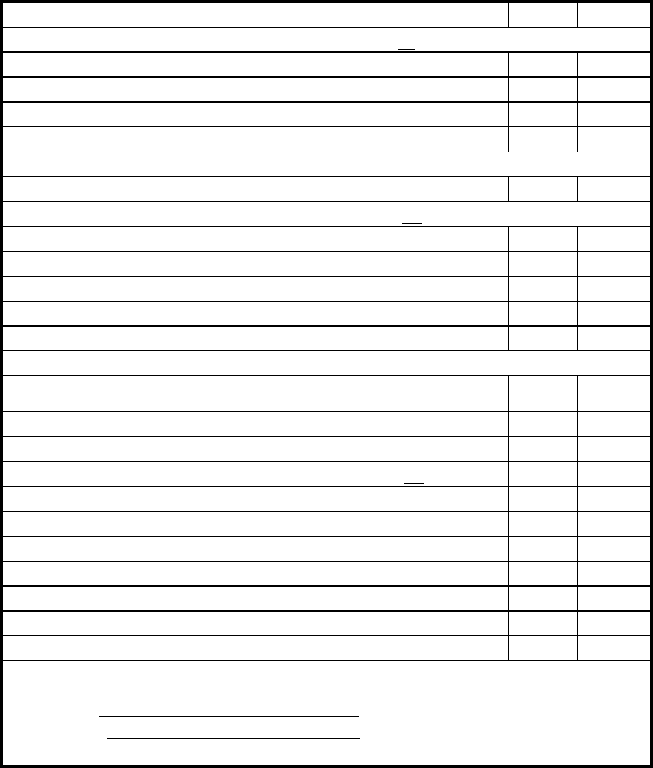
DOE-HDBK-1130-2007
Sample Checklist Attachment 2
117
ATTACHMENT 2 - Sample Grading Checklist for RW II
Name SSN/EMPLOYEE # COMPANY
Item Evaluated Pt Val Score
Pre-Job Preparation total 20 pts.
Selected proper PCs. 5
Selected and properly wear dosimetry. 5
** Donned PCs properly. 5
Recorded appropriate information. 5
Knowledge Level (See Attachment 5) total 20 pts.
Enter results of knowledge-level evaluation. 20
Radiological Work Practices total 20 pts.
** Minimized exposure while completing task. 4
Minimized Radioactive Waste. 4
Didn't walk thru contamination unnecessarily. 4
Didn’t spread contamination. 4
Chose low exposure route. 4
Emergency Response total 20 pts.
** Responded to alarm.
Exited area. 5
Notified RCT upon exit. 5
Exit Practices total 20 pts.
** Followed undress procedure. 3
Did not contaminate step-off pad. 3
** Did not contaminate self. 7
** Performed whole body survey properly. 5
Surveyed DRD. 1
Read DRD correctly upon exit. 1
100 pts.
I was instructed on the proper performance of all items for which my performance was unsatisfactory. I had the opportunity to ask questions. I understand
that failure of this exercise restricts me from entering the following areas:
Student
Date
Instructor
Date
Note: Comments on reverse.
** Indicates significant mistake. If not identified by student, deduct 21 points.
DOE-HDBK-1130-2007
Sample Job Scenario Attachment 3
118
ATTACHMENT 3 - Sample Job Scenario
Each facility/site should develop a job scenario and record it on a facility/site-specific RWP.
(Refer to DOE Radiological Control Technical Standard for guidance on the development of
RWPs.) The following is an example of a job scenario and radiological information.
JOB DESCRIPTION: Enter mock-up area and move the box that is located on
the floor in a lower dose rate area to the storage cabinet located in the
area. (A contamination area is located near the Job Site.)
LOCATION: Mock-up area, Building 2.
SPECIAL INSTRUCTIONS: RWP not valid for Contamination Area.
Avoid drum in Radiation Area.
RADIATION LEVELS: 80 mR/hr near drum on left side of work area.
25 mR/hr near cabinet and panel on north wall.
5 mR/hr near step-off pad area.
CONTAMINATION LEVELS: 5,000 dpm/100 cm
2
floor of Contamination
Area.
PROTECTIVE CLOTHING: None.
DOSIMETER(S): TLD and pocket dosimeter (self-reader).
DOE-HDBK-1130-2007
Sample Survey Map Attachment 4
119
ATTACHMENT 4 - Sample Survey Map
(Insert facility/site-specific sample survey map)
DOE-HDBK-1130-98
Sample Questions Attachment 5
120
ATTACHMENT 5 - Sample Questions
STATE PURPOSE OF ENTRY (Ask both questions.)
1. What is your job description?
2. Where is it located?
STATE RADIATION LEVELS IN THE AREA (include units) (Ask two questions.)
1. What is the highest dose rate in the radiological area?
2. Where is the highest dose rate in the radiological area?
3. What is the work area dose rate?
4. What is the lowest dose rate in the radiological area?
5. Where is the lowest dose rate in the radiological area?
STATE SPECIAL INSTRUCTIONS LISTED ON RWP (Ask one question.)
1. What are two of the special instructions?
2. What are the areas that you cannot enter?
STATE RESPONSE TO AREA RADIATION ALARM
1. A radiation monitor has alarmed. What should you do?
STATE RESPONSE TO COUNT RATE MONITOR INCREASE AND ALARMS (Ask both
questions.)
1. You hear increased clicks as you survey your ______________. What should you do?
2. The count rate monitor has alarmed. What should you do?

(Part 3 of 3)
Radiological Worker Training
Student’s Guide
Coordinated and Conducted
for
Office of Health, Safety and Security
U.S. Department of Energy
DOE-HDBK-1130-2007
ii
This page intentionally left blank.
DOE-HDBK-1130-2007
iii
Table of Contents
Page
MODULE 1: RADIOLOGICAL FUNDAMENTALS................................................................................ 1
MODULE 2: BIOLOGICAL EFFECTS ...................................................................................................14
MODULE 3: RADIATION DOSE LIMITS AND ADMINISTRATIVE CONTROL LEVELS............. 28
MODULE 4: ALARA PROGRAM........................................................................................................... 36
MODULE 5: PERSONNEL MONITORING PROGRAMS.....................................................................44
MODULE 6: RADIOLOGICAL ACCESS CONTROLS AND POSTINGS ........................................ 46
MODULE 7: RADIOLOGICAL EMERGENCIES ..................................................................................60
MODULE 8: HIGH/VERY HIGH RADIATION AREA TRAINING ...................................................... 65
MODULE 9: RADIOACTIVE CONTAMINATION CONTROL ...........................................................69
MODULE 10.1: PRACTICAL FACTORS FOR RADIOLOGICAL WORKER I...................................83
MODULE 10.2: PRACTICAL FACTORS FOR HIGH RADIATION AREAS ......................................89
MODULE 10.3: PRACTICAL FACTORS FOR RADIOLOGICAL WORKER II .................................91
DOE-HDBK-1130-2007
iv
This page intentionally left blank
DOE-HDBK-1130-2007
Radiological Worker Training Student’s Guide
Module 1: Radiological Fundamentals
1
Module 1: Radiological Fundamentals
Terminal Objective:
Given various radiological concepts, the participant will be able to define the fundamentals of
radiation, radioactive material, and radioactive contamination in accordance with the approved lesson
materials.
Enabling Objectives:
The participant will be able to select the correct response from a group of responses to verify his/her
ability to:
EO1 Identify the three basic particles of an atom.
EO2 Define radioactive material, radioactivity, radioactive half-life, and radioactive contamination.
EO3 Identify the units used to measure radioactivity and contamination.
EO4 Define ionization and ionizing radiation.
EO5 Distinguish between ionizing radiation and non-ionizing radiation.
EO6 Identify the four basic types of ionizing radiation and the following for each type:
a. Physical characteristics
b. Range
c. Shielding
d. Biological hazard(s)
e. Sources at the site
EO7 Identify the units used to measure radiation.
EO8 Convert rem to millirem and millirem to rem.
Instructional Aids:
1. Student Guide
2. Transparencies
3. Activities (as applicable)
4. Self-check quizzes (as applicable)
DOE-HDBK-1130-2007
Radiological Worker Training Student’s Guide
Module 1: Radiological Fundamentals
2
I. MODULE INTRODUCTION
A. Self Introduction
1. Name
2. Phone Number
3. Background
B. Module Overview
Nuclear science is truly a product of the 20th century. This module will discuss several
nuclear science topics at a basic level appropriate for the radiological worker. These concepts
are necessary for the worker to understand the nature of radiation and its potential effect on
health. The topics covered include basic particles of the atom, types of radiation, and the
definition of units used to measure radiation.
C. Objectives Review
D. Introduction
This module introduces the worker to basic radiological fundamentals and terms that are
common in the DOE complex. After learning the fundamentals of radiation, radioactive
material, and radioactive contamination, the worker will build from the basic to the more in-
depth concepts presented in the other modules.
DOE-HDBK-1130-2007
Radiological Worker Training Student’s Guide
Module 1: Radiological Fundamentals
3
II. MODULE OUTLINE
A. Atomic Structure
1. The basic unit of matter is the atom. The three basic particles of the atom are protons,
neutrons, and electrons. The central portion of the atom is the nucleus. The nucleus
consists of protons and neutrons. Electrons orbit the nucleus.
a. Protons
1) Protons are located in the nucleus of the atom.
2) Protons have a positive electrical charge.
3) The number of protons in the nucleus determines the element.
b. Neutrons
1) Neutrons are located in the nucleus of the atom.
2) Neutrons have no electrical charge.
3) Atoms of the same element have the same number of protons, but can have a
different number of neutrons.
4) Atoms which have the same number of protons but different numbers of
neutrons are called isotopes.
NOTE: Common notation for describing isotopes is to list the atomic symbol for
an element followed by its mass number. The mass number is the sum of
protons and neutrons. For example, tritium has 1 proton and 2 neutrons, and is
denoted as H-3.
5) Isotopes have the same chemical properties; however, the nuclear properties can
be quite different.
c. Electrons
1) Electrons are in orbit around the nucleus of an atom.
2) Electrons have a negative electrical charge.
4) This negative charge is equal in magnitude to the proton’s positive charge.

DOE-HDBK-1130-2007
Radiological Worker Training Student’s Guide
Module 1: Radiological Fundamentals
4
Basic Particles
3 Basic
Particles
Location
Charge
Comments
Protons
Nucleus
+
(positive)
Number of protons determines
the element. If the number of
protons changes, the element
changes.
Neutrons
Nucleus
No Charge
Atoms of the same element have
the same number of protons, but
can have a different number of
neutrons. This is called an
isotope.
Electrons
Orbit
nucleus
- (negative)
This negative charge is equal in
magnitude to the proton’s
positive charge.
2. Stable and unstable atoms
Only certain combinations of neutrons and protons result in stable atoms.
a. If there are too many or too few neutrons for a given number of protons, the nucleus
will not be stable.
b. The unstable atom will try to become stable by giving off excess energy. This
energy is in the form of particles or rays (radiation). These unstable atoms are
known as radioactive atoms.
3. Charge of the atom
The number of electrons and protons determines the overall electrical charge of the atom.
The term “ion” is used to define atoms or groups of atoms that have a net positive or
negative electrical charge.
a. No charge (neutral)
If the number of electrons equals the number of protons, the atom is electrically
neutral. This atom does not have a net electrical charge.
b. Positive charge (+)
If there are more protons than electrons, the atom is positively charged.
c. Negative charge (-)
If there are more electrons than protons, the atom is negatively charged.
DOE-HDBK-1130-2007
Radiological Worker Training Student’s Guide
Module 1: Radiological Fundamentals
5
B. Definitions and Units of Measure
1. Radioactive material
Radioactive material is any material containing unstable atoms that emit radiation.
Radiation means ionizing radiation: alpha particles, beta particles, gamma rays, X-rays,
neutrons, high-speed electrons, high-speed protons, and other particles capable of
producing ions. Radiation, as used in this part, does not include non-ionizing radiation,
such as radio waves or microwaves, or visible, infrared, or ultraviolet light.
2. Radioactivity
Radioactivity is the process of unstable (or radioactive) atoms becoming stable. This is
done by emitting radiation. This process over a period of time is referred to as
radioactive decay. A disintegration is a single atom undergoing radioactive decay.
3. Radioactivity units
Radioactivity is measured in the number of disintegrations radioactive material undergoes
in a certain period of time.
a. Disintegrations per minute (dpm)
b. Disintegrations per second (dps)
c. Curie (Ci)
One curie equals:
• 2,200,000,000,000 disintegrations per minute (2.2x10
12
dpm), or
• 37,000,000,000 disintegrations per second (3.7x10
10
dps), or
• 1,000,000 microcuries (1x10
6
µCi).
4. Radioactive half-life
Radioactive half-life is the time it takes for one half of the radioactive atoms present to
decay.
5. Radioactive contamination
Radioactive contamination is radioactive material that is uncontained and in an unwanted
place. (There are certain places where radioactive material is intended to be.)
Contamination is measured per unit area or volume.
· dpm/100 cm
2
· µCi/ml.
6. Ionization
Ionization is the process of removing electrons from neutral atoms.
a. Electrons will be removed from an atom if enough energy is supplied. The
remaining atom has a positive (+) charge. The ionized atoms may affect chemical
processes in cells. The ionizations may affect the cell’s ability to function normally.
DOE-HDBK-1130-2007
Radiological Worker Training Student’s Guide
Module 1: Radiological Fundamentals
6
b. The positively charged atom and the negatively charged electron are called an “ion
pair.”
c. Ionization should not be confused with radiation. Ions (or ion pairs) produced as a
result of the interaction of radiation with an atom allow the detection of radiation.
7. Ionizing radiation
Ionizing radiation is energy (particles or rays) emitted from radioactive atoms, and some
devices, that can cause ionization. Examples of devices that emit ionizing radiation are
X-ray machines, accelerators, and fluoroscopes.
a. It is important to note that exposure to ionizing radiation, without exposure to
radioactive material, will not result in contamination of the worker.
b. Radiation is a type of energy, and contamination is radioactive material that is
uncontained and in an unwanted place.
8. Non-ionizing radiation
a. Electromagnetic radiation that doesn’t have enough energy to ionize an atom is
called “non-ionizing radiation.”
b. Examples of non-ionizing radiation are radar waves, microwaves, and visible light.
C. The Four Basic Types of Ionizing Radiation
The four basic types of ionizing radiation of concern in the DOE complex are alpha particles,
beta particles, gamma or X rays, and neutrons.
1. Alpha particles
a. Physical characteristics
1) The alpha particle has a large mass and consists of two protons, two neutrons,
and no electrons.
2) It is a highly charged particle (charge of plus two) that is emitted from the
nucleus of an atom.
3) The positive charge causes the alpha particle (+) to strip electrons (-) from
nearby atoms as it passes through the material, thus ionizing these atoms.
b. Range
1) The alpha particle deposits a large amount of energy in a short distance of travel.
2) This large energy deposit limits the penetrating ability of the alpha particle to a
very short distance.

DOE-HDBK-1130-2007
Radiological Worker Training Student’s Guide
Module 1: Radiological Fundamentals
7
3) Range in air is about 1-2 inches.
c. Shielding
Most alpha particles are stopped by a few centimeters of air, a sheet of paper, or the
dead layer (outer layer) of skin.
d. Biological hazards
1) Alpha particles are not considered an external radiation hazard. This is because
they are easily stopped by the dead layer of skin.
2) Internally, the source of the alpha radiation is in close contact with body tissue
and can deposit large amounts of energy in a small volume of living body tissue.
e. Sources
(Insert facility/site-specific information.)
Table 1-2
Alpha Particles
Physical
Characteristics
· Large mass (2 protons, 2 neutrons, 0 electrons).
· +2 charge.
Range
· Very short (about 1-2 inches in air).
· Deposits large amount of energy in a short
distance of travel.
Shielding
· Few centimeters of air.
· Sheet of paper.
· Dead layer of skin (outer layer).
Biological
Hazards
· No external hazard (dead layer of skin will stop
alpha particles).
· Internally, the source of alpha radiation is in close
contact with body tissue. It can deposit large amounts
of energy in a small amount of body tissue.
Sources
Insert facility/site-specific information.
DOE-HDBK-1130-2007
Radiological Worker Training Student’s Guide
Module 1: Radiological Fundamentals
8
2. Beta particles
a. Physical characteristics
1) The beta particle has a small mass and is positively or negatively charged.
Positively charged beta particles are called positrons and have an electrical charge of
plus one. Negatively charged beta particles are high-energy electrons and have an
electrical charge of minus one.
2) A negatively charged beta particle is physically identical to an electron.
3) The beta particle ionizes target atoms due to the force between itself and the
electrons of the atom. Both have a charge of minus one.
b. Range
1) Because of its charge, the beta particle has a limited penetrating ability.
2) The range in air of beta particles depends on the energy of the beta particle. In the
case of tritium (H-3), the range is only an inch; in the case of phosphorous-32 (P-32)
or strontium-90 (Sr-90), the range is 20 feet in air.
c. Shielding
Beta particles are typically shielded by plastic, glass, or safety glasses.
d. Biological hazards
1) If ingested or inhaled, a beta emitter can be an internal hazard when the source of the
beta radiation is in close contact with body tissue and can deposit energy in a small
volume of living body tissue.
2) Externally, beta particles are potentially hazardous to the skin and eyes.
3) Provide facility/site-specific information on the additional risks or concerns from
high-energy beta sources (e.g., P-32, Y-90), as appropriate.
e. Sources
(Insert facility/site-specific information.)

DOE-HDBK-1130-2007
Radiological Worker Training Student’s Guide
Module 1: Radiological Fundamentals
9
Table 1-3
Beta Particles
Physical
Characteristics
· Small mass.
· -1 charge or + 1 charge.
Range
· Short distance (one inch to 20 feet).
Shielding
· Plastic.
· Glass.
· Safety glasses.
Biological
Hazard
· Internal hazard (this is due to short range).
· Externally, may be hazardous to skin and eyes.
Sources
Insert facility/site-specific information.
3. Gamma rays/X rays
a. Physical characteristics
1) Gamma/X-ray radiation is an electromagnetic wave (electromagnetic radiation) or
photon and has no mass and no electrical charge.
2) Gamma rays are very similar to X rays. The difference between gamma rays and X
rays is that gamma rays originate inside the nucleus and X rays originate in the
electron orbits outside the nucleus.
3) Gamma/X-ray radiation can ionize as a result of direct interactions with orbital
electrons.
b. Range
1) Because gamma/X-ray radiation has no charge and no mass, it has very high
penetrating ability.
2) The range in air is very far. It will easily go several hundred feet.
c. Shielding
Gamma/X-ray radiation is best shielded by very dense materials, such as lead. Water or
concrete, although not as effective as the same thickness as lead, are also commonly
used, especially if the thickness of shielding is not limiting.
d. Biological hazards
Gamma/X-ray radiation can result in radiation exposure to the whole body.

DOE-HDBK-1130-2007
Radiological Worker Training Student’s Guide
Module 1: Radiological Fundamentals
10
e. Sources
(Insert facility/site-specific information.)
Table 1-4
Gamma Rays/X-Rays
Physical
Characteristics
· No mass.
· No charge.
· Electromagnetic wave or photon.
· Similar (difference is the place of origin).
Range
· Range in air is very far.
· It will easily go several hundred feet.
· Very high penetrating power since it has no
mass and no charge.
Shielding
· Concrete.
· Water.
· Lead.
Biological
Hazard
· Whole body exposure.
· The hazard may be external and/or internal.
This depends on whether the source is
inside or outside the body.
Sources
Insert facility/site-specific information.
4. Neutrons
a. Physical characteristics
1) Neutron radiation consists of neutrons that are ejected from the nucleus.
2) A neutron has mass, but no electrical charge.
3) An interaction can occur as the result of a collision between a neutron and a nucleus.
The nucleus recoils due to the energy imparted by the neutron and ionizes other
atoms. This is called “secondary ionization.”
4) Neutrons may also be absorbed by a nucleus. This is called neutron activation. A
charged particle or gamma ray may be emitted as a result of this interaction. The
emitted radiation can cause ionization in other atoms.
b. Range
1) Because of the lack of a charge, neutrons have a relatively high penetrating ability
and are difficult to stop.

DOE-HDBK-1130-2007
Radiological Worker Training Student’s Guide
Module 1: Radiological Fundamentals
11
2) The range in air is very far. Like gamma rays, they can easily travel several hundred
feet in air.
c. Shielding
Neutron radiation is best shielded by materials with a high hydrogen content such as
water, concrete, or plastic.
d. Biological hazards
Neutrons are a whole body hazard due to their high penetrating ability.
e. Sources
(Insert facility/site-specific information.)
Table 1-5
Neutrons
Physical
Characteristics
· No charge.
· Has mass.
Range
· Range in air is very far.
· Easily can go several hundred feet.
· High penetrating power due to lack of charge
(difficult to stop).
Shielding
· Water.
· Concrete.
· Plastic (high hydrogen content).
Biological
Hazard
· Whole body exposure.
· The hazard is generally external.
Sources
Insert facility/site-specific information.
D. Units of Measure for Radiation
1. Roentgen (R)
a. Is a unit for measuring external exposure.
b. Defined only for effect on air.
c. Applies only to gamma and X rays.
d. Does not relate biological effects of radiation to the human body.
DOE-HDBK-1130-2007
Radiological Worker Training Student’s Guide
Module 1: Radiological Fundamentals
12
e. 1 R (Roentgen) = 1000 milliroentgen (mR).
2. Rad (Radiation absorbed dose)
a. A unit for measuring absorbed dose in any material.
b. Is defined for any material.
c. Applies to all types of radiation.
d. Does not take into account the potential effect that different types of radiation have on the
body.
e. 1 rad = 1000 millirad (mrad).
3. Rem (Roentgen equivalent man)
a. A unit for measuring equivalent dose.
b. Is the most commonly used unit.
c. Pertains to the human body.
d. Equivalent dose takes into account the energy absorbed (dose) and the biological effect
on the body due to the different types of radiation.
The Radiation Weighting Factor (RWF) is used as a multiplier to reflect the
relative amount of biological damage caused by the same amount of energy
deposited in cells by the different types of ionizing radiation. Alpha radiation
ionizes a lot of atoms in a very short distance and, for the same amount of energy
deposited as beta or gamma radiation, is more damaging.
Rem = rad x RWF.
Note: Prior to 2007, when DOE updated its dosimetric models and terminology,
DOE used a Quality Factor (QF). The quality factor was applied to the absorbed
dose at a point in order to take into account the differences in the effects of
different types of radiation. Now, for radiological protection purposes, the
absorbed dose is averaged over an organ or tissue and this absorbed average dose
is weighted for the radiation quality in terms of the radiation weighting factor.
Radiation Weighting Factors:
alpha = 20
beta = 1
gamma/x-ray = 1
neutron = 5-20(depending on the energy)
e. 1 rem = 1,000 millirem (mrem).

DOE-HDBK-1130-2007
Radiological Worker Training Student’s Guide
Module 1: Radiological Fundamentals
13
4. Radiation dose and dose rate
a. Radiation dose rate is the dose per time.
b. Example:
1) Radiation dose rate = dose/time.
2) Radiation equivalent dose rate = mrem/hr.
3) Radiation absorbed dose rate = mrad/hr.
Table 1-6
Radiation Units
Roentgen (R)
Rad
(Radiation Absorbed
Dose)
Rem
(Roentgen Equivalent
Man)
Unit for
measuring
exposure.
Unit for measuring
absorbed dose in any
material.
Unit for measuring dose
equivalence (most
commonly used unit).
Defined only for
effect on air.
Defined for any material.
Pertains to human body.
Applies only to
gamma and X-
ray radiation.
Applies to all types of
radiation.
Applies to all types of
radiation.
Does not relate
biological
effects of
radiation to the
human body.
Does not take into
account the potential
effect that different types
of radiation have on the
body.
Takes into account the
energy absorbed (dose)
and the biological effect
on the body due to the
different types of
radiation.
Equal doses of different
types of radiation (as
measured in rad) can
cause different levels of
damage to the body
(measured in rem).
III. SUMMARY
(Insert facility/site-specific information.)
IV. EVALUATION
DOE-HDBK-1130-2007
Radiological Worker Training Student’s Guide
Module 1: Radiological Fundamentals
14
(Insert facility/site-specific information.)
DOE-HDBK-1130-2007
Radiological Worker Training Student’s Guide
Module 2: Biological Effects
15
Module 2: Biological Effects
Terminal Objective:
Given various radiation doses and sources of radiation, identify natural and manmade sources of
radiation and the biological risks associated with radiation dose in accordance with lesson materials.
Enabling Objectives:
The participant will be able to select the correct response from a group of responses to verify his/her
ability to:
EO1 Identify the major sources of natural background and manmade radiation.
EO2 Identify the average annual dose to the general population from natural background and
manmade sources of radiation.
EO3 State the method by which radiation causes damage to cells.
EO4 Identify the possible effects of radiation on cells.
EO5 Define the terms “acute dose” and “chronic dose.”
EO6 State examples of chronic radiation dose.
EO7 Define the terms “somatic effect” and “heritable effect.”
EO8 State the potential effects associated with prenatal radiation dose.
EO9 Compare the biological risks from chronic radiation doses to health risks workers are
subjected to in industry and daily life.
Instructional Aids:
1. Student Guide
2. Transparencies
3. Activities (as applicable)
4. Self-check quizzes (as applicable)
DOE-HDBK-1130-2007
Radiological Worker Training Student’s Guide
Module 2: Biological Effects
16
I. MODULE INTRODUCTION
A. Self Introduction
1. Name
2. Phone Number
3. Background
B. Module Overview
The fact that ionizing radiation produces biological damage has been known for many years.
We have gained most of our knowledge of these effects since World War II.
In this module, we will discuss the potential for biological effects and risks due to ionizing
radiation and put these potential risks into perspective when compared to other occupations
and daily activities. With this information, it is hoped that employees will develop a healthy
respect for radiation rather than fear or disregard.
C. Objectives Review
D. Introduction
We know more about the biological effects of ionizing radiation than most other
environmental factors. Rather than just being able to base our information on animal studies,
we have a large body of information available regarding exposures to humans. There are four
major groups of people that have been exposed to significant levels of radiation.
The first group includes early radiation workers, such as radiologists. These workers received
large doses of radiation before the biological effects were recognized. Since that time,
standards have been developed to protect workers.
The second group is the more than 250,000 survivors of the atomic bombs dropped at
Hiroshima and Nagasaki. Some of these survivors received doses estimated to be in excess of
50,000 mrem.
The third group includes individuals who have been involved in radiation accidents.
The fourth and largest group of individuals are patients who have undergone radiation therapy
for cancer and other diseases.
DOE-HDBK-1130-2007
Radiological Worker Training Student’s Guide
Module 2: Biological Effects
17
II. MODULE OUTLINE
A. Sources of Radiation
We live in a radioactive world and always have. In fact, the majority of us will be exposed to
more ionizing radiation from natural background radiation than from our jobs.
1. Natural sources
There are several sources of radiation that occur naturally. The radiation emitted from
these sources is identical to the radiation that results from manmade sources.
The four major sources of naturally occurring radiation exposures are:
· Cosmic radiation
· Sources in the earth’s crust, also referred to as terrestrial radiation
· Sources in the human body, also referred to as internal sources
· Radon
a. Cosmic radiation (total average dose ~ 28 mrem/yr)
1) Cosmic radiation comes from the sun and outer space. It consists of positively
charged particles and gamma radiation.
2) At sea level, the average annual cosmic radiation dose is about 26 mrem.
3) At higher elevations, the amount of atmosphere shielding cosmic rays decreases;
therefore, the dose increases.
b. Sources in earth’s crust (terrestrial) (total average dose ~ 28 mrem/yr)
There are natural sources of radiation in the ground (i.e., rocks and soil).
1) Some of the contributors to terrestrial sources are the natural radioactive
elements radium, uranium, and thorium.
2) Many areas have elevated levels of terrestrial radiation due to increased
concentrations of uranium or thorium in the soil.
c. Internal (total average dose ~40 mrem/yr)
1) The food we eat and the water we drink contain trace amounts of natural
radioactive materials.
2) These naturally occurring radioactive materials deposit in our bodies and cause
internal exposure to radiation.
DOE-HDBK-1130-2007
Radiological Worker Training Student’s Guide
Module 2: Biological Effects
18
3) Some naturally occurring radioactive isotopes include Sodium-24 (Na-24),
Carbon-14 (C-14), Argon-41 (Ar-41), and Potassium-40 (K-40). Most of our
internal exposure comes from K-40.
d. Radon (total average dose ~ 200 mrem/yr)
1) Radon comes from the radioactive decay of uranium, which is naturally present
in the soil.
2) Radon is a gas. It can travel through the soil and enter through building
foundation cracks. The greatest concentrations of indoor radon are found in
basements.
3) Radon emits alpha radiation. It presents a hazard only when taken into the body
(e.g., when inhaled).
2. Manmade sources
The difference between manmade sources of radiation and naturally occurring sources is
the origin of the source, i.e., where the radiation is either produced or enhanced by human
activities.
The four top sources of manmade radiation exposures are:
· Tobacco products (cigarettes, cigars etc. although the tobacco is not manmade - the
products are)
· Medical radiation
· Building materials
· Domestic water supply (radon)
a. Tobacco products (average dose ~1300 mrem/yr)
b. Medical radiation sources (total average dose ~ 54 mrem/yr)
1) X rays (total average dose ~ 40mrem/yr)
a) X rays are similar to gamma rays; however, they originate outside the
nucleus.
b) A typical radiation dose from a chest X ray is about 10 mrem.
2) Diagnosis and therapy (total average dose ~14 mrem/yr)
In addition to X rays, radioactive materials and radioactive sources are used in
medicine for diagnosis and therapy.
DOE-HDBK-1130-2007
Radiological Worker Training Student’s Guide
Module 2: Biological Effects
19
c. Building materials (total average dose ~7 mrem/yr)
d. Domestic water supply (radon) (total average dose ~5 mrem/yr)
e. Other minor contributors
Other contributors to dose include consumer products, industrial sources, and
atmospheric testing of nuclear weapons.
4. Average annual dose
The average annual total effective dose to the general population (non-smokers) from
naturally occurring and manmade sources is about 360 mrem.
B. Effects of Radiation on Cells
The human body is made up of many organ systems. Each system is made up of tissues.
Specialized cells make up tissues. Ionizing radiation can potentially affect the normal
function of cells.
1. Biological effects begin with the ionization of atoms
a. The method by which radiation causes damage to human cells is by ionization of
atoms in the cells. It may also cause excitation. Excitation is where the radiation
deposits energy into an atom raising its energy level but not enough energy to eject a
bound electron. Atoms make up the cells that make up the tissues of the body. Any
potential radiation damage begins with damage to atoms.
b. A cell is made up of two principal parts, the body of the cell and the nucleus. The
nucleus is like the brain of the cell.
c. When ionizing radiation hits a cell, it may strike a vital part of the cell like the
nucleus or a less vital part of the cell, like the cytoplasm.
2. Cell sensitivity
Some cells are more sensitive than others to environmental factors such as viruses,
toxins, and ionizing radiation.
a. Actively dividing and non-specialized cells
1) Cells in our bodies that are actively dividing are more sensitive to ionizing
radiation.
DOE-HDBK-1130-2007
Radiological Worker Training Student’s Guide
Module 2: Biological Effects
20
2) Cells that are rapidly dividing include blood-forming cells, the cells that line our
intestinal tract, hair follicles, and cells that form sperm.
b. Less actively dividing and more specialized cells
Cells that divide at a slower rate or are more specialized (such as brain cells or
muscle cells) are not as sensitive to damage by ionizing radiation.
3. Possible effects of radiation on cells
Several things can happen when a cell is exposed to ionizing radiation. The following are
possible effects of radiation on cells.
a. There is no damage
b. Cells are damaged but are able to repair the damage and operate normally
1) The body of most cells is made up primarily of water. When ionizing radiation
hits a cell, it is most likely to interact with the water in the cell. One of the
byproducts of radiation-induced ionization of water is hydrogen peroxide.
Hydrogen peroxide can damage cell atomic structures.
2) Ionizing radiation can also hit the nucleus of the cell. The nucleus contains the
vital parts of the cell, such as chromosomes. The chromosomes determine cell
function. When chromosomes duplicate themselves, the chromosomes transfer
their information to new cells. Radiation may cause a change in the
chromosome that does not affect the cell.
3) Damage to chromosomes and other cell structures can be repaired. In fact, our
bodies repair a very large number of chromosome breaks every day (References
7 and 10).
3. Average annual dose
The average annual total effective dose to the general population from naturally occurring
and manmade sources is about 360 mrem.
c. Cells are damaged and operate abnormally
1) Cell damage may not be repaired or may be incompletely repaired. In that case,
the cell may not be able to function properly.
2) It is possible that a chromosome in the cell nucleus could be damaged but not be
repaired correctly. If the cell continues to reproduce, this is called a mutation
and may result in cancer.
d. Cells die as a result of the damage
DOE-HDBK-1130-2007
Radiological Worker Training Student’s Guide
Module 2: Biological Effects
21
At any given moment, thousands of our cells die and are replaced by normal
functioning cells. However, the radiation damage to a cell may be so extensive that
the cell dies prematurely.

DOE-HDBK-1130-2007
Radiological Worker Training Student’s Guide
Module 2: Biological Effects
22
C. Acute and Chronic Radiation Dose
Potential biological effects depend on how much and how fast a radiation dose is received.
Radiation doses can be grouped into two categories: acute and chronic dose.
1. Acute radiation doses
a. High doses of radiation received in a short period of time are called acute doses. The
body’s cell repair mechanisms are not as effective for damage caused by an acute
dose.
b. Acute doses to the whole body
After an acute dose, damaged cells may be replaced by new cells and the body may
repair itself, although this may take a number of months. Only in extreme cases,
such as with the Chernobyl firefighters (500 rem), would the dose be so high as to
make recovery unlikely.
c. Acute doses to only part of the body
1) X-ray machines
It is possible that radiation exposure may be limited to a part of the body, such as
the hands.
There have been accidents, particularly with X-ray machines, in which
individuals have exposed their fingers to part of the intense radiation beam. In
some of these cases, individuals have received doses of millions
of mrem to their
fingers, and some individuals have lost their finger or fingers. It is important for
individuals who work with X-ray or similar equipment to be trained in the safe
use of this equipment.
2) Radiation therapy
a) Radiation therapy patients receive high doses of radiation in a short period
of time, but generally only to a small portion of the body (not a whole body
dose).
b) The skin and limited tissue of these patients may receive significant doses,
but doses to the region of a tumor are many times higher.
c) Ionizing radiation is used to treat cancer in these patients because cancer
cells are rapidly dividing and therefore sensitive to ionizing radiation. Some
of the side effects of people undergoing radiation therapy are hair loss,
nausea, and tiredness.
DOE-HDBK-1130-2007
Radiological Worker Training Student’s Guide
Module 2: Biological Effects
23
d. Probability of a large acute dose
What is important to understand is that it takes a large acute dose of radiation before
any physical effect is seen. These acute doses have occurred in Hiroshima/Nagasaki,
and in a few radiation accidents, including Chernobyl. The possibility of a
radiological worker receiving a large acute dose of ionizing radiation on the job is
extremely low. Typically, radioactive materials are handled in small quantities that
do not produce a large amount of radiation. Where there is a potential for larger
exposures, many safety features are required.
2. Chronic radiation doses
A chronic radiation dose is typically a small amount of radiation received over a long
period of time. An example of a chronic dose is the dose we receive from natural
background every day of our lives. The body’s cell repair mechanisms are better able to
repair a chronic dose than an acute dose.
a. The body has time to repair damage because a smaller percentage of the cells need
repair at any given time.
b. The body also has time to replace dead or non-functioning cells with new, healthy
cells.
3. Biological effects of radiation exposure
Somatic effects refer to the effects radiation has on the individual receiving the dose.
Genetic effects refer to mutations due to radiation damage to the DNA of a cell. When
this change is in the DNA of parental reproductive cells, it is called a heritable effect.
a. Somatic Effects
Somatic effects can best be described in terms of prompt and delayed effects as
discussed below.
Prompt Effects
Although rare in the nuclear industry, large doses are typically acute
radiation doses representing serious overexposures. The biological effects
of large acute doses are as follows:

DOE-HDBK-1130-2007
Radiological Worker Training Student’s Guide
Module 2: Biological Effects
24
Table 2-1
Prompt Biological Effects
Dose (rad)
Effect
0-25
None detectable through
symptoms or routine blood tests.
25-100
Changes in blood.
100-300
Nausea, anorexia.
300-600
(450 rem is considered the dose
where 50% fatalities occur within
30 days with no medical help
(lethal dose - LD 50/30)
Diarrhea, hemorrhage, and
possible death
3) Delayed Effects
Delayed effects may result from either a single large acute overexposure or
from continuing low-level chronic exposure. Cancer in its various forms is
the most important potential delayed effect of radiation exposure. Other
effects noted include cataracts, life shortening and, for individuals exposed
in the womb, lower IQ test scores.
b. Heritable Effects
A heritable effect is a physical mutation or trait that is passed on to offspring. In
the case of heritable effects, the parental individual has experienced damage to
some genetic material in the reproductive cells and has passed the damaged
genetic material onto offspring.
1) Heritable effects from radiation have never been observed in humans but are
considered possible. They have been observed in studies of plants and
animals.
2) Heritable effects have not been found in the 77,000 Japanese children born
to the survivors of Hiroshima and Nagasaki (these are children who were
conceived after
the atom bomb -- i.e., heritable effects). Studies have
followed these children, their children, and their grandchildren.
DOE-HDBK-1130-2007
Radiological Worker Training Student’s Guide
Module 2: Biological Effects
25
4. Factors affecting biological damage due to exposure to radiation
a. Total dose
In general, the greater the dose, the greater the potential for biological effects.
b. Dose rate (how fast)
The faster the dose is delivered, the less time the body has to repair itself.
c. Type of radiation
For example, internally deposited alpha emitters are more damaging than beta or
gamma emitters for the same energy deposited. Alpha emitters deposit energy in
a very small mass, beta and gamma emitters deposit their energy over a larger
mass.
d. Area of the body that receives a dose
In general, the larger the area of the body that receives a dose, the greater the
biological effect.
Extremities are less sensitive than blood forming and other critical organs. That
is why the annual dose limit for extremities is higher than for a whole body dose
that irradiates internal organs.
e. Cell sensitivity
The most sensitive cells are those that are rapidly dividing. Examples include
blood cells, hair follicles, and the cells lining the gastrointestinal tract.
f. Individual sensitivity
Some individuals are more sensitive to environmental factors such as ionizing
radiation.
The developing embryo/fetus is the most sensitive, and children are more
sensitive than adults. This is due to their having large number s of rapidly
dividing cells.
In general, the human body becomes relatively less sensitive to ionizing
radiation with increasing age. The exception is that elderly people are more
sensitive than middle-aged adults due to the inability to repair damage as quickly
(less efficient cell repair mechanisms).
DOE-HDBK-1130-2007
Radiological Worker Training Student’s Guide
Module 2: Biological Effects
26
D. Prenatal Radiation Exposure
Although no effects were seen in Japanese children conceived after the atomic bomb, there
were effects seen in some children who were in the womb when exposed to the atomic bomb
radiation at Hiroshima and Nagasaki. Some of these children were born with a slightly
smaller head size, lower average birth weight, and increased incidence of mental retardation.
Some later showed lower IQ test scores and slower scholastic development, smaller physical
size, and increased incidence of behavioral problems.
1. Sensitivity of the fetus
Embryo/fetal cells are rapidly dividing, which makes them sensitive to many
environmental factors including ionizing radiation. The embryo/fetus is most susceptible
to developing adverse health effects if exposed during the time period of 8 - 15 weeks
after conception.
2. Factors for potential effects associated with prenatal exposures
Many chemical and physical (environmental) factors are suspected of causing or known
to have caused damage to a fetus, especially early in the pregnancy. Radiation, alcohol
consumption, exposure to lead, and heat, such as from hot tubs, are only a few such
factors.
E. Risks in Perspective
Current radiation protection standards and practices are based on the premise that any
radiation dose, no matter how small, can result in health effects such as cancer. Further, it is
assumed that these effects are produced in direct proportion to the dose received (i.e.,
doubling the radiation dose results in a doubling of the risk of the effect). These two
assumptions lead to a dose-response relationship, often referred to as the linear, no-threshold
model, for limiting health effects at very low radiation dose levels.
However, it should be noted that this is a conservative assumption made in the absence of
more conclusive evidence. Health effects (primarily cancer) have been observed in humans
only at doses in excess of 10 rem delivered at high dose rates. Below this dose, estimation of
adverse health effects is speculative. Risk estimates that are used to predict health effects in
exposed individuals or populations are based on epidemiological studies of well-defined
populations (e.g., the Japanese survivors of the atomic bombings in 1945 and medical
patients) exposed to relatively high doses delivered at high dose rates. It is generally accepted
that studies have not demonstrated adverse health effects in individuals exposed to small
doses (less than 10 rem) delivered over a period of many years.

DOE-HDBK-1130-2007
Radiological Worker Training Student’s Guide
Module 2: Biological Effects
27
1. Risk from exposures to ionizing radiation
a. No increases in cancer have been observed in individuals who receive a dose of
ionizing radiation at occupational levels. The possibility of cancer induction cannot
be dismissed even though an increase in cancers has not been observed. Risk
estimates have been derived from studies of individuals who have been exposed to
high levels of radiation.
b. The risk of cancer induction from radiation exposure can be put into perspective. This
can be done by comparing it to the normal rate of cancer death in today’s society. The
current rate of cancer death among Americans is about 20 percent. Taken from a
personal perspective, each of us has about 20 chances in 100 of dying of cancer. A
radiological worker who receives 25,000 mrem over a working life increases his/her risk
of cancer by 1 percent, or has about 21 chances in 100 of dying of cancer. A 25,000
mrem dose is a fairly large dose over the course of a working lifetime. The average
annual dose to DOE workers is less than 100 mrem, which leads to a working lifetime
dose (40 years assumed) of no more than approximately 4,000 mrem.
2. Comparison of risks
a. Table 2-2 compares the estimated days of life expectancy lost as a result of exposure
to radiation and other health risks.
The following information is intended to put the potential risk of radiation into
perspective when compared to other occupations and daily activities.
Table 2-2
Estimated Loss of Life Expectancy from Health Risks
Health Risk
Estimated Loss of Life Expectancy
Smoking 20 cigarettes a day 6 years
Overweight (by 15%) 2 years
Alcohol consumption (U.S. average) 1 year
Agricultural accidents 320 days
Construction accidents 227 days
Auto accidents 207 days
Home accidents 74 days
Occupational radiation dose (1 rem/y), from
age 18-65 (47 rem total) 51 days
(Note: the average DOE radiation worker receives less than 0.1
rem/yr)
All natural hazards

DOE-HDBK-1130-2007
Radiological Worker Training Student’s Guide
Module 2: Biological Effects
28
(earthquakes, lightning, flood) 7 days
Medical radiation 6 days
The estimates in Table 2-2 indicate that the health risks from occupational radiation doses are
smaller than the risks associated with normal day-to-day activities that we have grown to
accept.
b. Acceptance of a risk:
1) is a personal matter.
2) requires a good deal of informed judgment.
c. The risks associated with occupational radiation doses are generally considered acceptable as
compared to other occupational risks by most scientific groups who have studied them. There
are some scientific groups who claim that the risk is too high. DOE continues to fund and
review worker health studies to address these concerns.
III. SUMMARY
In summary, the estimated risk associated with occupation radiation dose is similar to other
routine occupational risks and much less than some risks widely accepted in society. The risk of
work in a radiation environment is considered within the normal occupational risk tolerance by
national and international scientific groups. However, acceptance of risk is an individual matter
and is best made with accurate information. A radiological worker should understand the risk of
working in a nuclear environment in relation to the risks of daily life and the risks presented by
work in other professions. The intent of this module is to give you the facts about radiation
exposure risks and provide you with an opportunity to ask questions about radiation risk. It is
hoped that understanding radiation risk and risk in general will help you to develop an informed
and healthy respect for radiation, and that your understanding will eliminate excessive fear of or
indifference to radiation.
IV. EVALUATION
(Insert facility/site-specific information.)
DOE-HDBK-1130-2007
Radiological Worker Training Student’s Guide
Module 3: Radiation Dose Limits and Administrative Control Levels
29
MODULE 3: RADIATION DOSE LIMITS AND ADMINISTRATIVE CONTROL
LEVELS
Terminal Objective:
Given various time frames and different parts of the body, identify the applicable DOE dose
limits, DOE administrative control levels, and facility/site-specific administrative control levels in
accordance with the lesson material.
Enabling Objectives:
The participant will be able to select the correct response from a group of responses to verify
his/her ability to:
EO1 State the purposes of administrative control levels.
EO2 Identify the DOE radiation dose limits, DOE recommended administrative control level,
and the facility/site administrative control level.
EO3 State the site policy concerning prenatal radiation exposure.
EO4 Identify the employee’s responsibilities concerning radiation dose limits and
administrative control levels.
Instructional Aids:
1. Student Guide
2. Transparencies
3. Activities (as applicable)
4. Self-check quizzes (as applicable)
DOE-HDBK-1130-2007
Radiological Worker Training Student’s Guide
Module 3: Radiation Dose Limits and Administrative Control Levels
30
I. MODULE INTRODUCTION
A. Self Introduction
1. Name
2. Phone Number
3. Background
B. Module Overview
This module will address DOE dose limits and administrative control levels.
C. Objectives Review
D. Introduction
DOE limits and administrative control levels have been established for the purpose of
restricting occupational radiation exposures to levels of acceptable risk.
II. MODULE OUTLINE
A. Basis for and Purpose of Radiation Dose Limits and Administrative Control Levels
1. Basis for DOE dose limits
a. DOE has established radiation dose limits for general workers. These limits are
based on guidance from national and international scientific groups and government
agencies, such as:
1) International Commission on Radiological Protection (ICRP)
2) National Council on Radiation Protection and Measurements (NCRP)
3) U.S. Environmental Protection Agency (EPA)
b. The radiation protection standards for all DOE workers are described in 10 CFR 835,
“Occupational Radiation Protection.” These regulations apply to DOE, its
contractors, and persons conducting DOE activities and include equivalent dose
limits.
2. Facility/site administrative control levels for general employees

DOE-HDBK-1130-2007
Radiological Worker Training Student’s Guide
Module 3: Radiation Dose Limits and Administrative Control Levels
31
The facility/site administrative control levels for workers are lower than the DOE limits
and are set to:
a. Ensure the DOE limits and control levels are not exceeded.
b. Help reduce individual and total worker population radiation dose (collective dose).
B. Dose Limits and Administrative Control Levels
In 2007 DOE updated its models for calculating dose to use newer models recommended by
international consensus groups. The impact of these changes is that some neutron exposures
will be assessed differently and intakes of radioactive material will be assessed using the
newer models. These models include revised terminology for some of the dosimetric terms.
The following table highlights the different terms.
Table 3-1 Updated Dosimetric Terms
Previous Dosimetric Term
DOE 2007 Amended Dosimetric Term
Committed effective dose equivalent
Committed effective dose
Committed dose equivalent
Committed equivalent dose
Cumulative total effective dose
equivalent
Cumulative total effective dose
Deep dose equivalent
Equivalent dose to the whole body
Dose equivalent
Equivalent dose
Effective dose equivalent
Effective dose
Lens of the eye dose equivalent
Equivalent dose to the lens of the eye
Quality factor
Radiation weighting factor
Shallow dose equivalent
Equivalent dose to the skin or
Equivalent dose to any extremity
Weighting factor
Tissue weighting factor
Total effective dose equivalent
Total effective dose

DOE-HDBK-1130-2007
Radiological Worker Training Student’s Guide
Module 3: Radiation Dose Limits and Administrative Control Levels
32
Table 3-2
Dose Limits and Controls
DOE Dose
Limit
rem/year
DOE
Recommendations
rem/year
Facility/site
Administrative
Control Level
rem/year
Whole body
5
2
facility/site-
specific
Extremity
50
N/A
facility/site-
specific
Skin & other
organs
50
N/A
facility/site-
specific
Lens of the eye
15
N/A
facility/site-
specific
Members of
the public
0.1
N/A
facility/site-
specific
Occupationally
exposed
minors
0.1 whole
body and 10%
of other above
limits
N/A facility/site-
specific
Declared
pregnant
worker
0.5/gestation
period
N/A
facility/site-
specific
NOTE: 1) The chart is based on limits and control levels for routine conditions. The limits and control
levels are also based on the sum of internal and external dose. External dose is from sources outside the
body. Internal dose is from sources inside the body. 2) The internal dose reported in a given calendar
year is actually the projected dose the individual will receive over the next 50 years from intakes in that
calendar year. Radioactive material may be inhaled, ingested, or absorbed through the skin or open
wound.
1. Whole body
a. Definition
The whole body extends from the top of the head down to just below the elbow and
just below the knee. This is the location of most of the blood-producing and vital
organs.

DOE-HDBK-1130-2007
Radiological Worker Training Student’s Guide
Module 3: Radiation Dose Limits and Administrative Control Levels
33
b. Limit and control levels
The DOE whole body dose limit is based on the sum of internal and external dose.
1) DOE radiation dose limit during routine conditions is 5 rem/year
.
2) Because DOE’s objective is to maintain personnel radiation dose well below the
regulatory limits, the DOE Radiological Control Technical Standard
recommends a DOE administrative control level during routine conditions of
2 rem/year
.
3) Facility/site administrative control level.
(Insert facility/site-specific information.)
2. Extremities
a. Definition
Extremities include the hands and arms below the elbow, and the feet and legs below
the knees.
b. Limit and control level
Extremities can withstand a much larger dose than the whole body because there are
no major blood-producing organs located here.
1) DOE radiation dose limit for extremities is 50 rem/year
.
2) Facility/site administrative control levels.
(Insert facility/site-specific information.)
3. Skin and other organs
a. DOE radiation dose limit for skin and other organs is 50 rem/year
.
b. Facility/site administrative control level
(Insert facility/site-specific information.)
4. Lens of the eye
a. DOE radiation dose limit for lens of the eye is 15 rem/year
.
b. Facility/site administrative control level.
(Insert facility/site-specific information.)

DOE-HDBK-1130-2007
Radiological Worker Training Student’s Guide
Module 3: Radiation Dose Limits and Administrative Control Levels
34
5. Declared pregnant worker: Embryo/fetus
After a female worker voluntarily notifies her employer in writing that she is pregnant,
she is considered a declared pregnant worker. For the purposes of radiological protection
of the fetus/embryo, DOE requires a special limit for dose to the fetus/embryo. In
addition, the DOE RCS recommends that the employer provide the option of a mutually
agreeable assignment of work tasks, with no loss of pay or promotional opportunity, such
that further occupational radiation exposure is unlikely during the remaining gestation
period.
• This declaration may be revoked, in writing, at anytime by the declared pregnant
worker.
b. DOE limit
For a declared pregnant worker who continues working as a radiological worker, the
following radiation dose limit will apply.
1) The dose limit for the embryo/fetus (during the entire gestation period) is 500
mrem.
a) Measures shall be taken to avoid substantial variation above the uniform
exposure rate necessary to meet the 500 mrem limit for the gestation period.
b) The DOE RCS recommends that efforts be made to avoid exceeding 50
mrem/month to the embryo/fetus of the declared pregnant worker.
2) If the dose to the embryo/fetus is determined to have already exceeded 500
mrem when a worker notifies her employer of her pregnancy, the worker shall
not be assigned to tasks where additional occupational radiation exposure is
likely during the remainder of the pregnancy.
b. Site policy
(Insert facility/site-specific information.)
c. Facility/site administrative control level
(Insert facility/site-specific information.)
6. Members of the public and occupationally exposed minors
a. DOE radiation dose limit is 100 mrem/year
. There is an additional limit for
occupationally expose minors of 10% of the other limits.
b. Facility/site administrative control levels
(Insert facility/site-specific information.)
DOE-HDBK-1130-2007
Radiological Worker Training Student’s Guide
Module 3: Radiation Dose Limits and Administrative Control Levels
35
C. Worker Responsibilities Regarding Dose Limits
1. It is each employee’s responsibility to comply with DOE dose limits and facility/site
administrative control levels.
2. If you suspect that dose limits or administrative control levels are being approached or
exceeded, you should notify your supervisor immediately.
3. (Insert facility/site-specific information.)
III. SUMMARY
(Insert Site Summary.)
IV. EVALUATION
(Insert facility/site-specific information.)
DOE-HDBK-1130-2007
Radiological Worker Training Student’s Guide
Module 4: ALARA Program
36
MODULE 4: ALARA PROGRAM
Terminal Objective:
Given different radiological conditions, identify the techniques for minimizing exposure to
radiation and radioactive material in accordance with lesson materials.
Enabling Objectives:
The participant will be able to select the correct response from a group of responses to verify
his/her ability to:
EO1 State the ALARA concept.
EO2 State the DOE/Site management policy for the ALARA program.
EO3 Identify the responsibilities of management, the Radiological Control Organization, and
the radiological worker in the ALARA Program.
EO4 Identify methods for reducing external and internal radiation dose.
EO5 State the pathways by which radioactive material can enter the body.
EO6 Identify methods a radiological worker can use to minimize radioactive waste.
Instructional Aids:
1. Student Guide
2. Transparencies
3. Activities (as applicable)
4. Self-check quizzes (as applicable)

DOE-HDBK-1130-2007
Radiological Worker Training Student’s Guide
Module 4: ALARA Program
37
I. MODULE INTRODUCTION
A. Self Introduction
1. Name
2. Phone Number
3. Background
B. Module Overview
This module is designed to inform the student of the concept of ALARA (As Low As
Reasonably Achievable). This module discusses radiation hazards. Methods for reducing
both external and internal doses from radiation and radioactive material are also discussed.
C. Objectives Review
D. Introduction
DOE establishes dose limits and administrative control levels for general employees.
However, radiological workers and their management strive to keep radiation dose well below
these limits. Radiological workers should always try to maintain their radiation dose A
s Low
A
s Reasonably Achievable (ALARA).
II. MODULE OUTLINE
A. ALARA Program
ALARA stands for As Low As Reasonably Achievable. ALARA is an approach to radiation
safety that strives to manage and control doses (both individual and collective) to the work
force and the general public to as low as is reasonable taking into account social, technical,
economic, practical, and public policy considerations.
1. ALARA concept
a. ALARA stands for A
s Low As Reasonably Achievable.
b. Because some risk, however small, exists from any radiation dose, all doses should
be kept ALARA. ALARA includes reducing both internal and external radiation
dose.
c. The ALARA concept is an integral part of all site activities that involve the use of
sources of ionizing radiation.
d. ALARA is the responsibility of all employees.
DOE-HDBK-1130-2007
Radiological Worker Training Student’s Guide
Module 4: ALARA Program
38
2. DOE Management Policy for the ALARA program
Personal radiation exposure shall be maintained As Low As Reasonably Achievable.
Radiation exposure to the work force and public shall be controlled such that:
· Radiation doses are well below regulatory limits.
· There is no radiation exposure without an overall benefit.
3. Site policy
(Insert facility/site-specific information.)
B. Responsibilities for the ALARA Program
The individual radiological worker is ultimately responsible for maintaining his/her radiation
dose ALARA. However, management and Radiological Control personnel also play an
important role in the ALARA program. The following are some of the responsibilities of the
three groups:
1. Management
(Insert facility/site-specific information.)
2. Radiological Control Organization
(Insert facility/site-specific information.)
3. Radiological workers
Each radiological worker is expected to demonstrate responsibility and accountability.
This is accomplished through an informed, disciplined, and cautious attitude toward
radiation and radioactivity.
(Insert facility/site-specific information.)
C. External and Internal Radiation Dose Reduction
Engineering controls should be the primary method to control exposure (e.g., enclosed
hoods). Administrative controls is the next method to control exposures (e.g., postings).
Personnel protective equipment is the last method (e.g., respirators).
DOE-HDBK-1130-2007
Radiological Worker Training Student’s Guide
Module 4: ALARA Program
39
1. Basic protective measures used to minimize external dose include:
· Minimizing time in radiation areas
· Maximizing the distance from a source of radiation
· Using shielding whenever possible
· Reducing the amount of radioactive material (source reduction)
a. Methods for minimizing time
Reducing the time spent in a field of radiation will lower the dose received by the
workers.
1) Plan and discuss the task thoroughly prior to entering the area. Use only the
number of workers actually required to do the job.
2) Have all necessary tools present before entering the area.
3) Use mock-ups and practice runs that duplicate work conditions.
4) Take the most direct route to the job site if possible and practical.
5) Never loiter in an area controlled for radiological purposes.
6) Work efficiently and swiftly.
7) Do the job right the first time.
8) Perform as much work outside the area as possible. When practical, remove
parts or components to areas with lower dose rates to perform work.
9) Do not exceed stay times. In some cases, the Radiological Control Organization
may limit the amount of time a worker may stay in an area due to various
reasons. This is known as “stay time.” If you have been assigned a stay time,
do not exceed this time.
10) (Insert facility/site-specific information.)
b. Methods for maximizing distance from sources of radiation
The worker should stay as far away as possible from the source of radiation.
1) Stay as far away from radiation sources as practical given the task assignment.
For point sources (such as valves and hot spots), the dose rate follows a principle
called the inverse square law. This law states that if you double the distance, the
DOE-HDBK-1130-2007
Radiological Worker Training Student’s Guide
Module 4: ALARA Program
40
dose rate falls to 1/4 of the original dose rate. If you triple the distance, the dose
rate falls to 1/9 of the original dose rate.
2) Be familiar with radiological conditions in the area.
3) During work delays, move to lower dose rate areas.
4) Use remote handling devices when possible.
5) (Insert facility/site-specific information.)
d. Proper uses of shielding
Shielding reduces the amount of radiation dose to the worker. Different materials
shield a worker from the different types of radiation.
1) Take advantage of permanent shielding, such as non-radiological
equipment/structures.
2) Use shielded containments when available.
3) Wear safety glasses/goggles to protect your eyes from beta radiation, when
applicable.
4) Temporary shielding (e.g., lead or concrete blocks) can only be installed when
proper procedures are used.
5) Temporary shielding will be marked or labeled with wording such as
“Temporary Shielding - Do Not Remove Without Permission from Radiological
Control.”
6) Once temporary shielding is installed, it cannot be removed without proper
authorization.
· When evaluating the use of shielding, the estimated dose saved is compared
to the estimated dose incurred during shield installation and removal.
7) (Insert facility/site-specific information.)
d. Source Reduction
Source reduction is another method of reducing radiation doses. Source reduction
often involves procedures such as flushing radioactive systems, decontamination, and
removal of contaminated items. This is done to reduce the amount of radioactive
materials present in/on a system because these materials can add to radiation levels in
an area.
DOE-HDBK-1130-2007
Radiological Worker Training Student’s Guide
Module 4: ALARA Program
41
2. Internal radiation dose reduction
a. Pathways
Internal dose is a result of radioactive materials being taken into the body.
Radioactive material can enter the body through one or more of the following
pathways:
1) Inhalation
2) Ingestion
3) Absorption through the skin
4) Absorption through wounds
b. Methods to reduce internal radiation dose
Reducing the potential for radioactive materials to enter the body is important. As
previously stated, install or use engineering controls followed by administrative
controls as the primary methods to control internal exposure. PPE is the last choice
for controlling internal exposure. In addition, the following are methods the worker
can use.
1) Wear respirators properly when required. Respirators should only be used
by personnel qualified to wear them.
2) Report all wounds or cuts (including scratches and scabs) to the appropriate
facility/site-specific organization before entering any area controlled for
radiological purposes.
3) Comply with the requirements of the controlling work documents.
4) Do not eat, drink, smoke, or chew in Radioactive Materials Areas,
Contamination Areas, High Contamination Areas, or Airborne Radioactivity
Areas, as dispersible radioactive materials may be present.
5) (Insert facility/site-specific information.)
3. Lessons Learned
Review lessons learned from your site or other sites to demonstrate what may be learned
from mistakes leading to excessive personnel exposures.
DOE-HDBK-1130-2007
Radiological Worker Training Student’s Guide
Module 4: ALARA Program
42
(Insert facility/site-specific information.)
D. Radioactive Waste Minimization
One of the potential consequences of working with radioactive materials is the generation of
radioactive waste. This radioactive waste must be properly disposed. Examples of
radioactive waste include:
· Paper
· Gloves
· Glassware
· Rags
· Brooms, mops
The ALARA concept also applies to minimizing radioactive waste. This will reduce
personnel exposure associated with the handling, packaging, storing, and disposing of
radioactive waste. This will also reduce the resultant costs. It is very important for each
radiological worker to minimize the amount of radioactive waste generated.
1. Methods to minimize radioactive waste
The following information identifies methods to minimize radioactive waste.
a. Minimize the materials used for radiological work.
1) Take only the tools and materials you need for the job into areas controlled for
radiological purposes. This is especially important for contamination areas.
2) Unpack equipment and tools in a clean area. This will help to avoid bringing
unnecessary material to the job site. This material can become radioactive waste
if it is contaminated.
3) Use tools and equipment that are identified for radiological work when possible.
(Add facility/site-specific information about where such tools are stored.)
4) Use only the materials required to clean the area. An excessive amount of bags,
rags, and solvent adds to radioactive waste.
5) Sleeve, or otherwise protect with a covering such as plastic, clean materials
brought into contaminated areas.
6) (Insert facility/site-specific information.)
b. Separate radioactive waste from nonradioactive waste.
DOE-HDBK-1130-2007
Radiological Worker Training Student’s Guide
Module 4: ALARA Program
43
1) Place radioactive waste in the containers identified for radioactive waste. Do not
place radioactive waste in nonradioactive waste containers.
2) Do not throw nonradioactive waste, or radioactive material that may be reused,
into radioactive waste containers.
4) (Insert facility/site-specific information.)
c. Separate compactable material from noncompactable material.
d. Minimize the amount of mixed waste generated. Mixed waste is waste that contains
both radioactive and hazardous materials.
e. Use good housekeeping techniques.
g. (Insert facility/site-specific information.)
III. SUMMARY
This module addressed key points for the implementation and success of the Site’s ALARA
Program. Responsibilities for all employees and methods to achieve the ALARA concepts were
also discussed.
IV. EVALUATION
(Insert facility/site-specific information.)
DOE-HDBK-1130-2007
Radiological Worker Training Student’s Guide
Module 5: Personnel Monitoring Programs
44
MODULE 5: PERSONNEL MONITORING PROGRAMS
Terminal Objective:
Given different personnel monitoring programs, identify the purpose, types, and worker
responsibilities for each in accordance with lesson material.
Enabling Objectives:
The participant will be able to select the correct response from a group of responses to verify
his/her ability to:
EO1 State the purpose and worker responsibilities for each of the external dosimeter devices
used at the site.
EO2 State the purpose and worker responsibilities for each type of internal monitoring method
used at the site.
EO3 State the methods for obtaining radiation dose records.
EO4 Identify worker responsibilities for reporting radiation dose received from other sites and
from medical applications.
Instructional Aids:
1. Student Guide
2. Transparencies
3. Activities (as applicable)
4. Self-check quizzes (as applicable)
DOE-HDBK-1130-2007
Radiological Worker Training Student’s Guide
Module 5: Personnel Monitoring Programs
45
I. MODULE INTRODUCTION
A. Self Introduction
1. Name
2. Phone Number
3. Background
B. Module Overview
The various types of personnel monitoring devices and the employee’s responsibilities
concerning each will be discussed.
C. Objectives Review
D. Introduction
External exposure results from radiation that comes from radioactive material outside of the
body. A “personnel dosimeter” is a device used to measure external dose. Internal dose is
radiation that comes from radioactive material within the body. The whole body counter,
chest counter, and bioassay sampling are methods for measuring internal dose.
Personnel monitoring for radiation dose involves assessing exposure due to external sources
and internal sources.
II. MODULE OUTLINE
A. External Dosimetry
A personnel dosimeter is a device used to measure radiation dose. Different types of external
dosimeters may be used. Radiological Control personnel determine which type(s) are needed.
The following information identifies the different types used at this facility/site.
1. Purpose
(Insert facility/site-specific information to describe purpose, and basic operation of each
type.)
2. Worker responsibilities for external dosimetry include the following:
a. Wear dosimeters when required.
Radiological Control personnel identify the requirements. Check signs and
radiological work permits (RWPs) for the requirements.
DOE-HDBK-1130-2007
Radiological Worker Training Student’s Guide
Module 5: Personnel Monitoring Programs
46
b. Wear dosimeters properly.
1) Primary dosimeters should be worn on the chest area. This area is on or between
the neck and the waist. Radiological control procedures or work authorizations
may also identify proper placement.
2) Supplement dosimeters are worn in accordance with site policy. This includes
pocket, electronic dosimeters, extremity dosimetry, or multiple dosimeter sets.
c. Take proper actions if dosimeter is lost, damaged, contaminated, or off-scale. If in
an area controlled for radiological purposes, take the following actions:
1) Place work activities in a safe condition.
2) Alert others.
3) Immediately exit the area.
4) Notify radiological control personnel.
d. Store the dosimeter in the proper storage location.
e. Return dosimeters for processing as directed. Personnel that fail to return dosimeters
may be restricted from continued radiological work.
f. Dosimeters issued from the permanent work site cannot be worn at another site.
g. (Insert facility/site-specific information.)
B. Internal Monitoring
Whole body counters, chest counters, and/or bioassay samples may be used to monitor
radioactive material in the human body. In some cases, the locations of radioactive material
may be determined. An internal dose estimate may be performed based on these
measurements.
1. Purpose of each type of internal monitoring.
(Insert facility/site-specific information.)
2. Worker responsibilities
(Insert facility/site-specific information.)
DOE-HDBK-1130-2007
Radiological Worker Training Student’s Guide
Module 5: Personnel Monitoring Programs
47
C. Methods for Obtaining Radiation Dose Records
1. Individuals who are monitored for exposure at DOE facilities have the right to request
reports of that exposure as follows:
a. Upon the request from an individual terminating employment, records of radiation
dose shall be provided by the DOE facility/site within 90 days. If requested, a
written estimate of radiation exposure received by the terminating employee shall be
provided at the time of termination.
b. Each individual required to be monitored for radiation exposure at a DOE
facility/site shall receive a report of that exposure on an annual basis.
c. Detailed information concerning any individual’s dose shall be made available to the
individual upon request of that individual.
d. When a DOE contractor is required to report to the Department, pursuant to
Departmental requirements for occurrence reporting and processing, any exposure of
an individual to radiation and/or radioactive material, or planned special exposure,
the contractor shall also provide that individual with a report on his/her exposure data
included therein. Such a report shall be transmitted at a time not later than the
transmittal to the Department.
2. Reporting radiation dose received from other facilities and medical applications
a. Notify Radiological Control personnel prior to and following any radiation dose
received at another facility/site so that dose records can be updated.
b. Notify Radiological Control of medical radioactive applications. This does not
include routine medical and dental X rays. This does include therapeutic and
diagnostic radio- pharmaceuticals.
(Insert facility/site-specific information.)
III. SUMMARY
(Insert facility/site-specific information.)
IV. EVALUATION
(Insert facility/site-specific information.)
DOE-HDBK-1130-2007
Radiological Worker Training Student’s Guide
Module 6: Radiological Access Controls and Postings
48
Module 6: Radiological Access Controls and Postings
Terminal Objective:
Given an area controlled for radiological purposes, the participant will be able to enter and exit the
area in accordance with radiological access controls and postings.
Enabling Objectives:
The participant will be able to select the correct response from a group of responses to verify
his/her ability to:
EO1 State the purpose of and information found on Radiological Work Permits (RWPs).
EO2 Identify the worker’s responsibilities in using Radiological Work Permits.
EO3 Identify the colors and symbol used on radiological postings.
EO4 State the radiological and disciplinary consequences of disregarding radiological
postings, signs, and labels.
EO5 Define the areas controlled for radiological purposes.
EO6 Identify the minimum or recommended requirements for entering, working in, and
exiting:
a. Radiological Buffer Areas
b. Radiation Areas
c. Radioactive Material Areas
d. Underground Radioactive Material Areas
e. Soil Contamination Areas
f. Fixed Contamination Areas
EO7 Identify the areas a Radiological Worker I trained person may enter.
EO8 Identify the purpose and use of personnel contamination monitors.
Instructional Aids:
1. Student Guide
2. Transparencies
3. Activities (as applicable)
4. Self-check quizzes (as applicable)
DOE-HDBK-1130-2007
Radiological Worker Training Student’s Guide
Module 6: Radiological Access Controls and Postings
49
I. MODULE INTRODUCTION
A. Self Introduction
1. Name
2. Phone Number
3. Background
B. Module Overview
Radiological Work Permits (RWP) used to control access into areas controlled for
radiological purposes will be addressed. In addition, radiological requirements for working in
these areas will be presented.
C. Objectives Review
D. Introduction
The previous modules discussed some important radiological topics from a theoretical
perspective. The current module will discuss the application of this theory to control
radiological work in a safe but efficient manner.
II. MODULE OUTLINE
A. Radiological Work Permits (RWPs)
1. Purpose of RWPs
RWPs may be used to establish radiological controls for entry into areas controlled for
radiological purposes. They serve to:
a. Inform workers of area radiological conditions.
b. Inform workers of entry requirements.
c. Provide a record relating radiation doses to specific work activities.
2. Types of RWPs
The type of RWP used will depend on the radiological conditions in the area.
a. General Radiological Work Permit
1) This should be used to control routine or repetitive activities such as tours and
inspections or minor work activities in areas with well characterized, stable
radiological conditions.
2) General RWPs should not be approved for periods longer than 1 year.
3) Examples of use
DOE-HDBK-1130-2007
Radiological Worker Training Student’s Guide
Module 6: Radiological Access Controls and Postings
50
(Insert facility/site-specific information.)
b. Job-specific radiological work permit
1) This should be used to control nonroutine operations or work in areas with
changing radiological conditions.
2) It should only remain in effect for the duration of a particular job.
3) Examples of use
(Insert facility/site-specific information.)
c. An alternate formal mechanism, such as written procedures, experiment
authorizations, or other written authorization, may be used in lieu of an RWP. The
alternate method should include the elements of an RWP.
3. Information found on the RWP
The RWP should include the following information:
a. Description of work.
b. Work area radiological conditions
This information may also be determined from area radiological survey
maps/diagrams or the radiological posting for that area.
c. Dosimetry requirements.
d. Pre-job briefing requirements.
Pre-job briefings generally consist of discussions among workers and supervisor(s)
concerning various radiological aspects of the job. The purpose of the briefings
should be to discuss radiological exposure and appropriate actions for unplanned
situations.
e. Required level of training for entry.
f. Protective clothing/equipment requirements.
g. Radiological Control coverage requirements and stay time controls, as applicable.
h. Limiting radiological condition that may void the permit.
i. Special dose or contamination reduction requirements.
j. Special personnel frisking requirements.
k. Technical work document to be used, as applicable.
DOE-HDBK-1130-2007
Radiological Worker Training Student’s Guide
Module 6: Radiological Access Controls and Postings
51
l. Date of issue and expiration.
m. Authorizing signatures and unique identifying designation or number.
4. Responsibilities of the worker when using an RWP
a. Workers must read and comply with the RWP requirements.
b. Workers must acknowledge they have read, understood, and agreed to comply with
the RWP prior to entering the area and after any revision to the RWP. This is done
by signature or through electronic means.
c. Radiological Control or a supervisor should be contacted prior to work if the RWP
appears to be incorrect or is difficult to understand.
d. Do not make substitutions for specified requirements.
e. Report to Radiological Control personnel if radiological controls are not adequate or
are not being followed.
B. Radiological Postings
1. Radiological postings are used to:
a. Alert personnel to the presence of radiation and radioactive materials.
b. Aid in minimizing personnel dose.
c. Prevent the spread of contamination.
In addition, 10 CFR 835, Subpart F, specifies requirements for personnel entry controls
for HR and VHR Areas.
2. Posting requirements
a. Areas and materials controlled for radiological purposes will be designated with a
magenta or black standard three-bladed radiological warning symbol (trefoil) on a
yellow background.
b. Fixed barriers such as walls, rope, tape, or chain will designate the boundaries of
posted areas. Where possible, the barriers will be yellow and magenta in color.
c. The barriers should be placed to clearly mark the boundary of the areas.
d. Entrance points to radiologically controlled areas should have signs or postings
stating the entry requirements, such as “Personnel Dosimeters, RWP and Respirator
Required.”
e. In some cases, more than one radiological condition may be present. The area shall
be posted to include all of the radiological conditions that are present.
DOE-HDBK-1130-2007
Radiological Worker Training Student’s Guide
Module 6: Radiological Access Controls and Postings
52
f. In areas of ongoing work activities, the dose rate and contamination levels (or ranges
of each) may be included in postings.
g. The posting will be placed where it is clearly visible to personnel.
3. Responsibilities of the worker
a. Before entering an area controlled for radiological purposes, read all of the signs.
Since radiological conditions can change, the signs will also be changed to reflect the
new conditions. A sign or posting that you saw one day may be replaced with a new
one the next day.
b. Obey any posted, written or oral requirements including “Exit,” “Evacuate,” “Hold
Point,” or “Stop Work Orders.” These requirements may be included in RWPs and
work procedures, and may come from Radiological Control personnel at the job site.
1) Hold points are specific times noted in a procedure, work permit, etc., where
work must stop for Radiological Control or other evaluations.
2) Stop Work Orders are usually a result of:
a) Inadequate radiological controls
b) Failure to implement radiological controls
c) Radiological hold point not being observed
d) Changing or unexpected conditions.
c. Report unusual conditions such as leaks, spills, or alarming area monitors to the
Radiological Control personnel.
d. Be aware of changing radiological conditions. Be aware that others’ activities may
change the radiological conditions in your area.
e. If any type of material used to identify a radiological hazard is found outside an area
controlled for radiological purposes, it should be reported to Radiological Control
personnel immediately.
4. Consequences of disregarding radiological postings, signs, and labels
a. It is each worker’s responsibility to read and comply with all the information
identified on radiological postings, signs, and labels.
b. Disregarding any of these or removing/relocating them without permission can lead
to:
1) Unnecessary or excessive radiation dose.
2) Personnel contamination.
DOE-HDBK-1130-2007
Radiological Worker Training Student’s Guide
Module 6: Radiological Access Controls and Postings
53
3) Disciplinary actions such as formal reprimand, suspension, or even termination.
C. Areas a RW I Trained Person Can Enter
The level of training a radiological worker has successfully completed determines the types of
areas he/she can enter.
1. Radiological Buffer Areas (RBAs)
RBAs are intermediate areas which DOE RCS recommends be established to prevent the
spread of radioactive contamination and to protect personnel from radiation exposure.
This area designation is not required by 10 CFR 835 and its use may vary from site to
site.
a. Posting Recommendations:
“CAUTION, RADIOLOGICAL BUFFER AREA”
b. Recommended requirements for unescorted entry should include:
1) Appropriate training, such as Radiological Worker I Training.
2) Personnel dosimetry, as appropriate.
3) (Insert facility/site-specific information.)
c. Recommended requirements for working in RBA
(Insert facility/site-specific information.)
d. Recommended requirements for exiting an RBA:
Personnel exiting a RBA containing a Contamination Area, High Contamination
Area, or Airborne Radioactivity Area should, at a minimum, perform a hand and foot
frisk.
1) General guidelines for handheld monitoring using a hand-held radioactive
contamination survey instrument include the following:
a) Verify the instrument is on, set to the proper scale, and within the
calibration date.
b) Verify instrument response and source check.
c) Ensure the audible function of the instrument is on and can be heard.
d) Determine the instrument background.
(Insert facility/site-specific information concerning acceptable background rates).
e) Survey hands before picking up the probe.
DOE-HDBK-1130-2007
Radiological Worker Training Student’s Guide
Module 6: Radiological Access Controls and Postings
54
f) Hold the probe approximately ½" from the surface being surveyed for
beta/gamma and ¼" for alpha radiation.
g) Move probe slowly over the surface, approximately 2" per second.
h) If the count rate increases during frisking, pause for 5 to 10 seconds over the
area to provide adequate time for instrument response. When scanning for
contamination there is a delay in instrument response and the cause of the
increased count rate might be back a short distance from where the
increased count rate was observed.
2) Alarm response for hand-held survey instrument
a) If contamination is indicated, remain in the area and notify the Radiological
Control personnel.
b) Minimize cross contamination. For example, put a glove on a contaminated
hand while waiting for the Radiological Control personnel to arrive.
3) Portal monitors
(Insert facility/site-specific information.)
2. Radiation Areas (RAs)
RAs are any areas accessible to individuals in which radiation levels could result in an
individual’s receiving an equivalent dose to the whole body in excess of 5 mrem in one
hour. This is established based on dose rates at 30 cm from the source of radiation or any
surface that the radiation penetrates.
a. Posting Requirements:
“CAUTION, RADIATION AREA”
Additionally, the posting may state:
“Personnel Dosimetry Required for Entry”
b. Minimum requirements for unescorted entry should be:
1) Appropriate training, such as Radiological Worker I Training.
2) Personnel dosimeter.
3) Worker’s signature on the RWP, as applicable.
4) (Insert facility/site-specific information.)
c. Minimum requirements for working in an RA
1) Don’t loiter in the area.
DOE-HDBK-1130-2007
Radiological Worker Training Student’s Guide
Module 6: Radiological Access Controls and Postings
55
2) Follow proper emergency response to abnormal situations.
3) Avoid hot spots.
Hot spots are localized sources of radiation or radioactive material normally
within facility/site piping or equipment. The radiation levels of hot spots exceed
the general area radiation level by more than a factor of 5 and are greater than
100 mrem per hour on contact.
Posting:
“Caution, Hot Spot”
4) (Insert facility/site-specific information.)
d. Minimum requirements for exiting a RA:
1) Observe posted exit requirements
2) Sign-out on RWP or equivalent, as applicable
3) Insert facility/site-specific information
3. Radioactive Materials Area (RMA)
RMA means an area, accessible to individuals, in which items or containers of
radioactive material exist and the total activity of rad-material exceeds ten times the
applicable value provided in 10 CFR 835 Appendix E.
a. Radioactive material may consist of equipment, components, or materials that have
been exposed to contamination or have been activated. Sealed or unsealed
radioactive sources are also included.
b. Radioactive material may be stored in drums, boxes, etc., and will be marked
appropriately.
c. Posting Requirements:
“CAUTION, RADIOACTIVE MATERIAL(S)”
d. Exceptions to posting requirements.
1) Areas may be excepted from the posting requirements for periods of less than 8
continuous hours when placed under continuous observation and control of an
individual knowledgeable of, and empowered to implement, required access and
exposure control measures.
2) The following areas may be excepted from the radioactive material area posting
requirements:
DOE-HDBK-1130-2007
Radiological Worker Training Student’s Guide
Module 6: Radiological Access Controls and Postings
56
a) Areas posted Radiation Area, High Radiation Area, Very High Radiation
Area, Airborne Radioactivity Area, Contamination Area, or High
Contamination Area
b) Areas in which each item or container of radioactive material is clearly and
adequately labeled in accordance with 10 CFR 835 such that individuals
entering the area are made aware of the hazard.
The radioactive material consists solely of structures or installed components
which have been activated.
d) Areas containing only packages received from radioactive material
transportation labeled and in a non-degraded condition need not be posted in
accordance with 10 CFR 835 until the packages are surveyed.
e. Minimum requirements for unescorted entry should include:
1) Appropriate training, such as Radiological Worker I Training.
2) For entry into Radioactive Material Areas where whole body dose rates exceed 5
mrem in one hour, the Radiation Area entry requirements will apply.
3) For entry into Radioactive Material Areas where removable contamination levels
exceed the specified DOE limits, the Contamination Area entry requirements
will apply.
4) (Insert facility/site-specific information.)
f. Minimum requirements for working in an RMA
(Insert facility/site-specific information.)
g. Minimum requirements for exiting an RMA
(Insert facility/site-specific information.)
4. Fixed Contamination Area (Recommended)
This area designation is recommended by the DOE RCS. It may be an area or equipment
that contains radioactive material that cannot be easily removed from surfaces by
nondestructive means, such as wiping, brushing, or laundering. This type of area
designation is not required by 10 CFR 835 and its use may vary from site to site.
a. Recommended Posting:
“CAUTION, FIXED CONTAMINATION”
b. Contact the Radiological Control Organization for entry and exit requirements.
DOE-HDBK-1130-2007
Radiological Worker Training Student’s Guide
Module 6: Radiological Access Controls and Postings
57
c. (Insert facility/site-specific information.)
5. Soil Contamination Areas for Work that Doesn’t Disturb the Soil (Recommended)
This area designation is recommended by the DOE RCS. It contains surface soil or
subsurface contamination levels that exceed the recommended DOE limits. This type of
area designation is not required by 10 CFR 835 and its use may vary from site to site.
a. Posting:
“CAUTION, SOIL CONTAMINATION AREA”
b. Contact the Radiological Control Organization for entry and exit requirements.
c. (Insert facility/site-specific information.)
6. Underground Radioactive Materials Areas (URMAS) Where an Individual is not Likely
to Receive a Dose > 0.1 rem in a Year (Recommended)
URMAS are area designations recommended by the DOE RCS. They are established to
indicate the presence of underground items that contain radioactive materials such as
pipelines, radioactive cribs, covered ponds, inactive burial grounds, and covered spills.
This type of area designation is not required by 10 CFR 835, and its use may vary from
site to site.
a. Posting:
“UNDERGROUND RADIOACTIVE MATERIALS”
Special instructions such as, "Consult with Radiological Control Organization before
Digging" or "Subsurface Contamination Exists" may be included.
b. General requirements:
1) An Underground Radioactive Materials Area may be exempt from the general
entry and exit requirements if individual doses do not exceed 100 mrem in a
year.
2) Contact the Radiological Control Organization prior to entry.
c. (Insert facility/site-specific information.)
D. Areas a RW I Trained Person May Not Enter (without additional training)
1. High Radiation Areas (HRAs)
DOE-HDBK-1130-2007
Radiological Worker Training Student’s Guide
Module 6: Radiological Access Controls and Postings
58
High radiation area means any area, accessible to individuals, in which radiation levels
could result in an individual receiving an equivalent dose to the whole body in excess of
0.1 rems (0.001 Sv) in 1 hour at 30 centimeters from the radiation source or from any
surface that the radiation penetrates.
a. Posting Requirements:
“CAUTION or DANGER, HIGH RADIATION AREA”
Additionally, the posting may state:
“Personnel Dosimetry Required for Entry”
b. Unescorted entry into this area requires appropriate training, such as RW II or RW I
with the High Radiation Area training module.
2. Very High Radiation Areas (VHRs)
A VHR is any area accessible to individuals in which radiation levels could result in an
individual receiving an absorbed dose in excess of 500 rad in one hour (rad is used
instead of rem for limits associated with very high doses and dose rates) at 1 meter from
the source or from any surface the radiation penetrates.
a. Posting Requirements:
“GRAVE DANGER, VERY HIGH RADIATION AREA”
3. Contamination Areas (CAs)
CAs are
any area, accessible to individuals, where removable surface contamination levels exceed or
are likely to exceed the removable surface contamination values specified in appendix D of 10 CFR
835, but do not exceed 100 times those values.
a. Posting Requirements:
“CAUTION, CONTAMINATION AREA”
b. Unescorted entry into this area requires appropriate training, such as RW II training.
4. High Contamination Areas (HCAs)
An HCA is an
area, accessible to individuals, where removable surface contamination levels
exceed or are likely to exceed 100 times the removable surface contamination values specified in
appendix D of 10 CFR 835.
a. Posting Requirements:
“CAUTION or DANGER, HIGH CONTAMINATION AREA”
DOE-HDBK-1130-2007
Radiological Worker Training Student’s Guide
Module 6: Radiological Access Controls and Postings
59
Additionally, the posting may state:
“RWP REQUIRED FOR ENTRY”
b. Unescorted entry into this area requires appropriate training, such as RW II training.
5. Airborne Radioactivity Areas (ARAs)
ARAs are those areas, accessible to individuals, where the concentration of airborne
radioactivity, above natural background, exceeds or is likely to exceed the specified
limits in 10 CFR 835.
a. Posting Requirements:
“CAUTION or DANGER AIRBORNE RADIOACTIVITY AREA”
Additionally, the posting may state:
“RWP REQUIRED FOR ENTRY”
b. Unescorted entry into this area requires appropriate training, such as RW II training.
III. SUMMARY
(Insert facility/site-specific information.)
IV. EVALUATION
(Insert facility/site-specific information.)
DOE-HDBK-1130-2007
Radiological Worker Training Student’s Guide
Module 7: Radiological Emergencies
60
Module 7: Radiological Emergencies
Terminal Objective:
Given a radiological emergency or alarm, identify the appropriate responses in accordance with
approved lesson materials.
Enabling Objectives:
The participant will be able to SELECT the correct response from a group of responses to verify
his/her ability to:
EO1 State the purpose and types of emergency alarms.
EO2 Identify the correct responses to emergencies and alarms.
EO3 State the possible consequences of disregarding radiological alarms.
EO4 State the site administrative emergency radiation dose guidelines.
Instructional Aids:
1. Student Guide
2. Transparencies
3. Activities (as applicable)
4. Self-check quizzes (as applicable)
DOE-HDBK-1130-2007
Radiological Worker Training Student’s Guide
Module 7: Radiological Emergencies
61
I. MODULE INTRODUCTION
A. Self Introduction
1. Name
2. Phone Number
3. Background
B. Module Overview
This module discusses off-normal and emergency situations and the appropriate response to
each. Radiological alarms associated with monitoring equipment will also be discussed.
C. Objectives Review
D. Introduction
Monitoring systems are used to warn personnel when off-normal radiological conditions exist.
Radiological workers must become familiar with these alarms and know the response to each.
These responses will help to minimize exposure and personal contamination during off-
normal conditions.
II. MODULE OUTLINE
A. Emergency Alarms and Responses
Equipment that monitors radiation dose rates and airborne radioactivity levels is placed
throughout DOE radiological facilities. It is essential for radiological workers to recognize
the equipment and the associated alarms and know the appropriate response.
1. Area Radiation Monitors
• Types and purpose
• Operational check (if appropriate)
• Alarms
• Appropriate response
(Insert facility/site-specific information.)
DOE-HDBK-1130-2007
Radiological Worker Training Student’s Guide
Module 7: Radiological Emergencies
62
2. Continuous Air Monitors
• Types and purpose
• Operational check (if appropriate)
• Alarms
• Appropriate response
(Insert facility/site-specific information.)
3. Disregard for Radiological Alarms
Disregarding any of these radiological alarms may lead to:
• Possible excessive radiation dose
• Unnecessary spread of contamination
• Unnecessary personal contamination
• Disciplinary action
B. Radiological Emergency Situations
Working in a radiological environment requires more precautionary measures than
performing the same job in a nonradiological setting. If an emergency arises during
radiological work, response actions may be necessary to ensure personnel safety.
1. Personnel injuries in areas controlled for radiological purposes.
(Insert facility/site-specific information.)
2. Situations that require immediate exit from an area controlled for radiological purpose.
(Insert facility/site-specific information.)
3. An accidental breach of a radioactive system or spill of radioactive material
a. For radioactive spills involving highly toxic chemicals, workers should immediately
exit the area without attempting to stop or secure the spill. They should then
promptly notify Industrial Hygiene or the Hazardous Material team and Radiological
Control personnel.
DOE-HDBK-1130-2007
Radiological Worker Training Student’s Guide
Module 7: Radiological Emergencies
63
For other spills:
• Stop or secure the operation causing the spill, if it can be done safely
• Warn others in the area and notify Radiological Control personnel
• Isolate the spill area, if possible
• Minimize individual exposure and contamination
• Secure unfiltered ventilation (fan, open windows, etc.) if you are qualified to do
so
C. Considerations in Rescue and Recovery Operations
1. In extremely rare cases, emergency exposure to high levels of radiation may be
necessary. This is done to rescue personnel or protect major property.
2. Rescue and recovery operations that involve radiological hazards can be very complex.
3. The type of response to these operations is generally left up to the official in charge of the
emergency situation. The official’s judgment is guided by many variables that include
determining the risk versus the benefit of an action and deciding how best to implement
the action.
4. No individual shall be required to perform a rescue action that might involve substantial
personal risk. All personnel selected to provide emergency response shall be trained
commensurate with the hazards in the area and required controls. They shall be briefed
beforehand on the known or anticipated hazards to which they shall be subjected.
5. The DOE guidelines for control of Emergency Exposure are as follows:

DOE-HDBK-1130-2007
Radiological Worker Training Student’s Guide
Module 7: Radiological Emergencies
64
Table 7-1
Guidelines for Control of Emergency Exposures
Dose
limit
1
(whole
body)
Activity performed
Conditions
5 rem
10 rem
25 rem
All activities
Protecting major property.
Lifesaving or protection of large
populations.
Where lower dose
limit is not
practicable.
>25 rem
Lifesaving or protection of large
populations.
Only on a voluntary
basis to personnel
fully aware of the
risks involved.
1
The lens of the eye dose guideline is three times the listed values.
The shallow dose guideline to the skin of the whole body and the
extremities is 10 times the listed values. These doses are in addition
to and accounted for separately from the doses received under the
limits in §§835.202 and 835.205.
6. Site administrative emergency dose guidelines for rescue and recovery operations.
(Insert facility/site-specific information.)
III. SUMMARY
(Insert facility/site-specific information.)
IV. EVALUATION
(Insert facility/site-specific information.)
DOE-HDBK-1130-2007
Radiological Worker Training Student’s Guide
Module 8: High/Very High Radiation Area Training
65
Module 8: High/Very High Radiation Area Training
Prerequisite: Core Academics (Modules 1-7)
Terminal Objective:
Given a High or Very High Radiation area sign, define the area and identify the requirements for entry
to High Radiation Areas in accordance with the lesson material.
Enabling Objectives:
The participant will be able to select the correct response from a group of responses to verify his/her
ability to:
EO1 Define “High Radiation Area” and “Very High Radiation Area.”
EO2 Identify sources and locations that may produce High Radiation Areas and Very High
Radiation Areas at the site.
EO3 State the minimum requirements for entering, working in, and exiting High Radiation Areas.
EO4 State the administrative and physical controls for access to High Radiation Areas.
Instructional Aids:
1. Student Guide
2. Transparencies
3. Activities (as applicable)
4. Self-check quizzes (as applicable)
DOE-HDBK-1130-2007
Radiological Worker Training Student’s Guide
Module 8: High/Very High Radiation Area Training
66
I. MODULE INTRODUCTION
A. Self Introduction
1. Name
2. Phone Number
3. Background
B. Module Overview
This module discusses information regarding entry, work in, and control of High Radiation
Areas and the materials and systems that can emit high radiation levels.
C. Objectives Review
D. Introduction
1. The High Radiation Area lesson plan familiarizes the participant with requirements for
entry, work in, and exit from High Radiation Areas.
2. Radiological Worker Modules 1-7 (core academic material) are a prerequisite for this
module. If prerequisite requirements are met, this module may be taught alone.
II. MODULE OUTLINE
A. High and Very High Radiation Area Definitions
1. High Radiation Area
A High Radiation Area is any area, accessible to individuals, in which radiation levels
could result in an individual receiving an equivalent dose to the whole body in excess of
0.1 rem (100 mrem), but less than or equal to 500 rad in one hour at 30 centimeters from
the radiation source or from any surface that the radiation penetrates.
2. Very High Radiation Area
A Very High Radiation Area is any area, accessible to individuals, in which radiation
levels could result in an individual receiving an absorbed dose in excess of 500 rads in
one hour at 1 meter from a radiation source or from any surface that the radiation
penetrates.
B. Signs and Postings
1. High Radiation Area
High Radiation Areas will be posted with a standard radiation symbol colored magenta
(or black) on a yellow background, reading:

DOE-HDBK-1130-2007
Radiological Worker Training Student’s Guide
Module 8: High/Very High Radiation Area Training
67
“CAUTION”
or
“DANGER
HIGH RADIATION AREA”
Additionally the posting may state:
“Personnel Dosimeter, Supplemental Dosimeters,
and RWP Required for Entry”
2.Very High Radiation Area
Very High Radiation Areas will be posted with a standard radiation symbol colored
magenta (or black) on a yellow background, reading:
“GRAVE DANGER,
VERY HIGH RADIATION AREA”
Additionally the posting may state:
“Special Controls Required for Entry”
Some HRAs and VHRAs only exist when machinery is energized, such as radiation
producing devices. For example, a posting could be:
“High Radiation Area When Warning Light is On”
“Controlled Area When Warning Light is Off”
3. Radiation sources
(Insert facility/site-specific information on radiation sources that can produce High/Very
High Radiation Areas and the location of each.)
Table 8-1
High and Very High Radiation Area
Definitions and sources (Objectives EO1 and EO2)
Sign
Definition
Sources
Insert HRA sign
> 100 mrem in 1 hour
This is taken at 30 centimeters from the
source of radiation or any surface that the
radiation penetrates.
(Insert facility/site-
specific sources and
locations.)
Insert VHRA sign
> 500 rad in 1 hour
This is taken at 100 centimeters from the
source of radiation, or any surface that the
radiation penetrates.
(Insert facility/site-
specific sources and
locations.)
DOE-HDBK-1130-2007
Radiological Worker Training Student’s Guide
Module 8: High/Very High Radiation Area Training
68
C. Entry, Work In, and Exit from High Radiation Areas
1. Minimum requirements for entering HRAs
a. Appropriate training (e.g., Radiological Worker I Training plus High Radiation Area
Training or Radiological Worker II Training).
b. Worker signature on the appropriate Radiological Work Permit (RWP).
c. Personal and supplemental dosimeter.
d. Survey meter(s) or dose rate indicating device available at the work area (may be
required for certain jobs).
e. Access control.
f. A radiation survey prior to first entry.
g. Notification of operations personnel.
h. Additional requirements where dose rates are greater than 1 rem in an hour. These
should include:
1) Determination of worker’s current dose.
2) Pre-job briefing, as applicable.
3) Review and determination by the RCO regarding the level of RC technician
coverage.
4) Access Points secured by control devices (required by 10 CFR 835).
i. Additional measures to ensure personnel are not able to gain unauthorized or
inadvertent access to Very High Radiation Areas.
j. (Insert facility/site-specific information.)
2. Minimum requirements for working in HRAs
a. Don’t loiter.
b. Practice ALARA.
c. (Insert facility/site-specific information.)
3. Minimum requirements for exiting HRAs
No controls shall be established in a Radiological Area that would prevent rapid
evacuation of personnel.
a. Sign out on RWP, as applicable.
DOE-HDBK-1130-2007
Radiological Worker Training Student’s Guide
Module 8: High/Very High Radiation Area Training
69
b. (Insert facility/site-specific information.)
D. Access Controls for High and Very High Radiation Areas
There are different controls that are used to prevent the inadvertent entry or unauthorized
access into Radiological Areas. The following identifies administrative and physical controls
that are used for HRAs.
1. Administrative controls
The following are administrative controls that may be used to control access to HRAs.
These are used in addition to physical controls.
a. Formal radiological reviews.
b. RWPs.
c. Pre-job briefings.
d. Procedures.
e. Postings.
f. Administrative control levels (ACLs).
g. (Insert facility/site-specific information.)
2. Physical controls
One or more of the following features should be used for each entrance or access point to
an HRA and shall be used for HRAs >1 rem in any one hour at 30 cm from the radiation
source or any surface the radiation penetrates.
It should be noted again that no controls shall be established in an HRA or VHRA that
would prevent rapid evacuation of personnel.
a. A control device that prevents entry or upon entry causes the radiation level to be
reduced below that level defining an HRA.
b. An automatic device that prevents use or operation of the radiation source.
c. A control device that energizes a visible or audible alarm.
d. Entryways that are locked. Maintain positive control over each entry.
e. Continuous direct or electronic surveillance.
f. A control device that will automatically generate audible and visual alarm signals to
alert personnel in the area before use or operation of the radiation source and in
sufficient time to permit evacuation of the area or activation of a secondary control
device that will prevent use or operation of the source.
DOE-HDBK-1130-2007
Radiological Worker Training Student’s Guide
Module 8: High/Very High Radiation Area Training
70
g. (Insert facility/site-specific information.)
3. Consequences of violating radiological signs or postings, or bypassing physical access
controls:
a. Equipment damage.
b. Personnel injury.
c. Excessive and unplanned personnel exposure.
d. Disciplinary action.
Access to VHRAs
Due to the extremely high dose rates in a VHRA, personnel access to these areas needs to be
strictly monitored and controlled. Additional training would be required, as well as enhanced
monitoring.
III. SUMMARY
(Insert facility/site-specific information.)
IV. EVALUATION
(Insert facility/site-specific information.)
DOE-HDBK-1130-2007
Radiological Worker Training Student’s Guide
Module 9: Radioactive Contamination Control
71
Module 9: Radioactive Contamination Control
Prerequisites: Core Academics - (Modules 1-7)
Terminal Objective:
Given different types of radioactive contamination, identify the methods used to control the spread
of radioactive contamination in accordance with lesson material.
Enabling Objectives:
The participant will be able to select the correct response from a group of responses to verify
his/her ability to:
EO1 Define fixed, removable, and airborne radioactivity.
EO2 State sources of radioactive contamination.
EO3 State the appropriate response to a spill of radioactive material.
EO4 Identify methods used to control radioactive contamination.
EO5 Identify the proper use of protective clothing.
EO6 Identify the purpose and use of personnel contamination monitors.
EO7 Identify the normal methods used for decontamination.
EO8 Define “Contamination,” “High Contamination,” and “Airborne Radioactivity Areas.”
EO9 Identify the minimum requirements for entering, working in, and exiting Contamination,
High Contamination, and Airborne Radioactivity Areas.
Instructional Aids:
1. Student Guide
2. Transparencies
3. Activities (as applicable)
4. Self-check quizzes (as applicable)
DOE-HDBK-1130-2007
Radiological Worker Training Student’s Guide
Module 9: Radioactive Contamination Control
72
I. MODULE INTRODUCTION
A. Self Introduction
1. Name
2. Phone Number
3. Background
B. Module Overview
This module is designed to inform the worker about radioactive contamination and discuss
methods used to control the spread of contamination.
C. Objectives Review
D. Introduction
Contamination control is one of the important aspects of radiological protection. Using
proper contamination control practices helps to ensure a safe working environment. It is
important for all employees to recognize potential sources of contamination and to use
appropriate contamination control methods.
II. MODULE OUTLINE
A. Comparison of Ionizing Radiation and Radioactive Contamination
1. Ionizing radiation
Energy (particles or rays) emitted from radioactive atoms or generated from machines
such as X-ray machines that can cause ionization (e.g., gamma rays, X rays, beta
particles, and other particles capable of ionizing atoms).
2. Radioactive contamination
Radioactive material is material that contains radioactive atoms. When radioactive
material is properly contained, it still emits radiation and may be an external dose hazard,
but it is not a contamination hazard. When radioactive material escapes its container, it is
then referred to as radioactive contamination, i.e., radioactive material in an undesired
location.
3. Radiation is energy; contamination is a material.
B. Types of Contamination
Radioactive contamination can be fixed, removable, or airborne.
1. Fixed contamination is contamination that cannot be easily removed from surfaces.
a. It cannot be removed by casual contact.
DOE-HDBK-1130-2007
Radiological Worker Training Student’s Guide
Module 9: Radioactive Contamination Control
73
b. It may be released when the surface is disturbed (buffing, grinding, using volatile
liquids for cleaning, cutting piping internally contaminated, etc.).
DOE-HDBK-1130-2007
Radiological Worker Training Student’s Guide
Module 9: Radioactive Contamination Control
74
c. Over time it may “weep,” leach, or otherwise become loose or removable.
2. Removable contamination is contamination that can easily be removed from surfaces.
Any object that comes in contact with it may become contaminated.
a. It may be transferred by casual contact, wiping, brushing, or washing.
b. Air movement across removable contamination could cause the contamination to
become airborne.
3. Airborne radioactivity is radioactive contamination suspended in air.
Table 9-1
Types of Radioactive Contamination
Types
Definitions (Objective EO1)
Fixed
Contamination
Cannot be removed by casual contact.
It may be released when the surface is disturbed
(buffing, grinding, using volatile liquids for
cleaning, cutting piping internally contaminated,
etc.).
Over time, may become loose or removable.
Removable
Contamination
May be transferred by casual contact.
Any object that makes contact with it may in turn
become contaminated.
Air movement across removable contamination may
cause the contamination to become airborne.
Airborne
Radioactivity
Airborne radioactivity is radioactive contamination
suspended in the air.
C. Radioactive Contamination
Radiological work is required in areas and in systems that are contaminated by design (e.g.,
maintenance of valves in radioactive fluid systems).
Regardless of the precautions taken, radioactive material will sometimes contaminate objects,
areas, and people.
1. Sources
The following are some sources of radioactive contamination.
a. Leaks or breaks in radioactive fluid systems.
b. Leaks or breaks in air-handling systems for radioactive areas.
c. Airborne radioactivity depositing on surfaces.
DOE-HDBK-1130-2007
Radiological Worker Training Student’s Guide
Module 9: Radioactive Contamination Control
75
d. Leaks or tears in radioactive material containers such as barrels, plastic bags or
boxes.
e. Another common cause of contamination is sloppy work practices. These may lead
to contamination of tools, equipment, and workers. Examples include:
1) Opening radioactive systems without proper controls.
2) Poor housekeeping in contaminated areas.
3) Excessive motion or movement in areas of higher contamination.
4) Improper usage of step-off pads and change areas.
5) Violation of contamination control ropes and boundaries.
f. Hot particles: Small, sometimes microscopic pieces of highly radioactive material
may escape containment. These pieces are known as “hot particles.”
1) Hot particles may be present when contaminated systems leak or are opened.
These particles may also be present when machining, cutting, or grinding is
performed on highly radioactive materials.
2) Hot particles can cause a high, localized radiation dose in a short period of time
if they remain in contact with skin.
2. Indicators of possible contamination:
Radiological workers should be aware of potential radioactive contamination problems.
Potential contamination problems should be reported to the Radiological Controls
Organization. Examples include:
a. Leaks, spills, or standing water that is possibly from a radioactive fluid system.
b. Damaged or leaking radioactive material containers.
c. Open radioactive systems with no observable controls.
d. Dust/dirt accumulations in radioactive contamination areas.
e. Torn or damaged tents and glove bags or containments on radioactive systems.
3. Radiological worker response to a spill of radioactive material
Each of the examples listed above may be a spill of radioactive material. Here is the
minimum response to a spill of radioactive material:
a. Stop or secure the operation causing the spill, if qualified.
b. Warn others in the area.
c. Isolate the area.
DOE-HDBK-1130-2007
Radiological Worker Training Student’s Guide
Module 9: Radioactive Contamination Control
76
d. Minimize exposure to radiation and contamination.
e. Secure unfiltered ventilation, if qualified to do so .
f. Notify Radiological Control personnel.
D. Contamination Control Methods
Every radiological worker should perform work in such a manner as to minimize the
generation of radioactive contamination and confine the spread of radioactive contamination
to the smallest area possible. By controlling contamination, the worker minimizes the
potential for internal exposure, and personnel contamination can be minimized. Examples of
methods used to control the spread of radioactive contamination follow:
1. Prevention
A sound maintenance program can prevent many radioactive material releases.
a. Establish a solid routine maintenance program for operating systems to minimize
failures and leaks that lead to contamination.
b. Repair leaks as soon as identified to prevent a more serious problem.
c. Establish adequate work controls before starting jobs.
d. During pre-job briefings, discuss measures that will help reduce or prevent
contamination spread. The agreed upon measures should be implemented by
workers at the job site.
e. Change protective gear (e.g., gloves) as necessary (typically as directed by
Radiological Control personnel) to prevent cross-contamination.
f. Stage areas to prevent contamination spread from work activities.
1) Cover work area to minimize cleanup afterward.
2) Cover piping/equipment below a work area to prevent dripping contamination
onto cleaner areas.
3) Cap contaminated pipes or systems when not in use.
g. Prepare tools and equipment to prevent contamination.
1) Bag or sleeve hoses and lines to prevent contamination.
2) Minimize the equipment and tools taken into and out of contamination areas.
3) Cover/tape tools or equipment used during the job to minimize decontamination
after the job (i.e., taping up a screwdriver before use).
i. Use good housekeeping practices; clean up during and after jobs.
DOE-HDBK-1130-2007
Radiological Worker Training Student’s Guide
Module 9: Radioactive Contamination Control
77
“Good Housekeeping” is a prime factor in an effective contamination control
program. Each radiological worker should keep his/her work area neat and clean to
control the spread of contamination.
i. Use standard contamination control procedures as established by the Radiological
Control Organization.
1) Do not violate contamination area ropes or barricades.
2) Frisk materials out of contamination areas as directed by site procedures.
3) Use change areas and step-off pads as directed.
4) Do not pass items out of contamination areas without following site procedures.
5) Be alert for potential violations to contamination control procedures.
j. Ensure ventilation systems are operating as designed (i.e., no unauthorized
modifications).
k. Radiological workers should always ensure that the proper entry, exit, and equipment
control procedures are used to avoid the spread of contamination. Comply with
procedures!!
2. Engineering controls
a. Ventilation
1) Systems and temporary spot ventilation (e.g., temporary enclosures with HEPA
filters) are designed to maintain airflow from areas of least contamination to
areas of most contamination (e.g., clean to contaminated to highly contaminated
areas).
2) A slight negative pressure is maintained on buildings/rooms/enclosures where
potential contamination exists.
3) High efficiency particulate air (HEPA) filters are used to remove radioactive
particles from the air.
b. Containment
Permanent and temporary containments are used for contamination control.
Examples include vessels, pipes, cells, glovebags, gloveboxes, tents, huts, and plastic
coverings.
3. Personal Protective Measures
Sometimes engineering controls cannot eliminate contamination. Personnel protective
measures, such as protective clothing and respiratory equipment, will be used at this
point.
a. Protective clothing
DOE-HDBK-1130-2007
Radiological Worker Training Student’s Guide
Module 9: Radioactive Contamination Control
78
1) Protective clothing is required for entering areas containing contamination and
airborne radioactivity levels above specified limits to prevent personnel
contamination.
2) The amount and type of protective clothing required is dependent on work area
radiological conditions and nature of the job.
3) Personal effects such as watches, rings, jewelry, etc., should not be worn.
4) Full protective clothing generally consists of:
a) Coveralls.
b) Cotton liners.
c) Rubber gloves.
d) Shoe covers.
e) Rubber overshoes.
f) Hood.
NOTE: Cotton glove liners may be worn inside rubber gloves for comfort, but
should not be worn alone or considered as a layer of protection against
contamination.
5) Proper use of protective clothing
a) Inspect protective clothing for rips, tears, or holes prior to use. If you find
damaged protective clothing, discard properly.
b) Supplemental and multiple dosimeters should be worn as prescribed by the
Radiological Control Organization.
c) After donning protective clothing, proceed directly from the dress-out area
to the work area.
d) Avoid getting coveralls wet. Wet coveralls provide a means for
contamination to reach the skin/clothing.
e) Contact Radiological Control personnel if clothing becomes ripped, wet, or
otherwise compromised.
b. Respiratory protection equipment
This is used to prevent the inhalation of radioactive materials. This training course
DOES NOT qualify a worker to wear respiratory protection equipment.
E. Contamination Monitoring Equipment
DOE-HDBK-1130-2007
Radiological Worker Training Student’s Guide
Module 9: Radioactive Contamination Control
79
1. Purpose
Contamination monitoring equipment is used to detect radioactive contamination on
personnel and equipment.
2. Types and uses
Hand Held Contamination Monitor:
a. Verify instrument is in service, set to proper scale, and has functioning audio
equipment.
b. Note background count rate at frisking station.
c. Frisk hands before picking up the probe.
d. Hold probe approximately ½ inch from surface being surveyed for beta/gamma and
¼ inch for alpha.
e. Move probe slowly over surface, approximately 2 inches per second.
f. Perform frisk as follows:
1) Head (pause at mouth and nose for approximately 5 seconds).
2) Neck and shoulders.
3) Arms (pause at each elbow).
4) Chest and abdomen.
5) Back, hips, and seat of pants.
6) Legs (pause at each knee).
7) Shoe tops.
8) Shoe bottoms (pause at sole and heel).
9) Personal and supplemental dosimetry.
g. The whole body survey should take at least 2-3 minutes.
h. Carefully return the probe to holder. The probe should be placed on the side or face
up to allow the next person to monitor.
i. If the count rate increases during frisking, pause for 5-10 seconds over the area to
provide adequate time for instrument response. When scanning for contamination
there is a delay in instrument response and the cause of the increased count rate
might be back a short distance from where the increased count rate was observed
j. Take appropriate action if contamination is indicated:

DOE-HDBK-1130-2007
Radiological Worker Training Student’s Guide
Module 9: Radioactive Contamination Control
80
1) Remain in the area.
2) Notify Radiological Control personnel.
3) Minimize cross-contamination (e.g., put a glove on a contaminated hand).
F. Decontamination
Decontamination is the removal of radioactive materials from locations where it is not
wanted. If removable contamination is discovered, decontamination is the normal means of
control.
1. Personnel decontamination
a) Normally accomplished using mild soap and lukewarm water per radiological control
organization instructions.
b) More aggressive decontamination techniques are performed under the guidance of
the Radiological Controls Organization.
2. Equipment and area decontamination
Equipment and area decontamination is the removal of radioactive materials from tools,
equipment, floors, and other surfaces in the work area.
NOTE: In some situations, decontamination is not possible.
a. Economic considerations
: Cost of time and labor to decontaminate the location may
outweigh the hazards of the contamination present.
b. Radiological conditions: Radiation dose rates or other radiological conditions may
present hazards which exceed the benefits of decontamination. The decontamination
activity may not be ALARA, in that it costs, rather than saves personnel dose.
c. Hazardous conditions
: The physical or chemical conditions in the area may prevent
entry for decontamination purposes.
G. Types of Contamination Areas
1. Definitions and posting requirements
a. Contamination Area
A Contamination Area is an area where removable contamination levels exceed or
are likely to exceed the limits specified in 10 CFR 835 Appendix D, but do not
exceed 100 times these levels. Posting requirements include:
“CAUTION, CONTAMINATION AREA"
b. High Contamination Area
DOE-HDBK-1130-2007
Radiological Worker Training Student’s Guide
Module 9: Radioactive Contamination Control
81
A High Contamination Area is an area where contamination levels exceed or are
likely to exceed 100 times the Contamination Area limits. Posting requirements
include:
“DANGER or CAUTION, HIGH CONTAMINATION AREA”
Additionally, the posting may state:
“RWP REQUIRED FOR ENTRY”
c. Airborne Radioactivity Area
An Airborne Radioactivity Area is an area where airborne radioactivity exceeds
specified limits. Posting requirements include:
“CAUTION OR DANGER, AIRBORNE RADIOACTIVITY AREA”
additionally the posting may state:
“RWP REQUIRED FOR ENTRY”
2. Minimum requirements for entering Contamination, High Contamination, and Airborne
Radioactivity Areas without an escort.
a. Appropriate training, such as Radiological Worker II training.
b. Personnel dosimetry, as appropriate.
c. Protective clothing and respiratory protection as specified in the RWP.
d. Worker's signature on the RWP, as applicable.
e. Pre-job briefings, as applicable.
f. (Insert facility/site-specific information.)
3. Minimum requirements for working in Contamination, High Contamination, and
Airborne Radioactivity Areas.
a. Avoid unnecessary contact with contaminated surfaces.
b. Secure equipment (lines, hoses, cables, etc.,) to prevent them from crossing in and
out of contamination areas.
c. When possible, wrap or sleeve materials, equipment, and hoses.
d. Place contaminated materials in appropriate containers when finished.
e. Do not touch exposed skin surfaces. High levels of skin contamination can cause a
significant skin dose. It may also lead to internal contamination with radioactive
material.
f. Avoid stirring contamination as it could become airborne.
DOE-HDBK-1130-2007
Radiological Worker Training Student’s Guide
Module 9: Radioactive Contamination Control
82
g. Do not smoke, eat, drink, or chew. Do not put anything in your mouth.
h. Exit immediately if a wound occurs or if your protective clothing is compromised
(e.g., becomes wet, torn, or otherwise compromised.)
i. (Insert facility/site-specific information.)
4. Minimum requirement for exiting Contamination, High Contamination, and Airborne
Radioactivity Areas.
a. Exit only at step-off pad.
b. Remove protective clothing carefully. Follow posted instructions.
c. Frisk or be frisked for contamination when exiting a contaminated area. If personal
contamination is found, stay in the area, notify the Radiological Control Technician,
and minimize the potential for cross contamination.
d. Survey all tools and equipment prior to removal from the area.
e. Observe RWP and control point guidelines.
f. Use proper techniques to remove protective clothing.
g. Do not smoke, eat, drink, or chew.
h. Do not put anything in your mouth.
i. When exiting, perform a whole-body frisk at the location provided by the
Radiological Control Organization. If personal contamination is found, stay in the
area, notify the Radiological Control Technician, and minimize the potential for
cross-contamination (e.g., place a glove over a contaminated hand).
H. Lessons Learned
(Insert facility/site-specific information.)
III. SUMMARY
(Insert facility/site-specific information.)
IV. EVALUATION
(Insert facility/site-specific information.)
DOE-HDBK-1130-2007
Radiological Worker Training Student’s Guide
Module 10.1: Practical Factors for Radiological Worker I
83
Module 10.1: Practical Factors for Radiological Worker I
Prerequisites: Successful completion of a written examination based on modules 1-7 must be
accomplished prior to the evaluation of the practical factors.
NOTE: This module may be taught prior to the written examination, but the
student should not be evaluated until he/she successfully completes the written
examination.
Terminal Objective:
Given an RWP, a simulated radiological area, and the necessary materials and tools, the student
will enter, work in, and exit the area using ALARA techniques in accordance with Radiological
Control procedures.
Enabling Objectives:
Given an RWP, a simulated radiological area, and applicable materials, the student will:
EO1 Identify and comply with RWP requirements.
EO2 Record appropriate information on the RWP.
EO3 Select and wear dosimeter(s) as per RWP.
EO4 Enter a simulated area and perform a specified task/job using ALARA techniques.
EO5 Respond to abnormal radiological conditions and alarms.
EO6 Monitor for personnel contamination in accordance with posted instructions.
Instructional Aids:
1. Student Guide
2. Attachments to Module 10.3 (Instructor use only)
• Attachment 1 - Instructions for Evaluators
• Attachment 2 - Sample Grading Checklist
• Attachment 3 - Sample Job Scenario
• Attachment 4 - Sample Survey Map
• Attachment 5 - Sample Questions
DOE-HDBK-1130-2007
Radiological Worker Training Student’s Guide
Module 10.1: Practical Factors for Radiological Worker I
84
I. MODULE INTRODUCTION
A. Self Introduction
1. Name
2. Background
B. Module Overview
1. Demonstration/activities
The practical factors module consists of various activities/demonstrations that are led by
the instructor, but participation by the entire class is encouraged. These activities are
provided as a practice session for the student before he/she is evaluated.
This module will:
a. Provide the radiological worker with “hands-on” training.
b. Apply the basic knowledge and skills obtained from the theory portions of
Radiological Worker Training that are required to enter and exit Radiological Buffer
Areas and Radiation Areas at the site.
c. Review good radiological work practices.
d. Review lessons learned (when applicable) from on-site and off-site occurrences.
2. Evaluation
Upon completion of the “hands on” training, each student shall demonstrate the ability to
enter, work in, and exit a simulated Radiological Buffer Area/Radiation Area following
facility/site-specific guidelines. The scope of the practical evaluation shall cover the topics as
discussed in Attachment 1.
C. Objectives Review
1. Terminal objective
2. Enabling objectives
D. Introduction
Prior knowledge of radiological conditions can reduce the potential for personnel radiation
dose. Using proper radiological techniques and information provided by Radiological Control
personnel will help ensure a safe working environment for all employees.
DOE-HDBK-1130-2007
Radiological Worker Training Student’s Guide
Module 10.1: Practical Factors for Radiological Worker I
85
II. MODULE OUTLINE
This module allows each student to practice identifying requirements for entering, working in, and
exiting a simulated Radiological Buffer Area and Radiation Area using ALARA techniques. The
instructor should correct errors or answer student questions.
A. Review an Appropriate Radiological Work Permit (RWP)
Each worker must review an RWP to identify the specific requirements and special
instructions for the job.
· Suggest: Using a facility/site-specific RWP and, if applicable, a survey map, conduct a
pre-job brief. Have students review the RWP and survey map and answer questions
regarding each.
· Suggest: Conduct small group activity where one group can give the brief to the rest of
the class or provide a questionnaire and have groups complete.
· Incorporate lessons learned occurrences, if applicable. (Demonstrate or discuss
occurrence.)
B. Record the Appropriate Information on the RWP
After reviewing the RWP and identifying the applicable requirements, workers must record
the appropriate information.
· Suggest: Using a facility/site-specific RWP, have students practice completing
appropriate information on RWP sign-in sheet.
· Incorporate lessons learned occurrences, if applicable. (Demonstrate or discuss
occurrence.)
C. Select and Wear Required Dosimeter(s)
Radiological Control personnel identify the dosimeter requirements necessary for entry on the
RWP. Supplemental pocket dosimeters should be worn near the primary dosimeter.
· Suggest: Using a facility/site-specific RWP, have students select dosimeter(s) per RWP
and properly wear the device.
· Incorporate lessons learned occurrences, if applicable. (Demonstrate or discuss
occurrences.)
D. Enter Simulated Area and Demonstrate ALARA Techniques
· Suggest: From this point on, the instructor should demonstrate entering, performing tasks
,and exiting simulated area using the techniques listed in the lesson plan. This
demonstration should cover sections D and E of this lesson plan. After instructor has
completed demonstration, if practical, have student do the same.
· Incorporate lessons learned occurrences, if applicable (Demonstrate or discuss
occurrences).
DOE-HDBK-1130-2007
Radiological Worker Training Student’s Guide
Module 10.1: Practical Factors for Radiological Worker I
86
Once the worker has donned the dosimeter(s) and recorded the appropriate information on the
RWP sign in sheet, he/she should proceed directly to the work area. At a minimum, the
following ALARA techniques should be used.
1. Take only the necessary tools and equipment into a Radiological Buffer Area or a
Radiation Area.
2. Read and comply with all posted instructions.
3. Perform work safely and efficiently.
4. Use time, distance, and shielding.
a. When possible, maximize distance from higher levels of radiation.
b. When possible, stay in areas that have lower levels of radiation.
c. Do not loiter in the Radiation Area or Radiological Buffer Area.
5. Use good housekeeping techniques.
6. Advise Radiological Control personnel of any unusual conditions or situations that may
alter the status of the job or the work area.
· Suggestion: During demonstration, pre-stage abnormal situations and discuss
appropriate response.
· Abnormal situations or conditions may include off-scale dosimeter, spill of water,
shielding that has slipped, posting that is different from pre-job brief information,
etc.
7. Take appropriate actions to radiological alarms. Be familiar with location of area
monitors.
8. Personnel and equipment must be monitored.
E. Monitor for Contamination
Immediately upon exiting an RBA that contains a Contamination, High Contamination Area,
or Airborne Radioactivity Area, you are required to monitor for contamination.
· Suggest: Using facility/site-specific posted instructions and contamination control
techniques, monitor for contamination. If practical, have students do the same.
· Incorporate lessons learned occurrences, if applicable (demonstrate or discuss
occurrences).
DOE-HDBK-1130-2007
Radiological Worker Training Student’s Guide
Module 10.1: Practical Factors for Radiological Worker I
87
III. SUMMARY
The practical factor exercise provided an opportunity for each student to practice the skills
required to safely perform work within a simulated radiological area.
IV. EVALUATION
A. Review Evaluation Rules/Process
Each facility/site must develop a practical factors evaluation. Incorporating the requirements
of the DOE Radiological Control Standard is recommended, and facility/site-specific
procedures should also be included (guidance for conducting a practical factors evaluation is
contained in Attachment 1).
1. Review areas to be evaluated
Students should be evaluated based on:
a. Pre-job preparation
1) Wearing of dosimeter(s).
2) Compliance with RWP, work documents. Understanding of RWP requirements
and survey data.
3) Compliance with facility/site-specific entry procedures.
b. Job or task performance
1) Minimization of dose.
2) Compliance with facility/site-specific procedures and RWP requirements.
3) Response to abnormal situation(s) – alarm/condition.
c. Exiting simulated area
1) Compliance with facility/site-specific procedures.
2) Self-monitoring technique.
2. Explain acceptable role-playing during evaluation
a. Student responsibilities
Students are expected to conduct themselves as though the evaluation was in an
actual radiological area (e.g., chewing gum is not permitted).
b. Instructor interface/responsibilities
Evaluators have two main roles during the evaluation:
DOE-HDBK-1130-2007
Radiological Worker Training Student’s Guide
Module 10.1: Practical Factors for Radiological Worker I
88
1) The primary role is to evaluate whether student performs the entire scenario in
accordance with pre-established criteria.
2) The secondary role is to role-play as Radiological Control specialist, supervisor,
co-worker, etc., to relay information that is necessary for the role-playing
evaluation.
B. Review Pass/Fail Criteria
(Guidance for establishing scoring criteria is contained in Attachment 1.)
C. Provide Students With Necessary Documentation/ Materials for Evaluation
Once the facility/site-specific pass/fail criteria has been reviewed, provide the following
materials at a minimum for the evaluation:
1. RWP.
2. Work procedure or task assignment (scenario). (Attachment 2 is an example.)
3. Survey map of area (optional). (Attachment 4 is an example.)
4. Dosimeter(s).
5. Any other applicable item(s).
DOE-HDBK-1130-2007
Radiological Worker Training Student’s Guide
Module 10.2: Practical Factors for High Radiation Areas
89
Module 10.2: Practical Factors for High Radiation Areas
Prerequisites: The instructional material of this module may be presented prior to the written
examination; however, participants must pass a written examination based on the
High Radiation Areas module before being evaluated in accordance with the
guidelines of this module.
Terminal Objective:
Given an RWP, a simulated High Radiation Area and the necessary materials and tools, the
student will enter, work in, and exit the area using ALARA techniques in accordance with
Radiological Control procedures.
Enabling Objectives:
Given an RWP, a simulated radiological area, and applicable materials, the student will:
EO1 Identify and comply with RWP requirements.
EO2 Record appropriate information on the RWP.
EO3 Select and wear appropriate dosimeter(s) as per RWP.
EO4 Enter a simulated High Radiation Area and perform a specified task/job using ALARA
techniques.
EO5 Respond to abnormal radiological conditions and alarms.
EO6 Demonstrate proper exit from a simulated High Radiation Area.
Instructional Aids:
1. Student Guide
2. Attachments to Module 10.3 (Instructor use only)
• Attachment 1 - Instructions for Evaluators
• Attachment 2 - Sample Grading Checklist
• Attachment 3 - Sample Job Scenario
• Attachment 4 - Sample Survey Map
• Attachment 5 - Sample Questions
DOE-HDBK-1130-2007
Radiological Worker Training Student’s Guide
Module 10.2: Practical Factors for High Radiation Areas
90
I. MODULE INTRODUCTION
A. Self Introduction
1. Name
2. Phone Number
3. Background
B. Module Overview
1. Demonstration/activities
The practical factors module consists of various activities/demonstrations that are led by
the instructor, but participation by the entire class is encouraged. These activities are
provided as a practice session for the participant before he/she is evaluated.
This module will:
a. Provide the Radiological Worker with "hands-on" training.
b. Apply the knowledge obtained from the theory portions of High Radiation Area
training.
c. Review good radiological work practices for dose control.
d. Review lessons learned (as applicable) from on-site and off-site occurrences.
2. Evaluation
Upon completion of the presentation, each participant shall demonstrate the ability to
enter, work in, and exit a simulated High Radiation Area following facility/site-specific
guidelines. The scope of the practical evaluation shall cover the topics as discussed in
Attachment 1.
C. Objectives Review
D. Introduction
Prior knowledge of radiological conditions can reduce unnecessary personnel exposure.
Using proper radiological techniques and information provided by Radiological Control
personnel will help ensure a safe working environment for all employees.
Each site shall develop a practical factors evaluation for High Radiation Areas (as applicable).
The site may choose to incorporate the High Radiation Area practical factor into the
Radiological Worker I practical factors, or develop a separate practical factors lesson for High
Radiation Areas.
DOE-HDBK-1130-2007
Radiological Worker Training Student’s Guide
Module 10.2: Practical Factors for High Radiation Areas
91
II. MODULE OUTLINE
Entry, Work, and Exit Requirements
The instructor should evaluate student knowledge of requirements. Suggested items to be
evaluated follow:
A. Identify High Radiation Area signs.
B. State special controls on RWP.
C. State area radiation levels (with appropriate units).
D. State facility/site-specific administrative control levels.
E. Select dosimetry in accordance with RWP.
F. Wear dosimetry in accordance with procedures.
G. Perform pre-operational checks (as appropriate) on survey meter and/or dose rate indicating
device.
H. Record appropriate information on RWP prior to entry.
I. Verify current radiation survey prior to first entry.
J. Enter only areas designated on RWP.
K. Maximize distance from higher radiation areas.
L. Do not loiter.
M. State appropriate actions to take when a radiation area monitor alarms.
N. Record appropriate information on RWP upon exit.
III. SUMMARY
The practical factor exercise provided an opportunity for each student to practice the skills
required to safely perform work within a simulated High Radiation Area.
IV. EVALUATION
C. Review Evaluation Rules/Process
Each facility/site must develop a practical factors evaluation. Incorporating the requirements
of the DOE Radiological Control Technical Standard and facility/site-specific procedures is
recommended (guidance for conducting a practical factors evaluation is contained in
Attachment 1).
DOE-HDBK-1130-2007
Radiological Worker Training Student’s Guide
Module 10.2: Practical Factors for High Radiation Areas
92
1. Review the skills to be evaluated for each student:
a. Pre-job preparation
1) Donning of dosimeter(s).
2) Compliance with RWP, work documents. Understanding of RWP requirements
and survey data.
3) Compliance with facility/site-specific entry procedures.
b. Job or task performance
1) Minimization of dose.
2) Compliance with facility/site-specific procedures and RWP requirements.
3) Response to abnormal situation(s) – alarm/condition.
c. Exiting simulated area
1) Compliance with facility/site-specific procedures.
2) Self-monitoring technique.
2. Explain acceptable role-playing during evaluation
a. Student responsibilities
Students are expected to conduct themselves as though the evaluation was in an
actual radiological area (e.g., chewing gum is not permitted).
D. Review Pass/Fail Criteria
(Guidance for establishing scoring criteria is contained in Attachment 1.)
C. Provide Students With Necessary Documentation/ Materials for Evaluation
Once the facility/site-specific pass/fail criteria has been reviewed, provide the
following materials at a minimum for the evaluation:
1) RWP.
2) Work procedure or task assignment (scenario). (Attachment 2 is an example.)
3) Survey map of area (optional). (Attachment 4 is an example.)
4) Dosimeter(s).
5) Any other applicable item(s).
DOE-HDBK-1130-2007
Radiological Worker Training Student’s Guide
Module 10.3: Practical Factors for Radiological Worker II
93
Module 10.3: Practical Factors for Radiological Worker II
Prerequisites: The instructional material of this module may be presented prior to the written
examination; however, students must pass a written examination based on the
modules 1-9 before being evaluated in accordance with the guidelines of this
lesson.
Terminal Objective:
Given an RWP, a simulated radiological area and the necessary materials and tools, the student
will enter, work in, and exit the area using contamination control and ALARA techniques in
accordance with Radiological Control procedures.
Enabling Objectives:
Given an RWP, a simulated radiological area, and applicable materials, the student will:
EO1 Identify and comply with RWP requirements.
EO2 Record appropriate information on the RWP.
EO3 Select and don protective clothing and dosimeter(s) as per RWP.
EO4 Enter a simulated area and perform a specific task using contamination control and
ALARA techniques.
EO5 Respond to abnormal radiological conditions and alarms.
EO6 Remove protective clothing and dosimeter(s) in accordance with facility/site-specific
instructions.
EO7 Monitor for personnel contamination in accordance with facility/site-specific instructions.
Instructional Aids:
1. Student Guide
2. Attachments (Instructor use only)
• Attachment 1 - Instructions for Evaluators
• Attachment 2 - Sample Grading Checklist
• Attachment 3 - Sample Job Scenario
• Attachment 4 - Sample Survey Map
• Attachment 5 - Sample Questions
DOE-HDBK-1130-2007
Radiological Worker Training Student’s Guide
Module 10.3: Practical Factors for Radiological Worker II
94
I. MODULE INTRODUCTION
A. Self Introduction
1. Name
2. Phone Number
3. Background
B. Module Overview
1. Demonstration/activities
The practical factors unit consists of various activities/demonstrations that are led by the
instructor, but participation by the entire class is encouraged. These activities are
provided as a practice session for the student before he/she is evaluated.
This module WILL:
a. Provide the radiological worker with “hands-on” training.
b. Apply the basic knowledge and skills obtained from the theory portions of
Radiological Worker Training that are required to enter and exit radiological areas at
the site.
c. Review good radiological work practices for contamination control and dose control.
d. Review lessons learned (when applicable) from on-site and off-site occurrences.
2. Evaluation
Upon completion of the “hands on” training, each student shall demonstrate the ability to
enter, work in, and exit a simulated radiological area following facility/site-specific
guidelines. The scope of the practical evaluation shall cover the topics as discussed in
Attachment 1.
C. Introduce Objectives
1. Terminal objective.
2. Enabling objectives.
D. Introduction
Prior knowledge of radiological conditions and proper use of protective clothing can reduce
the potential for personnel radiation dose and contamination. Using proper radiological
techniques and information provided by Radiological Control personnel will help ensure a
safe working environment for all employees.
DOE-HDBK-1130-2007
Radiological Worker Training Student’s Guide
Module 10.3: Practical Factors for Radiological Worker II
95
II. MODULE OUTLINE
This module allows each student to practice identifying work requirements for entering, working
in, and exiting a simulated radiological area using contamination control and ALARA techniques.
The instructor should correct errors or answer student questions.
A. Review an appropriate Radiological Work Permit (RWP)
Each worker must review an RWP to identify the specific requirements and special
instructions for the job.
• Suggest: Using a facility/site-specific RWP and, if applicable, a survey map, conduct a
pre-job brief. Have students review the RWP and survey map and answer questions
regarding each.
• Suggest: 1) Conduct small group activity where one group can give the brief to the rest of
the class. 2) Provide a questionnaire and have groups complete it.
• Incorporate lessons learned occurrences. (Demonstrate or discuss occurrence.)
B. Record the Appropriate Information on the RWP sign in sheet
After reviewing the RWP and identifying the applicable requirements, workers must record
the appropriate information.
• Suggest: Using a facility/site-specific RWP, have students practice completing
appropriate information on RWP sign in sheet.
• Incorporate lessons learned occurrences. (Demonstrate or discuss occurrence.)
C. Select Required Dosimeter(s) and Protective Clothing
• Suggest: Using a facility/site-specific RWP, have students select dosimeter(s) and
protective clothing as per RWP.
• Incorporate lessons learned occurrences. (Demonstrate or discuss occurrences.)
1. Dosimetry requirements
Radiological Control personnel identify the dosimeter requirements necessary for entry
on the RWP.
a. Supplemental pocket dosimeters should be worn outside the protective clothing,
accessible to the worker.
b. Workers should protect dosimeter from contamination by placing in a coverall
pocket or in plastic bags or pouches.
2. Protective clothing
Protective clothing is provided for all employees who enter contamination areas.
a. Effective use of protective clothing will minimize skin and personal contamination.
DOE-HDBK-1130-2007
Radiological Worker Training Student’s Guide
Module 10.3: Practical Factors for Radiological Worker II
96
b. The required clothing will be identified on the RWP.
D. Don Protective Clothing and Dosimeter(s)
Once the radiological worker has obtained appropriate items, he/she must properly don the
protective clothing. Workers should inspect protective clothing prior to use for tears, holes,
or split seams that would diminish protection. Any defective items should be replaced with
intact protective clothing.
• Suggest: Using facility/site-specific posted instructions, the instructor should don the
protective clothing. If practical have students do the same.
• Incorporate lessons learned and occurrences, if applicable. (Demonstrate or discuss
occurrences.)
E. Enter Simulated Area and Demonstrate Contamination Control and ALARA Techniques
Once the worker has donned protective clothing and recorded the appropriate information on
the RWP, he/she should proceed directly to the work area. At a minimum, the following
contamination control and ALARA techniques should be used.
• Suggest: The instructor should demonstrate entering, performing tasks, and exiting
simulated area using the techniques listed in the lesson plan. This demonstration should
cover sections E through G of this lesson plan. After instructor has completed
demonstration, if practical, have student do the same.
• Incorporate lessons learned occurrences. (Demonstrate or discuss occurrences.)
1. Take only the necessary tools and equipment into a Radiological Buffer Area or a
Contamination Area.
2. Read and comply with all posted instructions.
3. While in a radiological area, do not touch any uncovered portions of the body.
4. Perform work safely and efficiently.
5. Use time, distance, and shielding.
a. When possible, maximize distance from higher radiation areas.
b. When possible, stay in areas that have lower contamination levels.
c. Do not loiter in the area.
d. Avoid hot spots.
6. Change outer gloves when instructed by Radiological Control personnel, or periodically
while working with contaminated or highly contaminated equipment.
7. Use good housekeeping techniques.
DOE-HDBK-1130-2007
Radiological Worker Training Student’s Guide
Module 10.3: Practical Factors for Radiological Worker II
95
8. Advise Radiological Control personnel of any unusual conditions or situations that may
alter the status of the job or the work area.
• Suggest: During demonstration, pre-stage abnormal situations, and discuss
appropriate response.
• Unusual situations or conditions may include off-scale dosimeter, spill of water,
shielding that has slipped, posting that is different from pre-job brief information,
etc.
9. Take appropriate actions to radiological alarms. Be familiar with location of area
monitors.
10. Personnel and equipment must be monitored.
F. Remove Protective Clothing and Dosimeter(s)
Once the job has been completed, the worker should proceed directly to the step-off pad area
and follow facility/site-specific instructions.
1. General requirements
a. Protective clothing should be removed without spreading contamination and, in
particular, without contaminating the skin.
b. Workers should be instructed not to touch the skin or place anything in the mouth
during protective clothing removal.
c. Posted instructions for protective clothing removal should be posted adjacent to the
step-off pad.
2. Removal process
• Suggest: Using facility/site-specific posted instructions, remove protective clothing
and dosimeter(s). If practical, have students do the same.
• Incorporate lessons learned occurrences. (Demonstrate or discuss occurrences.)
G. Monitor for Contamination
Immediately upon exiting Contamination, High Contamination, Airborne Radioactivity Areas
or prior to exiting a Radiological Buffer Area that contains these areas, monitoring for
contamination is required.
• Suggest: Using facility/site-specific instructions and contamination control techniques,
monitor for contamination. If practical, have students do the same.
• Incorporate lessons learned occurrences. (Demonstrate or discuss occurrences.)
DOE-HDBK-1130-2007
Radiological Worker Training Student’s Guide
Module 10.3: Practical Factors for Radiological Worker II
96
III. SUMMARY
The practical factor exercise provided an opportunity for each student to practice the skills
required to safely perform work within a simulated radiological area.
IV. EVALUATION
A. Review Evaluation Rules/Process
Each facility/site must develop a practical factors evaluation incorporating the requirements
of the DOE Radiological Control Standard and facility/site-specific procedures. (Guidance
for conducting a practical factors evaluation is contained in Attachment 1.)
1. Review areas to be evaluated
Student should be evaluated based on:
a. Pre-job preparation
1) Wearing of protective clothing and dosimeter(s).
2) Compliance with RWP, work documents. Understanding of RWP requirements
and survey data.
3) Compliance with facility/site-specific entry procedures.
b. Job or task performance
1) Minimization of dose.
2) Contamination control practices.
3) Compliance with facility/site-specific procedures and RWP requirements.
4) Response to abnormal situation(s) – alarm/condition.
c. Exiting simulated area
1) Undress procedure (techniques and sequence).
2) Contamination control practices.
3) Compliance with facility/site-specific procedures.
4) Self-monitoring technique.
2. Explain acceptable role-playing during evaluation
DOE-HDBK-1130-2007
Radiological Worker Training Student’s Guide
Module 10.3: Practical Factors for Radiological Worker II
97
a) Student responsibilities
Students are expected to conduct themselves as though the evaluation was in an
actual radiological area (e.g., chewing gum is not permitted).
b) Instructor interface/responsibilities
Evaluators have two main roles during the evaluation:
1) The primary role is to evaluate whether the student performs the entire
scenario in accordance with pre-established criteria.
2) The secondary role is to role-play as Radiological Control specialist,
supervisor, co-worker, etc., to relay information important to the conduct of
the evaluation. A list of questions permitted for the evaluation should be
identified in the lesson plan to ensure consistency within the training group
(see Attachment 5 for sample questions).
B. Review Pass/Fail Criteria
(Guidance for establishing scoring criteria is contained in Attachment 1.)
C. Evaluation
Provide students with necessary documentation/materials for evaluation.
Once the facility/site-specific pass/fail criteria has been reviewed, provide the following
materials for the evaluation:
1. RWP.
2. Work procedure or task assignment (scenario). (Attachment 2 is an example.)
3. Survey map of area (optional). (Attachment 4 is an example.)
4. Protective clothing.
5. Dosimeter(s).
6. Any other applicable item(s).
DOE-HDBK-1130-2007
Radiological Worker Training Student’s Guide
95
This page intentionally left blank.

DOE-HDBK-1130-2007
Radiological Worker Training Student’s Guide
96
Review Activities:
DOE
Ops Offices Preparing Activity:
NNSA AL
HS CH DOE HS-11
EM ID
ER NV Project Number:
FM OR
LM RL TRNG-0052
NE SR
NS
PR
SA
Area/Site Offices
National Laboratories
Amarillo BNL
Ashtabula LANL
Carlsbad LLNL
Columbus PNNL
Fernald Sandia
Kansas City FNL
Kirtland SRNL
Los Alamos
West Valley
Y-12
