DetectaGene™ Green CMFDG lacZ Gene Expression KitMP 02920
Revised: 25–February–2001
Product Information
DetectaGene™ Green CMFDG lacZ Gene Expression Kit (D-2920)
For Detecting
ββ
ββ
β-Galactosidase Activity in Living Cells
Storage upon receipt:
• –20°C
• Protect from light
Abs/Em: 492/517 nm for reaction product
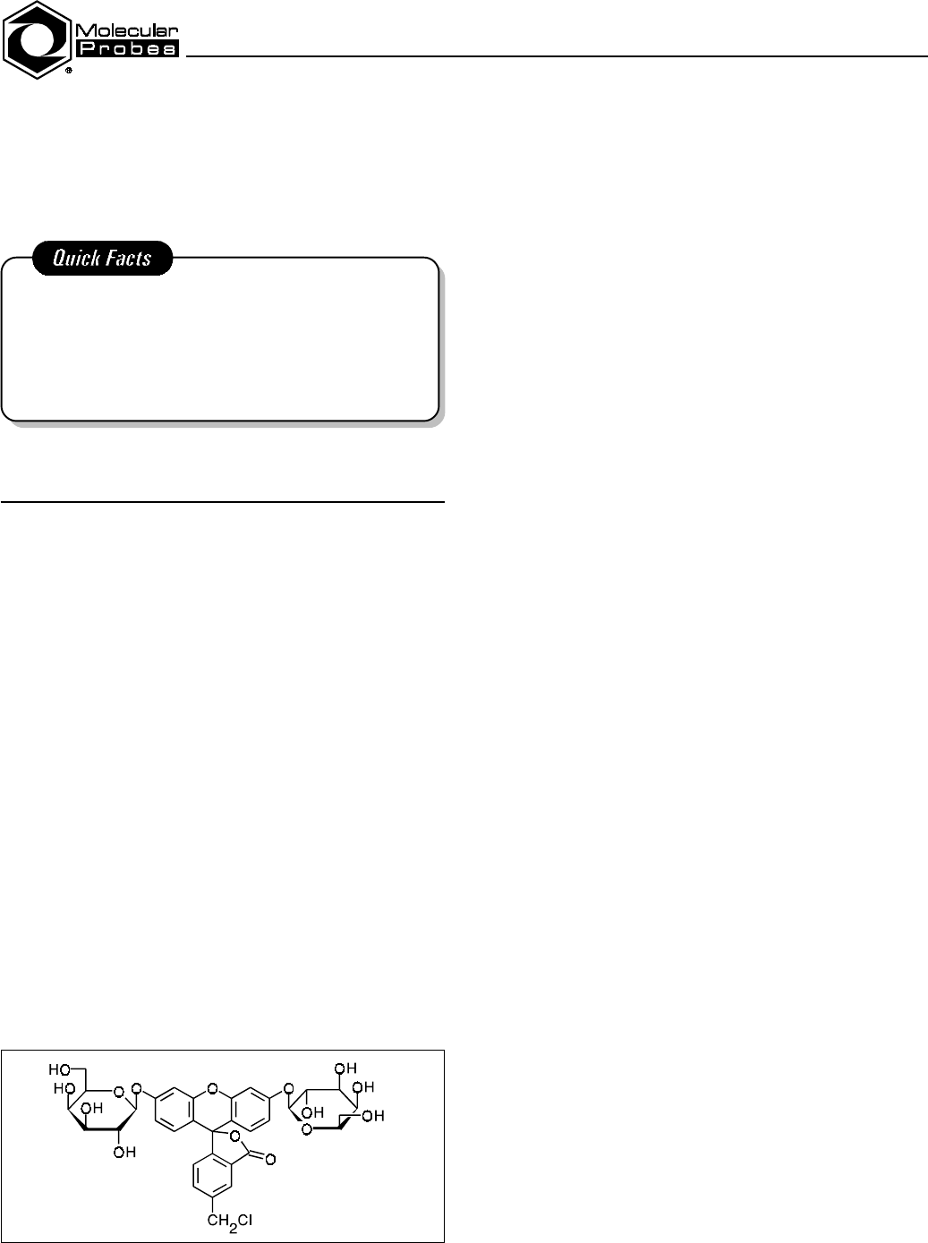
Figure 1. Structure of CMFDG.
Introduction
The Escherichia coli β-D-galactosidase gene (lacZ) is an im-
portant reporter gene for detecting the expression of recombinant
genes in animal cells. Once reporter genes are fused with other
genes or genomic regulatory elements, the resulting DNA con-
structs can be introduced into cells of interest and the reporter
gene product assayed. In present analytical techniques, tran-
scription from the transfected promoter is monitored by RNA
analysis or by the detection of an encoded protein product. Typi-
cally, reporter genes encode enzymes not ordinarily found in the
type of cell being studied, and their unique activity is monitored
to determine the degree of transcription of the foreign genetic
material. The E. coli lacZ gene has been extensively studied and
utilized for this purpose.
5-Bromo-4-chloro-3-indolyl galactopyranoside (X-gal) is
commonly used for detection of genes fused in frame with the
lacZ gene. When X-gal is cleaved, an intensely blue halo-
genated indoxyl derivative is formed that is effective for visual
identification of transformed cells. However, the cleavage prod-
uct of X-gal is nonfluorescent and toxic to viable cells and there-
fore not useful for fluorescence-activated cell sorting analysis.
For this reason, the fluorescent β-galactosidase substrate, fluo-
rescein di-β-
D-galactopyranoside (FDG), has been used for a
highly sensitive flow cytometric β-galactosidase assay.
1,2
Under
physiological conditions, however, the fluorescent hydrolysis
product (fluorescein) leaks quickly from the lacZ-positive cells
after enzymatic cleavage. To retard leakage, the cells must be
maintained in conditions that reduce cell viability prior to
β-galactosidase detection.
To overcome the limitations of these substrates, scientists at
Molecular Probes have developed the DetectaGene Green
CMFDG lacZ Gene Expression Kit with a unique β-galacto-
sidase substrate that yields a bright green fluorescent product
with greatly improved cellular retention. The fluorogenic sub-
strate in our DetectaGene Green lacZ Gene Expression Kit is
5-chloromethylfluorescein di-β-
D-galactopyranoside (CMFDG,
Figure 1). This substrate has been designed to react with intra-
cellular glutathione, a ubiquitous tripeptide, through a glu-
tathione S-transferase–mediated reaction. In lacZ-positive cells,
the CMFDG–glutathione adduct is subsequently converted to a
bright green fluorescent product. Because peptides do not
readily cross cellular membranes, the resulting fluorescein–
glutathione adduct is well retained, even in cells that have been
incubated for 18 hours in fresh medium at 37°C.
The DetectaGene Green Kit also includes stock solutions of
phenylethyl β-
D-thiogalactopyranoside (PETG), chloroquine
diphosphate, propidium iodide and verapamil. PETG is a com-
petitive inhibitor of β-galactosidase that can be added to termi-
nate reactions prior to analysis. Chloroquine may be used to
raise lysosomal pH and thereby inhibit the interfering endo-
genous lysosomal β-galactosidase activity present in some mam-
malian cells. Propidium iodide is useful for identifying dead
cells in the population; this dye permeates damaged plasma
membranes of dead cells and results in a red fluorescent nuclear
stain. Verapamil, when added to the medium, can greatly en-
hance the signals obtained following intracellular hydrolysis of
CMFDG by β-galactosidase; verapamil apparently blocks the
efflux of fluorescent products produced from the reaction.
3
In
addition, the DetectaGene Green Kit includes a vial of our
Influx™ pinocytic cell-loading reagent.
The Influx pinocytic cell-loading reagent provides a conven-
ient, rapid and simple procedure for loading CMFDG into live
cells. With the Influx reagent, CMFDG can be introduced into
many cells simultaneously without significantly altering normal
cell function. In general, the Influx reagent provides a more
gentle cell-loading method than the typical cell-loading tech-
niques of microinjection, electroporation, hypotonic shock or
scrape loading, which are all physically disruptive to cells. Sci-
entists at Molecular Probes have found using the Influx reagent
to be the best method for loading CMFDG into live cells.
The Influx cell-loading technique is based on the osmotic
lysis of pinocytic vesicles, a technique introduced by Okada and
Rechsteiner.
4
Briefly, compounds to be loaded are mixed at
high concentration with a hyperosmotic medium, allowing the
DetectaGene™ Green CMFDG lacZ Gene Expression Kit2
material to be carried into the cells via endocytosis. The cells are
then transferred to a hypotonic medium, which results in the re-
lease of trapped material from the pinocytic vesicles within the
cells, filling the cytosol with the compound (Figure 2). Park and
colleagues showed that endosomal compartments containing the
hypertonic loading medium do not fuse with lysosomes.
5
There-
fore, materials introduced into cells by the Influx cell-loading
technique are not exposed to lysosomal enzymes. Furthermore,
lysosomal components are not released into the cytosol as a con-
sequence of the procedure.
Materials
Contents
••
••
• DetectaGene Green substrate reagent (Component A),
100 µL of 10 mM 5-chloromethylfluorescein di-β-
D-
galactopyranoside (CMFDG) in 1:1 (v/v) water/
dimethylsulfoxide (DMSO)
••
••
• PETG (Component B), 1 mL of 50 mM phenylethyl β-
D-
thiogalactopyranoside in water
••
••
• Chloroquine (Component C), 1 mL of 30 mM chloroquine
diphosphate in water
••
••
• Propidium iodide (Component D), 1 mL of 150 µM
propidium iodide in water
••
••
• Verapamil (Component E), 1 mL of 100 mM verapamil
hydrochloride in 1:1 (v/v) water/DMSO
••
••
• Influx pinocytic cell-loading reagent (Component F),
one plastic tube containing an optimized mixture of polyeth-
ylene glycol (PEG) and sucrose crystals
Storage and Handling
The stock CMFDG reagent is stable for several months if
stored frozen and protected from light. To reduce decomposition
of this reagent during freezing and thawing, we recommend that
you divide the reagent into several small aliquots and store at
-20°C. Do not keep the CMFDG working solution at elevated
temperatures for extended periods, as spontaneous hydrolysis
may occur. Note: The presence of a pronounced yellow color in
the CMFDG reagent or observation of an unusually high fluores-
cent background in the cells may indicate deterioration of the
reagent.
The other reagents included in this kit are also stable for
several months when stored at -20°C. Minimize exposure to
light. The Influx cell-loading reagent may be stored at room
temperature.
Experimental Protocols
The DetectaGene Green CMFDG lacZ Gene Expression Kit
can be used for either flow cytometric analysis or fluorescence
imaging of β-galactosidase–containing cells. The following pro-
tocols are suggested as basic methods for loading CMFDG into
live cells and detecting fluorescence in a flow cytometer or fluo-
rescence microscope. Protocol I describes the use of the Influx
reagent for loading CMFDG into cells. Protocol II describes the
use of hypotonic shock to load cells, and Protocol III describes a
method for direct loading of cells. Also described are methods
for using the competitive inhibitor, PETG, to slow or completely
block β-galactosidase activity, methods for using chloroquine to
lower the background from endogenous lysosomal β-galactosi-
dase activity, which is present in some cells, and methods for
using verapamil to help prevent the efflux of fluorescent reaction
products and thereby boost the signal obtained. These protocols
should serve as guidelines and may require modification based
on specific experimental requirements.
The DetectaGene Green CMFDG lacZ Gene Expression Kit
provides sufficient reagents for 50–200 assays when the
CMFDG is loaded into cells in suspension, or 10–25 assays
when the CMFDG is loaded into cells adhering to coverslips.
Protocol I: Loading Cells Using Influx Reagent
Preparation of Solutions
Hypertonic Loading Medium
1.1 Prewarm 5 mL culture medium, without serum, to 37°C.
1.2 Add 4.7 mL of warm medium to the plastic tube containing
the Influx pinocytic cell loading reagent (Component F).
1.3 Place the tube in very hot water (~80°C) for 2 minutes.
1.4 Vortex several times over a 5 minute period to completely
dissolve the PEG and sucrose crystals.
1.5 Remove the cap and add 250 µL of serum (the type required
for your cell line) and 50 µL of 1 M HEPES buffer, pH 7.4, or
other suitable buffer, to the tube.
1.6 Replace the cap and mix by vortexing several times.
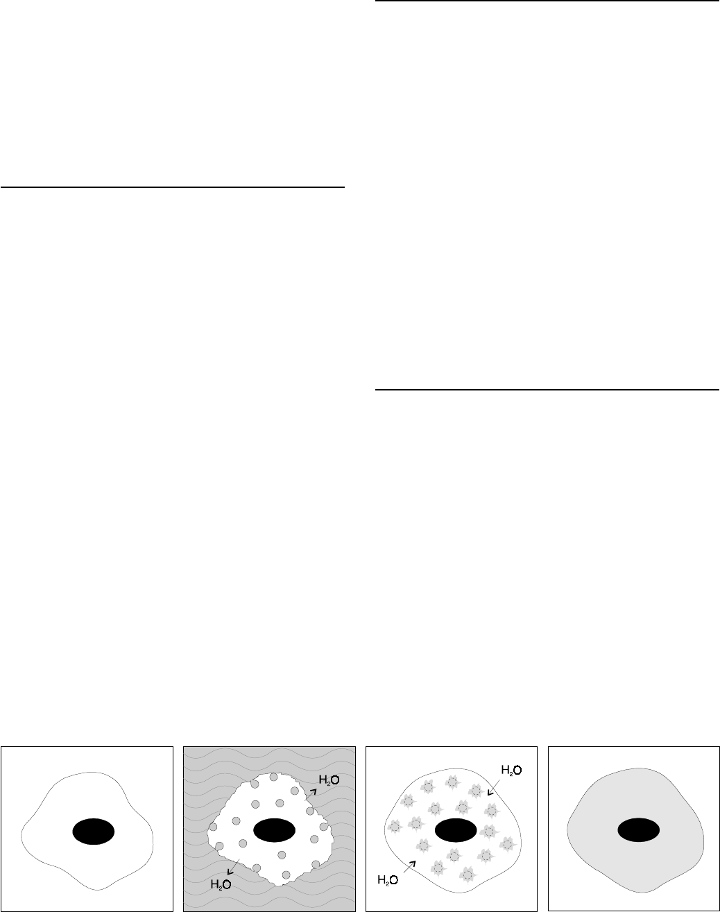
Figure 2. Principle of the Influx reagent cell-loading method. Cultured cells (A) are exposed to Influx Hypertonic Loading Medium containing the
CMFDG, which is carried into the cells via pinocytic vesicles (B). When the cells are placed in Hypotonic Lysis Medium, the pinocytic vesicles burst
(C), releasing CMFDG into the cytosol (D).
ABCD
DetectaGene™ Green CMFDG lacZ Gene Expression Kit3
1.7 Maintain the Hypertonic Loading Medium at the optimal
temperature for your cell line. CMFDG should be added to the
Hypertonic Loading Medium immediately prior to use. DO NOT
dilute the Hypertonic Loading Medium to less than 70% strength
when adding the compound to be loaded into the cells.
Note: The Hypertonic Loading Medium may be filter-sterilized
following steps 1.4 or 1.6. We recommend using a 0.8/0.2 µm
Supor
®
Acrodisc
®
PF syringe sterilization filter (Gelman Sci-
ences #4187). Filter-sterilized Hypertonic Loading Medium may
be stored at 4°C for later use.
Hypotonic Lysis Medium
Prepare Hypotonic Lysis Medium by combining culture me-
dium, without serum, and sterile deionized water in a 6:4 ratio.
The volume required per loading will vary from 3–10 mL de-
pending upon the method used.
Recovery Medium
Prepare 10 mL of culture medium supplemented with serum
(i.e. the growth medium used for your cell line).
Loading Cells in Suspension Using the Influx Reagent
2.1 For each sample, prewarm at least 20 µL of Hypertonic
Loading Medium containing 1 mM CMFDG to the ideal growth
temperature for your cell type. In addition, prewarm 3 mL of
Hypotonic Lysis Medium and ~2 mL of Recovery Medium, as
well as all glassware. The following protocol assumes that the
ideal temperature is 37°C.
2.2 Use trypsin or EDTA to remove cells from the surface of cul-
ture dishes or flasks, or use cells that are naturally in suspension
(note A).
2.3 Wash the cells to remove the trypsin or EDTA by suspending
the cells in medium and then pelleting the cells by centrifugation.
2.4 Resuspend the cells in a 1 mL volume of fresh medium so
that the cell density is no higher than 1 × 10
6
cells per mL.
Transfer the cell suspension to a sterile 1.5 mL microfuge tube.
2.5 Pellet the cells by centrifugation in a microfuge for 2 minutes
at 2000 rpm.
2.6 Carefully remove the supernatant solution. Make sure to re-
move as much of the supernatant solution as possible to mini-
mize dilution of the Hypertonic Loading Medium, which will be
added next.
2.7 Add 20 µL of prewarmed Hypertonic Loading Medium con-
taining 1 mM CMFDG. Gently resuspend the cells by tapping
on the tube. If desired, the suspension medium may contain
200 µM verapamil to inhibit the efflux of the fluorescent product
(note B).
2.8 Incubate the cells at 37°C for 10 minutes.
2.9 Quickly, but gently, add 1 mL of Hypotonic Lysis Medium to
the cell suspension, then transfer the suspension to a separate
5 mL tube containing 2 mL of Hypotonic Lysis Medium.
2.10 Aliquot the cell suspension between two 1.5 mL microfuge
tubes, then incubate the cells for 1.5 minutes at 37°C. Longer
exposure to the Hypotonic Lysis Medium may result in blebbing
of the cell membranes and loss of cell viability.
2.11 Pellet the cells by centrifugation in a microfuge for 2 min-
utes at 2000 rpm.
2.12 Quickly, but carefully, remove the supernatant.
2.13 Add at least 1 mL of Recovery Medium to each microfuge
tube and resuspend the cells.
2.14 Allow 30 minutes prior to observing the cells. Alternatively
you can plate them immediately onto fresh coverslips, culture
dishes or flasks for future examination. If desired, 1.5 µM
propidium iodide may be included in the Recovery Medium to
facilitate the identification of dead cells (note C).
Loading Adherent Cells Using the Influx Reagent
3.1 Prewarm at least 100 µL of Hypertonic Loading Medium
containing 1 mM CMFDG to the ideal growth temperature for
your cell type. In addition, prewarm ~10 mL of Hypotonic Lysis
Medium and 10 mL of Recovery Medium, as well as all glass-
ware. The following protocol assumes that the ideal temperature
is 37°C.
3.2 Using sterile forceps, remove a coverslip from the culture
dish in which the cells were grown.
3.3 Touch the edge of the coverslip to a sterile Kimwipe
®
to
remove excess media.
3.4 Place the coverslip cell-side up in a staining dish (a 60 or
100 mm–tissue culture dish with a lid). To ensure that the cover-
slip does not adhere to the dish, we recommend using a “pedes-
tal,” e.g. resting the coverslip on the inverted top removed from a
1.5 mL microfuge tube or on a 10 mm–diameter O-ring, steril-
ized with ethyl alcohol.
3.5 Quickly, but gently, pipet 100 µL of the prewarmed Hyper-
tonic Loading Medium, containing 1 mM CMFDG, onto a cor-
ner of the coverslip so that the viscous Hypertonic Loading
Medium will displace the small amount of residual medium
without significantly diluting the loading solution.
3.6 Place the lid on the staining dish and incubate the coverslip
at 37°C for 10 minutes.
3.7 Using sterile forceps, quickly, but gently, lift the coverslip
and remove the excess Hypertonic Loading Medium by touching
an edge of the coverslip to a sterile Kimwipe. As an alternative,
gently remove staining solution by tipping coverslip and pipet-
ting off the solution.
3.8 Place the coverslip vertically in a coverslip staining jar filled
with at least 7 mL of prewarmed Hypotonic Lysis Medium, mak-
ing certain that the coverslip is fully submerged.
3.9 Incubate the coverslip for only 2 minutes in the Hypotonic
Lysis Medium. Longer exposure to the Hypotonic Lysis Medium
may result in blebbing of the cell membranes and loss of cell
viability.

DetectaGene™ Green CMFDG lacZ Gene Expression Kit4
3.10 Using sterile forceps, quickly, but gently, remove the cover-
slip from the Hypotonic Lysis Medium. Touch an edge of the
coverslip to a sterile Kimwipe to remove excess medium.
3.11 Submerge the coverslip in ~10 mL of prewarmed Recovery
Medium in a new coverslip staining jar or staining dish. If de-
sired, 1.5 µM propidium iodide may be included in the Recovery
Medium to facilitate the identification of dead cells (note C).
3.12 Allow the cells on the coverslip to recover at 37°C for at
least 30 minutes before observing in the microscope.
Protocol II: Loading Cells by Hypotonic Shock
Preparation of Solutions
Make up 10 mL of Staining Medium. A typical staining
medium is phosphate-buffered saline (PBS), 4% (v/v) fetal calf
serum and 10 mM HEPES, pH 7.2.
Loading Cells in Suspension by Hypotonic Shock
4.1 Centrifuge the cells to obtain a cell pellet and aspirate the
supernatant (note A). Resuspend the cells in Staining Medium
(prepared as described above) and draw the sample through a
pipet to obtain a single cell suspension. Filter out any cell
clumps with a nylon screen. Centrifuge the cells again and
remove the supernatant.
4.2 Resuspend the cells in Staining Medium to approximately
10
7
cells/mL (note D) and pipet 100 µL into a centrifuge tube.
If inhibition of endogenous β-galactosidase is desired, prepare
Staining Medium with 1 mM chloroquine diphosphate (freshly
diluted from the 30 mM stock solution (Component C);
concentrations greater than 1 mM may be deleterious to cells
(note E). Proceed to step 4.3 immediately, or put the cells on ice.
4.3 Pre-warm the tube containing 100 µL of the cells in a 37°C
water bath for 10 minutes, or for 30 minutes when inhibiting en-
dogenous β-galactosidase with chloroquine diphosphate.
4.4 Immediately before use, prepare 100 µL of 200 µM CMFDG
substrate working solution in deionized water from the 10 mM
stock solution (Component A) (notes F and G). Warm the solu-
tion at 37°C for about 10 minutes.
4.5 Combine 100 µL of the pre-warmed CMFDG substrate
working solution with the 100 µL of pre-warmed cells from step
4.3. Mix rapidly and thoroughly. Return the sample to the 37°C
water bath for 2 minutes. Note: The optimal working concen-
tration of the CMFDG substrate must be determined experi-
mentally. The recommended 200 µM working concentration
suggested may have to be varied based on the method of loading
(note G) and the level of β-galactosidase activity in cells.
4.6 Stop the CMFDG loading at the end of 2 minutes by adding
1.8 mL of Staining Medium to the 200 µL volume of CMFDG
and cell suspension.
4.7 Wash the cells by centrifugation and resuspend them in
2.0 mL of Staining Medium. If desired, 1.5 µM propidium io-
dide may be included in the Staining Medium to facilitate the
identification of dead cells (note C).
4.8 Keep the cells under normal culture conditions for 30 min-
utes to allow for turnover of the substrate prior to analysis.
Note: At any point after the termination of loading, you may
inhibit further intracellular hydrolysis of the substrate by treat-
ment with PETG (see note H).
Loading Adherent Cells by Hypotonic Shock
5.1 Grow cells on coverslips according to normal tissue culture
procedures. Use cells at a 40% to 70% confluency for best
results (note A). If inhibition of endogenous β-galactosidase is
desired, prepare Staining Medium with 1 mM chloroquine
diphosphate (freshly diluted from the 30 mM stock solution
(Component C)); concentrations greater than 1 mM may be
deleterious to cells
(note E).
5.2 Immediately before use, dilute the CMFDG substrate stock
reagent (Component A) to 400 µM using a 1:1 mixture of deion-
ized water and Staining Medium. Warm the substrate solution at
37°C for 10 minutes. A 100 µL volume will be used for each
coverslip.
5.3 Rinse the cells once with a physiological saline solution,
such as Hank’s balanced salt solution or PBS.
5.4 Place the coverslip with adherent cells in a petri dish. Apply
100 µL of the substrate solution to the coverslip and incubate the
sample at room temperature for 1 minute.
5.5 Stop the CMFDG loading by flooding the petri dish with
Staining Medium. If desired, 1.5 µM propidium iodide may be
included in the Staining Medium to facilitate the identification of
dead cells (note C). Note: Do not remove the substrate solution
before flooding the cells with medium, as this will often wash
away many of the cells.
5.6 Return the cells to the 37°C incubator and allow the cells to
recover for 1–3 hours. Note: At any point after the termination
of loading, you may inhibit furthur intracellular hydrolysis of the
substrate by treatment with PETG (note H).
5.7 Mount the cells in Staining Medium on a slide. Seal and
view immediately. For flow cytometric assay, treat adherent cells
with trypsin in phosphate buffer until they can be removed from
the plate by gentle agitation. Afterwards, remove the trypsin by
washing in Staining Medium. Centrifuge the cell suspension,
aspirate the supernatant and resuspend the cells in Staining
Medium.
Protocol III: Direct Loading of Cells
The following simple procedure has been developed using the
mouse fibroblast CRE BAG 2 cell line, an NIH 3T3-derived cell
line that stably expresses lacZ-encoded β-galactosidase under the
control of a murine leukemia virus promoter. The procedure may
be generally applicable to other cell lines.
6.1 Culture the cells in suitable growth medium (e.g. Dulbecco’s
modification of Eagle’s Minimal Essential Medium (DMEM)
supplemented with 10% fetal bovine serum (FBS), 50 µg/mL
gentamicin, 300 µg/mL
L-glutamine and 10 mM HEPES,
pH 7.4), in a humidified atmosphere of 5% CO
2
in air.

DetectaGene™ Green CMFDG lacZ Gene Expression Kit5
Subculture every 2 to 3 days by trypsinization using 0.05%
trypsin and 0.02% EDTA in phosphate-buffered saline (PBS)
(note A).
6.2 Prior to an experiment, trypsinize the cells, collect by cen-
trifugation and resuspend at a density of 10
6
to 10
7
cells per mL
(note D) in pre-warmed culture medium supplemented with
10% FBS and containing at 50–200 µM CMFDG. A 100 µL
volume is usually sufficient for analysis. If desired, the suspen-
sion medium may contain 200 µM verapamil to inhibit the efflux
of fluorescent product (note B).
6.3 Incubate the cell suspension at 37°C for the desired time
interval, typically 10–60 minutes. Following incubation, the
cells should be placed on ice, or diluted into ice-cold PBS in
order to increase the volume, and then analyzed promptly. Alter-
natively, the turnover of CMFDG can be inhibited by the addition
of PETG (note H).
6.4 For analysis by flow cytometry, propidium iodide can be
diluted into the cell suspension to attain a final concentration of
1.5 µM to facilitate the identification of dead cells (note C), so
that they may be eliminated electronically from the analysis.
Analysis
Flow Cytometry
Set up and calibrate the flow cytometer to detect fluorescein,
propidium iodide and forward scatter according to standard pro-
cedures. Use unstained cells of the same type you are analyzing
to set the background autofluorescence compensation (note I).
Imaging
ββ
ββ
β
-Galactosidase Activity
Fluorescence is detected using standard fluorescein or FITC
filter sets.
Notes
[A] Keep the cells as healthy as possible. Endogenous lysosomal
β-galactosidase activity increases dramatically if the cells are
abused or allowed to reach confluency (see note E on inhibition
of endogenous β-galactosidase activity with chloroquine diphos-
phate).
[B] Verapamil (Component E), an inhibitor of plasma mem-
brane–resident drug efflux systems is effective in reducing the
efflux of fluorescent products produced from the action of
β-galactosidase on the CMFDG substrate.
3
Inclusion of verap-
amil at 100–200 µM in the staining and post-staining media can
result in substantially improved detection of β-galactosidase
activity. Concentrations of verapamil above 200 µM may be
toxic to cells, and the ideal concentration of verapamil for a par-
ticular application may need to be determined experimentally.
[C] Propidium iodide (Component D) is impermeant to the
plasma membrane and selectively labels the nuclei of dead cells
with red fluorescence. Prepare dye solution by diluting the
150 µM propidium iodide stock solution (Component D)
100-fold to obtain a 1.5 µM solution.
[D] Staining results are not critically dependent on the cell
concentration. Staining patterns are essentially the same
using cell concentrations ranging from 10
5
cells/mL to
5 × 10
7
cells/mL.
[E] Some mammalian cells have endogenous lysosomal β-
galactosidase that can interfere with accurate measurement of
lacZ expression. The endogenous activity can be selectively
depressed by pre-incubating cells with the weak base, chloro-
quine (Component C).
[F] For bacterial cells or yeast, the cell wall restricts the swelling
induced by osmotic loading, thus preventing CMFDG entry.
Brief (1–3 minute) hypertonic shrinking of the cell membrane
within the wall, followed by a 2-minute hypotonic loading of
CMFDG can correct this difficulty with entry.
[G] The loading procedure described in steps 4.4 and 5.2 sub-
jects cells to hypotonic shock in order to facilitate entry of the
substrate. This treatment may not be necessary for some cell
types. For loading under isotonic conditions, prepare the
CMFDG working solution in the staining medium instead of the
deionized water and increase the incubation time from 2 to about
30 minutes, or see Protocol III.
[H] Competitive inhibition of β-galactosidase by PETG (Compo-
nent B) can be used to terminate CMFDG turnover prior to
analysis. After terminating CMFDG loading (steps 4.6, 5.5 or
6.3), select a time interval between zero and 60 minutes (zero
time for cells with high lacZ expression levels, 60 minutes for
cells with low lacZ expression levels) and add an aliquot of the
50 mM PETG stock reagent to yield a final PETG concentration
of 1 mM. Mix thoroughly. PETG is a competitive, reversible in-
hibitor of E. coli β-galactosidase in mammalian cells. It is hy-
drophobic and can readily cross the cell membrane to inhibit
β-galactosidase. Because it has a low K
i
(3 × 10
-6
M), very little
PETG is required to inhibit the reaction. In addition, PETG is
not hydrolyzed by the enzyme, which simplifies its influence on
the kinetics.
[I] Some endogenous constituents of cells give rise to broad
bandwidth autofluorescence when excited by the argon-ion laser.
It is essential to compensate for autofluorescence in order to
accurately measure low levels of β-galactosidase activity. Cor-
rection for the autofluorescence component of the emission sig-
nal is typically based on the proportionality of measured
autofluorescence at one wavelength to that at another wave-
length.

DetectaGene™ Green CMFDG lacZ Gene Expression Kit6
References
1. Proc Natl Acad Sci USA 85, 2603 (1988); 2. Cytometry 12, 291 (1991); 3. Cytometry 28, 36 (1997); 4. Cell 29, 33 (1982); 5. J Cell Physiol 135, 443 (1988).
Product List Current prices may be obtained from our Web site or from our Customer Service Department.
Cat # Product Name Unit Size
D-2920 DetectaGene
TM
Green CMFDG lacZ Gene Expression Kit ....................................................................................................... 1 kit
I-14402 Influx
TM
pinocytic cell-loading reagent *makes 10 x 5 mL* ....................................................................................................... 1 set
Contact Information
Further information on Molecular Probes' products, including product bibliographies, is available from your local distributor or directly from Molecular
Probes. Customers in Europe, Africa and the Middle East should contact our office in Leiden, the Netherlands. All others should contact our Technical Assis-
tance Department in Eugene, Oregon.
Please visit our Web site — www.probes.com — for the most up-to-date information
Molecular Probes, Inc.
PO Box 22010, Eugene, OR 97402-0469
Phone: (541) 465-8300 • Fax: (541) 344-6504
Customer Service: 7:00 am to 5:00 pm (Pacific Time)
Phone: (541) 465-8338 • Fax: (541) 344-6504 • [email protected]
Toll-Free Ordering for USA and Canada:
Order Phone: (800) 438-2209 • Order Fax: (800) 438-0228
Technical Assistance: 8:00 am to 4:00 pm (Pacific Time)
Phone: (541) 465-8353 • Fax: (541) 465-4593 • [email protected]
Molecular Probes Europe BV
PoortGebouw, Rijnsburgerweg 10
2333 AA Leiden, The Netherlands
Phone: +31-71-5233378 • Fax: +31-71-5233419
Customer Service: 9:00 to 16:30 (Central European Time)
Phone: +31-71-5236850 • Fax: +31-71-5233419
Technical Assistance: 9:00 to 16:30 (Central European Time)
Phone: +31-71-5233431 • Fax: +31-71-5241883
Molecular Probes’ products are high-quality reagents and materials intended for research purposes only. These products must be used by, or directly
under the supervision of, a technically qualified individual experienced in handling potentially hazardous chemicals. Please read the Material Safety Data
Sheet provided for each product; other regulatory considerations may apply.
Several of Molecular Probes’ products and product applications are covered by U.S. and foreign patents and patents pending. Our products are not
available for resale or other commercial uses without a specific agreement from Molecular Probes, Inc. We welcome inquiries about licensing the use of our
dyes, trademarks or technologies. Please submit inquiries by e-mail to [email protected]. All names containing the designation ® are registered with the
U.S. Patent and Trademark Office.
Copyright 2001, Molecular Probes, Inc. All rights reserved. This information is subject to change without notice.
