2
THE INTERNATIONAL JOURNAL OF ESTHETIC DENTISTRY
VOLUME 11 • NUMBER 2 • SUMMER 2016
CLINICAL RESEARCH
The use of a standardized
gray reference card in dental
photography to correct the effects of
ve commonly used diffusers on the
color of 40 extracted human teeth
Sascha Hein, MDT
Private Dental Laboratory
Michael Zangl, MDT
Private Dental Laboratory
HEIN/ZANGL
CLINICAL RESEARCH
Correspondence to: Sascha Hein, MDT
Dentaltechnik Christ & Hein GmbH, Karl-Benz-Str. 25, 86825 Bad Wörishofen, Germany.
Tel.: 0049 8247 5320 Fax.: 0049 8247 31965; E-mail: [email protected]

3
THE INTERNATIONAL JOURNAL OF ESTHETIC DENTISTRY
VOLUME 11 • NUMBER 2 • SUMMER 2016
HEIN/ZANGL
Abstract
Objective: The aim of this in vitro study
was to investigate the color changes of
human teeth caused by five different dif
-
fuser materials commonly used in dental
photography, as well as software influ
-
ence, and to confirm whether the use
of a standardized gray reference card
is effective in correcting these color
changes during digital postproduction.
Materials and method: Forty extracted
human teeth were obtained from a spe
-
cialized oral surgery practice in Cham,
Germany. Five commonly used diffuser
materials were chosen to be investi
-
gated, which included: polyethylene
(PET), White Frost photographic paper,
LumiQuest polyamide (nylon) mater
-
ial, 80 gsm white printing paper, and
3M linear polarizing filter sheet used for
cross polarization. A digital single-lens
reflex camera (Canon EOS5DMKII) was
used, together with a twin flash suitable
for macrophotography (Canon MT-24EX
Macro Twin Lite). Images were tethered
into Adobe Lightroom CC using the
RAW format. A standardized gray refer
-
ence card (WhiBal, Michael Tapes De-
sign) was used for exposure calibration
and white balancing. Classic Color Me
-
3
THE INTERNATIONAL JOURNAL OF ESTHETIC DENTISTRY
VOLUME 11 • NUMBER 2 • SUMMER 2016
ter software (Ricci Adams, version 1.6
(122)) was used to obtain CIE L*a*b*
values of the specimens before and af
-
ter white balancing and exposure cor-
rection.
Results: All diffusers caused visually
perceivable color changes on the ex
-
tracted teeth: White Frost (∆E* 1.24;
sd 0.47), 80 gsm printing paper
(∆E* 2.94; sd 0.35), LumiQuest polyam
-
ide (∆E* 3.68; sd 0.54), PET (∆E* 6.55;
sd 0.41), and 3M linear polarizing fil
-
ter sheet (∆E* 7.58; sd 1.00). The use
of a standardized gray reference card
(WhiBal) could correct these values be
-
low the visually perceivable threshold:
White Frost (∆E* 0.58; sd 0.36), 80 gsm
printing paper (∆E* 0.93; sd 0.54), Lu
-
miQuest polyamide (∆E* 0.66; sd 0.58),
PET (∆E* 0.59; sd 0.33), and 3M linear
polarizing filter sheet (∆E* 0.53; sd 0.42).
Significance: The use of a standard
-
ized gray reference card with specified
CIEL*a*b* values should be considered
when diffusers are used in dental pho
-
tography in order to reveal the color of
preoperative situations (ie, shade docu
-
mentation) and document postoperative
results accurately.
(Int J Esthet Dent 2016;11:XXX–XXX)

4
THE INTERNATIONAL JOURNAL OF ESTHETIC DENTISTRY
VOLUME 11 • NUMBER 2 • SUMMER 2016
CLINICAL RESEARCH
wavelengths will pass through. Due to
absorption, only specific wavelengths
that are characteristic of the material will
be transmitted. Hence, all the power that
is transmitted is concentrated in a few
narrow wavelength regions,
7
causing
large color distortions, since they affect
both the Correlated Color Temperature
(CCT) and the Color Rendering Index
(CRI) of the emitted light, making it diffi
-
cult to judge shade differences between
a shade tab or dental restoration and the
surrounding natural dentition on a digi
-
tal image. Preliminary relative irradiance
measurements of five commonly used
diffuser materials which were included
in this study, using a radiospectrometer
(Sekonic C-700, Sekonic), in conjunc
-
tion with a commonly used electronic
flash (Canon MT-24EX Macro Twin Lite),
revealed that different diffuser mater
-
ials did indeed influence CCT and CRI,
but only slightly. However, the visually
perceivable effects appeared notice
-
able in the digital images, suggesting
that software interpretation might play
a significant role (Fig 1). The use of a
standardized gray reference card prom
-
ises to overcome this limitation through a
remapping process of the original RAW
image to a defined standard. However,
natural teeth are heavy light scatter
-
ers, and irradiation with an intermittent
spectral power distribution may affect
their color within the threshold of visual
perception. The aim of this study was
to determine the effects that five com
-
monly used diffuser materials have on
tooth color, to identify their origin, and
to determine if the use of a gray refer
-
ence card is effective in correcting these
changes.
Introduction
The use of dental photography plays an
increasingly important role in everyday
dental practice as an effective tool for
communication between the dental sur
-
gery and the dental laboratory. Modern
digital single-lens reflex (DSLR) camer
-
as are in common use to document im-
portant restorative aspects such as the
preoperative situation, the tooth shade,
the final result, and long-term perfor
-
mance.
1
Photographic documentation
for purely medical purposes requires lit
-
tle more than basic equipment, such as
a DSLR camera paired either with a ring
or twin flash.
2
In the field of esthetic den-
tistry, however, elaborate assemblies
are often used to depict the restorative
process and especially the final result in
a rather “emotional” way, with the use of
various bouncers and diffusers and ad
-
justable brackets.
3
On the other hand,
cross polarized photography is a useful
method to reveal intrinsic shade varia
-
tions of natural teeth for the purpose of
shade analysis.
4
This is achieved with
the help of a linear polarizing filter sheet
that is placed over the electronic flash
in an orientation which is perpendicular
to that of another linear polarizing filter
simultaneously placed over the lens, re
-
sulting in the exclusion of diffuse light
and specular reflection from the labial
surface of natural teeth and dental res
-
torations alike.
5
Clinical experience has shown that
in vivo photographs of natural dentition
routinely show significant color altera
-
tions of teeth and soft tissue when cer-
tain types of diffusers are used.
6
When
a diffuser is placed in front of an illu
-
minant (ie, an electronic flash), not all
5
THE INTERNATIONAL JOURNAL OF ESTHETIC DENTISTRY
VOLUME 11 • NUMBER 2 • SUMMER 2016
HEIN/ZANGL
Materials and method
Camera set-up
A digital single-lens reflex camera (Can-
on EOS 5D MKII) was used, together
with a twin flash suitable for macro pho
-
tography (Canon MT-24EX Macro Twin
Lite) (Figs 2 and 3). Images were teth
-
ered into Adobe Lightroom CC using
the RAW format and a USB 2.0 cable.
The screen (Cinema Display, Apple)
was calibrated using a spectrophotom
-
eter (ColorMunki, Pantone). The working
distance between the front of the lens
and the labial surface of one randomly
selected test specimen was varied to
achieve life-size magnification (1:1) at a
constant distance of 130 mm, as would
be the case in a clinical situation, and
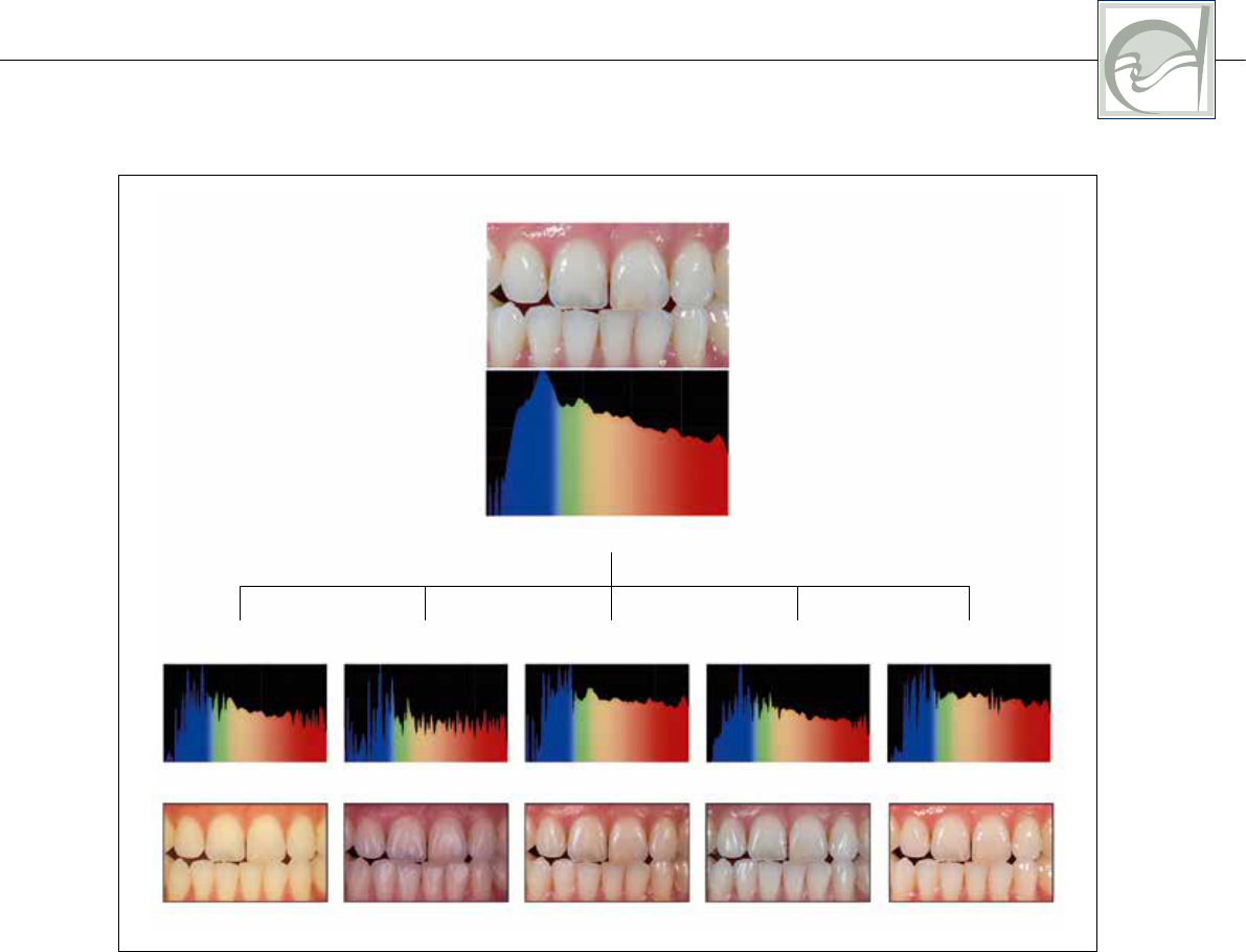
Fig 1 Relative irradiance measurements reveal small influences on the Color Rendering Index (CRI)
and Correlated Color Temperature (CCT) caused by five diffusers when placed in front of an electronic
flash. Note that a variance in the rendering of the images occurs due to software interpretation of the DSLR
camera.
Canon MT-24EX Macro Twin Lite
6400 K (CRI Ra 100)
Linear polarizer
Printing paper
(80 gsm)
LumiQuest SoftBox White Frost Polyethylene PET
CRI Ra 97.2 CRI Ra 96.0 CRI Ra 97.6 CRI Ra 97.6CRI Ra 92.4
5864 K 5775 K 6108 K 5407 K5971 K
380 780
380 780 380 780 380 780 380 780 380 780
CLINICAL RESEARCH
6
THE INTERNATIONAL JOURNAL OF ESTHETIC DENTISTRY
VOLUME 11 • NUMBER 2 • SUMMER 2016
hence this value was chosen as the
standard distance for all measurements.
Using the camera’s manual mode, ex
-
posure time and aperture were set to a
constant value of 125/sec and f32. The
twin flash normally operates with four
AA Mignon batteries (Energizer Ultimate
Lithium, +AA 1.5V, 3000mAh). However,
preliminary tests showed noticeable var
-
iations in flash intensity after a few meas-
urement cycles due to battery depletion
and increased recycle time. In order to
overcome these limitations, a compact
battery pack which normally holds eight
additional AA Mignon batteries (Canon
CP-E4) was modified to be attached to
a 12 V, 1500 mAh direct current trans
-
former (Yumatron, Model NT6), ensuring
steady flash intensity and short recycling
times (< 5 s). The camera was attached
to a microcomputer (Stack Shot, Cogn
-
isys) that was programmed to trigger the
camera shutter nine times in a row, with
a precisely timed interval of 15 s.
Diffuser materials
Five commonly used diffuser materials
were chosen for the study: polyethyl
-
ene (PET), White Frost photographic
paper (ProTech Lighting), Mini SoftBox
polyamide (nylon) material (LumiQuest),
80gsm white printing paper, and 3M lin
-
ear polarizing filter sheet used for cross
polarization. The materials were cut into
squares and attached to a set of cus
-
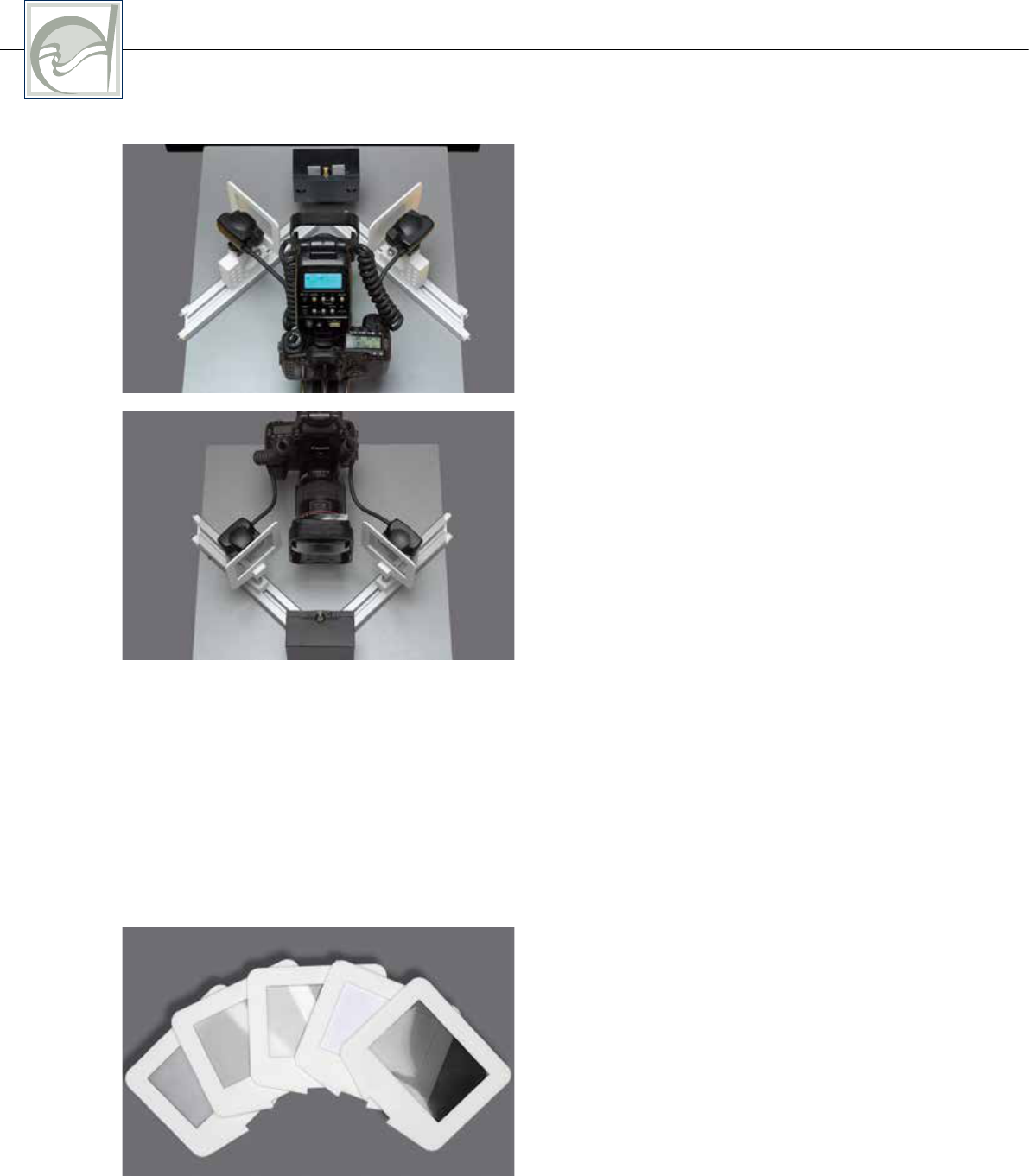
Figs 2 and 3 The experimental setup consisted
of a DSLR camera paired with a twin flash com-
monly used for dental photography. In order to mini-
mize specular reflection from the labial surface of
the teeth, both electronic flash guns were arranged
in two azimuthal illumination angles correspond-
ing to 0 degrees/45 degrees geometry using two
aluminum rails and two custom 3D-printed variable
sled assemblies.
Fig 4 Five commonly used diffuser materials were
chosen for the study. They included polyethylene
(PET), White Frost photographic paper, LumiQuest
polyamide (nylon) material, 80gsm white printing
paper, and 3M linear polarizing filter sheet used for
cross polarization. Each frame had an open window
of 80mmx55mm.

HEIN/ZANGL
7
THE INTERNATIONAL JOURNAL OF ESTHETIC DENTISTRY
VOLUME 11 • NUMBER 2 • SUMMER 2016
tom-made frame holders with an open
window of 80 mmx55 mm (Fig 4).
Specimen assembly
In order to minimize specular reflection
from the labial surface of the teeth, both
electronic flash guns were arranged in
two azimuthal illumination angles cor
-
responding to 45 degrees/0 degrees
geometry using two aluminum rails and
two custom 3D-printed variable sled
assemblies. The specimen holder con
-
sisted of a square block made of mela-
mine, which was designed to hold one
extracted tooth in its middle which could
be exchanged and repositioned pre
-
cisely using a round ABB grid pattern
(LEGO). An adjustable slot allowed the
attachment of two square pieces of the
gray reference card in the same vertical
plane as the tooth specimen. The dis
-
tance from each of the diffusers to the
labial surface of a randomly chosen test
specimen was 150 mm.
Specimen preparation
Forty-four extracted, unrestored teeth
were delivered to the dental labora
-
tory already stored in a 0.9% solution of
thymol. The teeth had been previously
cleaned and pumiced before visual in
-
spection for suitability was carried out.
Four specimens were discarded be
-
cause they showed severe signs of
damage from the extraction surgery. The
remaining 40 teeth (Table 1) were mildly
sandblasted with 50 µm aluminum ox
-
ide to remove the surface gloss from the
enamel in order to avoid specular reflec
-
tion that could obstruct color measure-
ments. The tips of the roots were cut
off before they were attached to round
ABB grid patterns (LEGO) using super
-
glue gel and accelerator spray, while
fixing them in a perpendicular position
using the sample holder for guidance.
The specimens were numbered and re
-
turned to a jar that contained 0.9% thy-
mol solution to preserve their color.
White balance reference card
A standardized white balance refer-
ence card (WhiBal, Michael Tapes De-
sign) was used. This particular product
was chosen because of its even reflec
-
tance and its defined color coordinates
(CIE L*75; a*0; b*0). The manufactur
-
er claims a chromaticity accuracy of
∆C*< 0.71 (a*±0.5; b*±0.5) (Fig 5). In
a previous investigation, triple measure
-
ments of 14 individual new WhiBal cards
were carried out with a spectrophotom
-
eter (ColorMunki) to confirm this claim
(∆C*0.29). One WhiBal card was ran
-
domly chosen and cut into two squares
to be used on either side of the tooth dur
-
ing the entire measurement sequence.
Table 1 Forty extracted human teeth were obtained from a specialized oral surgery practice in Cham,
Germany, and deemed suitable for inclusion in the study
Tooth 14 16 17 18 22 23 24 26 27 28 31 33 36 37 38 41 44 46 47
Quantity 4 1 4 2 1 1 1 1 1 5 2 2 4 3 2 1 1 3 1
CLINICAL RESEARCH
8
THE INTERNATIONAL JOURNAL OF ESTHETIC DENTISTRY
VOLUME 11 • NUMBER 2 • SUMMER 2016
Custom white balance was carried out
using the camera’s menu function and
one WhiBal card, which was positioned
in the same horizontal plane and dis
-
tance as the tooth specimen.
Measurement sequence
Each measurement sequence com-
menced with a hydrated tooth in place
and two squares of WhiBal cards to the
left and right of it, with two empty frame
holders in front of each electronic flash.
The first four photographs were taken
in this way, followed by one photograph
each using five different diffuser ma
-
terials: PET, White Frost, LumiQuest,
80 gsm printing paper, and 3M linear
polarizing sheet. In order to obtain im
-
ages with increased tonality and dy-
namic range, as well as reduced noise,
the concept of “exposing to the right”
(ETTR) was used, with red–green–blue
(RGB) values distributed predominantly
to the right of the exposure histogram.
8
The camera ISO was set to a value of
100, and the flash intensity to a value of
½ (half), except for the 80 gsm printing
paper and linear polarizing sheet, due
to the noticeable attenuation of luminous
flux. For adequate comparability with the
standard, an adjustment of the ISO to a
value of 200, as well as an adjustment of
the flash intensity to a value of 1/1 (full)
was required with this particular group.
Each complete measurement cycle took
a total of 135 s.
Digital image development
and color measurements
The first image was immediately dis-
carded since it only served to empty
the flash capacitor. The following three
Fig 5 The color accuracy of 14 new gray reference cards (WhiBal) was measured and confirmed to be
within the margin of error claimed by the manufacturer (∆C*<0.71; (a*±0.5; b*0.5)).
a b c
L*
75.00
75.32
75.63
75.95
76.26
76.58
76.89
77.21
77.53
77.84
76.53
a* b*
-0.50
-0.42
-0.34
-0.26
-0.18
-0.11
-0.03
0.05
0.13
0.21
0.29
0.37
0.45
-0.14
-0.50
-0.42
-0.34
-0.26
-0.18
-0.11
-0.03
0.05
0.13
0.21
0.29
0.37
0.45
-0.25
HEIN/ZANGL
9
THE INTERNATIONAL JOURNAL OF ESTHETIC DENTISTRY
VOLUME 11 • NUMBER 2 • SUMMER 2016
images taken with no diffuser were ex-
posure balanced by moving the cursor
over the gray area of the WhiBal card.
Due to the camera’s custom white bal
-
ance, it was merely necessary to adjust
the exposure of the image until the lu
-
minosity value of the gray card in the
photograph matched L*75. The values
for chromaticity (a*+b*) were within the
threshold of ±0.5 each time, as claimed
by the manufacturer. The same proced
-
ure was carried out with the five photo-
graphs taken with each diffuser material.
Color Meter Classic software (Ricci Ad
-
ams, version 1.6 (122)) was used to lo-
cate an area in the middle of each tooth.
The measurement window was adjusted
to the maximum size possible within the
boundary of the tooth in order to meas
-
ure CIEL*a*b* color coordinates. Once
this position was locked, values for each
tooth were copied and pasted into a
spreadsheet (Numbers (version 3.5),
Apple) (Fig 6). In order to determine the
standard error caused by subtle varia
-
tions in flash intensity, the first three sets
of color coordinates from the gray card
where averaged and compared with the
ideal value of L*75; a*0; b*0. If ∆E* was
< 1.0, the measurement sequence was
included in the study; if the value was
∆E* > 1.0, the tooth was to be measured
again (which was never the case). Once
the color coordinates for the five differ
-
ent diffuser groups were recorded, white
balancing was carried out by choosing
the eyedropper tool in Adobe Lightroom
CC and clicking on a randomly chosen
gray area in close proximity to the tooth.
In most cases it was then necessary to
adjust the exposure values again to ob
-
tain L*75; a*0; b*0, before the color coor-
dinates of the tooth could be recorded.
Results
∆E* was calculated as described in the
CIE prescriptions:
∆E*
ab
= √(L*
2
- L*
1
)
2
+ (a*
2
- a*
1
)
2
+ (b*
2
- b*
1
)
2
All diffuser materials caused visually per-
ceivable color changes on the extracted
teeth. The values for the different diffus
-
er materials before and after white bal-
ance correction can be seen in Figure 7:
White Frost (∆E* 1.24; sd 0.47), 80 gsm
printing paper (∆E* 2.94; sd 0.35), Lu
-
miQuest polyamide (∆E* 3.68; sd 0.54),
PET (∆E* 6.55; sd 0.41), and 3M linear
polarizing filter sheet (∆E* 7.58; sd 1.00).
The use of a standardized gray refer
-
ence card (WhiBal) could correct these
values below the visually perceivable
threshold (Fig 8): White Frost (∆E* 0.58;
sd 0.36), 80 gsm printing paper
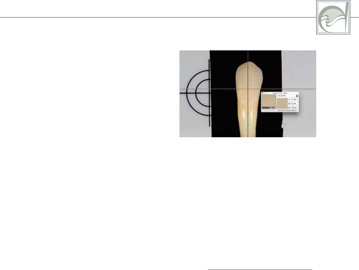
Fig 6 Color measurements were carried out using
Classic Color Meter software in the middle of each
tooth. The measurement window was adjusted to
the maximum size possible within the boundary of
the tooth to measure CIEL*a*b* color coordinates.
Once this position was locked, values for each tooth
were recorded in the exact same position.
CLINICAL RESEARCH
10
THE INTERNATIONAL JOURNAL OF ESTHETIC DENTISTRY
VOLUME 11 • NUMBER 2 • SUMMER 2016
(∆E* 0.93; sd 0.54), LumiQuest polyam-
ide (∆E* 0.66; sd 0.58), PET (∆E* 0.59;
sd 0.33), and 3M linear polarizing filter
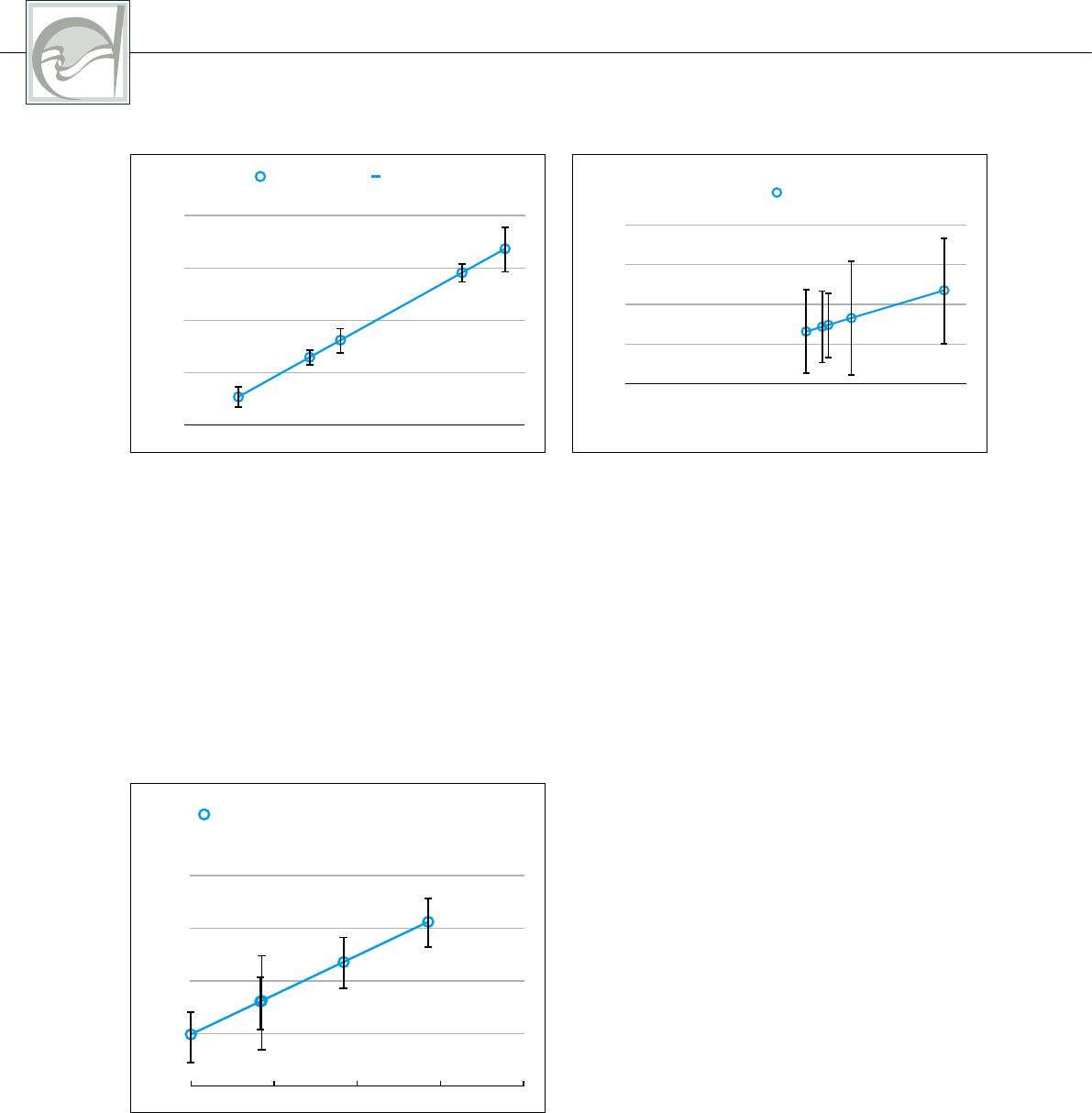
Fig 7 Color changes of 40 extracted teeth caused
by five diffuser materials commonly used in den-
tal photography. White Frost (∆E* 1.24; sd 0.47),
80gsm printing paper (∆E* 2.94; sd 0.35), Lumi-
Quest polyamide (∆E*3.68; sd0.54), PET (∆E*6.55;
sd0.41), and linear polarizing filter sheet (∆E*7.58;
sd1.00).
∆E
0.00
2.25
Trend 1
4.50
6.75
9.00
2 4 6 8
y = x
R
2
= 1
∆E
1.24
∆E
2.94
∆E
3.68
∆E
6.55
∆E
7.58
Fig 8 The use of a standardized gray reference
card (WhiBal) could correct the color changes be-
low the visually perceivable threshold: White Frost
(∆E*0.58; sd0.36), 80gsm printing paper (∆E*0.93;
sd0.54), LumiQuest polyamide (∆E*0.66; sd0.58),
PET (∆E*0.59; sd0.33), and linear polarizing filter
sheet (∆E*0.53; sd0.42).
∆E
0.00
0.40
0.80
1.20
1.60
0 1
y = x + 1.986E-16
R
2
= 1
∆E
0.53
∆E
0.66
∆E
0.93
Fig 9 The average exposure compensation that
was required during digital postproduction was:
White Frost (EV-0.20; sd 0.106), 80gsm printing
paper (EV-0.38; sd0.114), LumiQuest polyamide
(EV-0.55; sd0.108), PET (EV-0.55; sd0.197), and
linear polarizing filter sheet (EV-0.70; sd0.116).
-0.90
-0.68
Average exposure adjustment
-0.45
-0.23
0.00
-0.7 -0.525 -0.35 -0.175
y = x
R
2
= 1
-0.20
0
-0.38
-0.55
-0.70
sheet (∆E* 0.53; sd 0.42). The aver-
age exposure compensation that was
required during digital postproduction
is illustrated in Figure 9: White Frost
(EV -0.20; sd 0.106), 80 gsm print
-
ing paper (EV -0.38; sd 0.114), Lumi-
Quest polyamide (EV -0.55; sd 0.108),
PET (EV -0.55; sd 0.197), and 3M lin
-
ear polarizing filter sheet (EV -0.70;
sd 0.116).
Discussion
During the era of film photography, so
called “gray cards” (ie, Kodak) were
used in conjunction with the camera’s
light metering system (TTL) to determine
the correct exposure for objects illumi
-
nated by continuous light sources like
the sun. With the arrival of digital photog
-
raphy, it became necessary to use white
balance reference cards, which in their
HEIN/ZANGL
11
THE INTERNATIONAL JOURNAL OF ESTHETIC DENTISTRY
VOLUME 11 • NUMBER 2 • SUMMER 2016
general appearance were similar to the
older gray cards but darker, in order to
correct the color cast of digital images,
either by defining a custom white bal
-
ance value using the camera’s menu, or
during postproduction using software.
9
The protocol that has been put forward
here is an adapted, simplified version
of the one suggested by Meng et al,
10
which combines the correction of white
balance with exposure correction to a
defined standard.
The use of a white balance refer
-
ence card was indeed effective in
compensating the changes in tooth
color caused by different diffuser ma
-
terials (∆E*
min
0.53-∆E*
max
0.93) and
software interpretation. Every diffuser
caused characteristic tooth chroma
-
Figs 10a and b Changes of tooth color caused by different diffuser materials (a) before, and (b) after
white balancing, using a standardized gray reference card.
Fig 11 Average change
of chromaticity caused by
different diffuser materials.
∆a*
0.00
2.00
4.00
6.00
8.00
White Frost
1.28
∆b*
Printing paper
(80 gsm)
LumiQuest
SoftBox
Polyethylene
PET
Linear
polarizer
0.51
0.72
0.30
2.96
2.57
6.20
2.86
7.81
1.33
Fig 12 The average tooth color found in this study
showed little deviation.
0.00
20.00
Average tooth color
40.00
60.00
80.00
0 20 40 60
y = x+8.205E-15
R
2
= 1
L* 74.78
80
a* 3.78
b* 16.53
a b

CLINICAL RESEARCH
12
THE INTERNATIONAL JOURNAL OF ESTHETIC DENTISTRY
VOLUME 11 • NUMBER 2 • SUMMER 2016
ticity changes, which can be seen in
Figures 10 and 11. The ranking of ∆E*
values after white balancing correlated
well with the ranking of CRI values for
each diffuser (Fig
12), suggesting that
software interpretation plays the most
significant role in the visually perceiv
-
able alteration of tooth color before
white balancing.
11
The average tooth color found in this
study showed little deviation (Table 2).
This result corresponds generally well
with those of other studies,
12
but in par-
ticular with one in vivo study by Gozalo-
Diaz et al, which utilized a similar experi
-
mental setup and equipment, and which
found a similar average tooth color value
to that found in this study (∆C* 2.99).
13
This supports the suggestions by ear
-
lier authors
14-18
that digital cameras can
be used confidently for quantification of
tooth colors.
The closest match to conventional
shade guide systems was the shade
1C (∆E* 1.90) from the Ivoclar PE shade
guide system, which is made of hard
acrylic, followed by Vita 3M shade 2R2.5
(∆E* 2.14), and Ivoclar PE shade 1A
(∆E* 2.31) (Table 3).
A basic protocol for practical use in
the dental surgery and dental laboratory
is provided in Figures 13 to 15.
Table 2 The ranking of ∆E* values after white balancing correlated well with the ranking of CRI values for
each diffuser, suggesting that software interpretation plays the most significant role in the visually perceiv-
able alteration of tooth color before white balancing
Diffuser material CRI ∆E*
Linear polarizer
97.2 0.53
White Frost
97.6 0.58
PET
97.6 0.59
LumiQuest SoftBox
96.0 0.66
Printing paper (80 gsm)
92.4 0.93
Table 3 The closest match to conventional shade guide systems
L* a* b* Shade ∆E*
72.960 4.336 16.527 1C
1.90
73.928 3.865 14.571 2R2.5
2.14
73.094 3.381 15.003 1A
2.31
HEIN/ZANGL
13
THE INTERNATIONAL JOURNAL OF ESTHETIC DENTISTRY
VOLUME 11 • NUMBER 2 • SUMMER 2016
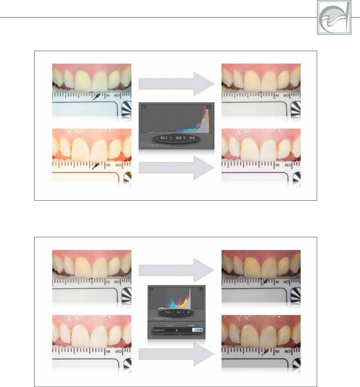
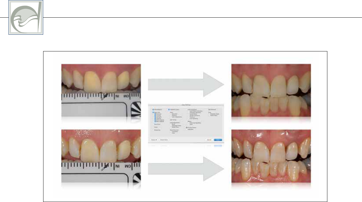
Fig 13 White balancing procedure: After importing the RAW file into Adobe LightroomCC, the color
picker tool is selected to click on a randomly selected area of the white balance reference card ideally
located in the center of the image. This will neutralize chromaticity values a* and b* towards 0 (±0.5).
white balance
white balance
Fig 14 Exposure balancing procedure: The color picker tool is held steadily over the central area of the
white balance reference card while the exposure value is adjusted simultaneously until the L* value is as
close as possible to the known L* value of the gray reference card (ie, L*75 WhiBal).
exposure balance
exposure balance
CLINICAL RESEARCH
14
THE INTERNATIONAL JOURNAL OF ESTHETIC DENTISTRY
VOLUME 11 • NUMBER 2 • SUMMER 2016
Fig 15 The adjustments during white balancing and exposure balancing can be copied and pasted to
achieve synchronicity among images which were obtained with the same type of diffusor.
copy and paste
copy and paste
Conclusion
Within the limitations of this study, the
use of a white balance reference card
with known color coordinates can be
recommended when diffusers are used
for dental photography in daily practice
to record color accurate images, espe
-
cially for shade communication and for
documentation of clinical results.

15
THE INTERNATIONAL JOURNAL OF ESTHETIC DENTISTRY
VOLUME 11 • NUMBER 2 • SUMMER 2016
HEIN/ZANGL
References
1. Bengel W. Mastering Digital
Dental Photography. Quin-
tessence, 2006:2–6.
2. Ahmad I. Digital dental
photography. Part 4: choos-
ing a camera. Br Dent J
2009;206:575–581.
3. Ahmad I. Digital dental pho-
tography. Part 5: lighting. Br
Dent J 2009;207:13–18.
4. Bazos P, Magne P. Bio-
Emulation: biomimetically
emulating nature utilizing
a histoanatomic approach;
visual synthesis. Int J Esthet
Dent 2014;9:330–352.
5. Gordon P, Wander P. Spe-
cialised equipment for dental
photography. Br Dent J
1987;162:346–359.
6. Ahmad I. Digital dental
photography. Part 8: intra-
oral set-ups. Br Dent J
2009;207:151–157.
7. Berns RS. Billmeyer and
Salzman’s Principles of Color
Theory, ed 3. New York: John
Wiley and Sons, 2000:4.
8. Martinec E. “Noise, Dynamic
Range, and Bit Depth in
Digital SLRs”, 2008. http://
theory.uchicago.edu/~ejm/
pix/20d/tests/noise/noise-p3.
html. Accessed 17 Septem-
ber 2015.
9. Bengel W. Mastering Digital
Dental Photography. Quin-
tessence, 2006:148–149.
10. Meng J, Kontogiorgos E,
Newby M. An Alternative
Method of Shade Selection
for Indirect Dental Res-
torations: A Case Report.
http://www.mtmengs.net/
images/joe/Articles/Joe%20
Meng-%20Poster%20-Ameri-
can%20Adademy%20of%20
Fixed%20Prosthodontics%20
2008.pdf. Accessed 10 May
2015.
11. Snow SR. Assessing and
Achieving Accuracy in Digi-
tal Dental Photography. CDA
Journal 2009;37.
12. Bosch JJ, Coops JC. Tooth
color and reflectance as
related to light scattering and
enamel hardness. J Dent
Res 1995;74:374–380.
13. Gozalo-Diaz D, Johnston
WM, Wee AG. Estimating
the color of maxillary cen-
tral incisors based on age
and gender. J Prosthet Dent
2008;100:93–98.
14. Cal E, Sonugelen M, Guneri
P, Kesercioglu A, Kose
T. Application of a digital
technique in evaluating the
reliability of shade guides. J
Oral Rehabil 2004;31:483–
491.
15. Jarad FD, Russel MD, Moss
BW. The use of digital imag-
ing for colour matching and
communication in restora-
tive dentistry. Br Dent J
2005;199:43–49.
16. Luo W, Westland S, Ellwood
R, Petty I. Uncertainties in
tooth colour measurement
using digital camera. In:
Proceedings of the 30th
International Congress of
Imaging Science, Rochester,
2006:582–584.
17. Reno EA, Lapujade P, Poore
CM, Crisanti MM, Miller JM,
Anastasia MK. Reproducibil-
ity of a digital imaging meth-
od for measuring tooth color.
J Dent Res 2002;81(special
issue A):2726.
18. Smith RN, Collins LZ, Naeeni
M, et al. The in vitro and in
vivo validation of mobile
non-contact camera-based
digital imaging system for
tooth colour measurement. J
Dent 2008;36(suppl 1):S15–
S20.
