Agarose
Bead
O
N
H
Bead
NH2
NH2
H2N
H2N
+
O
O
O
O
O
N
Thermo Scientific Pierce Protein
Purification Technical Handbook
Version 2

Introduction for Protein Purification 1-5
Purification Accessories – Protein Extraction,
Binding and Elution Buffers 6-11
Protease and Phosphatase Inhibitors 8
for Protein Purification
Buffers for Protein Purification 9
Spin Cups and Columns 10
Disposable Plastic and Centrifuge Columns 11
Fusion Protein Purification 12-21
His-Tagged Protein Purification Resin 14-15
Cobalt Resin, Spin Columns and 16-17
Chromatography Cartridges
High-quality purification of GST-fusion proteins 18-19
GST- and PolyHis-Tagged Pull-Down Assay Kits 20-21
Covalent Coupling of Affinity Ligands
to Chromatography Supports 22-35
Covalent Immobilization of Ligands 22-25
Products for Immobilizing Ligands 26-29
through Primary Amines
Products for Immobilizing Ligands 30-32
through Sulfhydryl Groups
Products for Immobilizing Ligands 33
through Carbonyl Groups
Products for Immobilizing Ligands 34-35
through Carboxyl Groups
IP/Co-IP 36-45
Immunoprecipitation 36
Traditional Methods vs. 37
Thermo Scientific Pierce Innovations for Co-IP
Approaches to Co-IP Free of Antibody Interference 37
Optimization Parameters in IP and Co-IP 38
Evaluating a Co-IP-Captured Interaction 38
IP and Co-IP Kits 39-45
Protein Enrichment 46-57
Phosphoprotein Enrichment Kits 46-47
SH2 Domain Phosphotyrosine Capture Kits 48-50
Use of Titanium Dioxide for 51-52
Phosphopeptide Enrichment
Pierce Fe-NTA Phosphopeptide Enrichment Kit 53-54
Cell Surface Protein Isolation Kit 55
Glycoprotein Isolation Kits 56
Ubiquitin Enrichment Kit 57
Antibody Purification 58-67
Overview 58
Immobilized Protein L, Protein A, Protein G 59-61
and Protein A/G
IgG Binding and Elution Buffers for 62
Protein A, G, A/G and L
Melon Gel Purification Products 63
Thiophilic Gel Antibody Purification 64
IgM and IgA Purification 65
Avidin:Biotin Binding 66-69
Biotin-binding Proteins 66
Immobilized Avidin Products 67
Immobilized Streptavidin Products 67
Immobilized NeutrAvidin Products 68
Immobilized Monomeric Avidin and Kit 68
Immobilized Iminobiotin and Biotin 69
FPLC Cartridges 70-77
Overview 70
His-tagged Protein FPLC Purification 71-72
GST-Tagged Protein FPLC Purification 72
Antibody FPLC Purification 73-75
Phosphoprotein FPLC Purification 75
Biotinylated Protein FPLC Purification 76
Protein Desalting 77
Affinity Supports 78-80
Table of Contents
To order, call 800-874-3723 or 815-968-0747. Outside the United States, contact your local branch office or distributor.
1
To order, call 800-874-3723 or 815-968-0747. Outside the United States, contact your local branch office or distributor.
1
General Protein Purification Techniques
Protein purification is essential for a host of biochemical appli-
cations. However, with thousands of proteins each displaying
unique characteristics, it is important to develop a strategy for
purification that delivers the correct yield, purity and activity
needed for downstream applications. For low resolution/high yield
protein purification, methods such as fractional precipitation using
salts such as ammonium sulfate exist. For applications requiring
the highest purity and relatively small amounts of protein, affinity
purification techniques can be chosen to selectively extract a
target protein from the complex mixture of proteins found in cell
or tissue extracts. Table 1 summarizes a few general strategies.
Each of these protein purification techniques requires specific
buffers (mobile phase), chromatography resins (solid phase) and
column accessories. These three components can be used in a
variety of different configurations, based on the scale of purifica-
tion and available equipment. Common formats include:
Batch purification
Mixing the mobile and stationary phases in a conical tube and
separating the two via centrifugation. Batch method purification
can be performed at any scale. However, it is most commonly
reserved for microcentrifuge tube scale purifications involving
10-200μL of resin. In batch method purification, wash and elution
fractions are separated from the resin after centrifuging to pellet
the resin beads. The liquid cannot be removed completely because
some of it is contained within the volume of porous bead pellet.
Consequently, a portion of each fraction about equal to the volume
of resin used is left behind in the pellet, making washes and elution
somewhat inefficient.
Gravity flow chromatography
Passively adding the mobile phase to
packed columns, without mechanically
increasing the flow rate. Gravity flow com-
monly uses 1mL- to 5mL-packed columns
set-up on a bench or in a chromatography
refrigerator. Larger columns can be packed
to support larger scale protein purification. However, the weight of
the resin in the column must be considered to prevent damage to
resin at the bottom of the column. Gravity flow allows for extended
binding, washing and elution times, which is ideal for samples with
low binding affinity.
Introduction to Protein Purification
Table 1. Summary of protein purification techniques.
Resolution/ purity Technique Protein Yield Description
Low
Ammonium sulfate precipitation High Fractional precipitation of proteins based on their solubility in salt solutions of varying saturation
Hydrophobic interaction chromatography High Separation of proteins based in their surface hydrophobicity
Size exclusion chromatography (SEC) Medium Separation of proteins based on molecular size
Medium
Ion exchange chromatography Medium Separation based on protein charge at a particular pH
Gel electrophoresis Medium Two-step separation of protein based on size using polyacrylimide gel electrophoresis
(SDS-PAGE) and charge using isoelectric focusing. Note: This is a denaturing method that results
in a complete loss of protein activity.
Proteome fractionation Medium Enrichment of classes of proteins, such as:
• phosphoproteins using immobilized metal affinity chromatography (IMAC)
• glycoproteins using immobilized lectins
• nitrosylated or palmitylated proteins using the biotin switch assay
• organelle-specific proteomes using cellular fractionation techniques
High
Affinity purification Low Selective purification using affinity tags attached to the target gene, such as polyhistidine (6xHis),
glutathione S-transferase (GST) or maltose binding protein (MBP)
Immunoprecipitation Lowest Antibody-based extraction of a single protein species, typically from cell or tissue lysatesh
2
For more information, or to download product instructions, visit www.thermoscientific.com/pierce
Spin cup purification
Separating the mobile and stationary
phases using centrifugation in packed spin
tubes with filters that retain resin in column.
Spin purification can also be performed
with spin plates, where each well of a
96-well microtiter plate has a filter base
and is packed with the appropriate chromotagraphy resin. The
spin cup purification method provides improved efficiency of wash
and elution steps compared to the batch method. Centrifugation
separates the liquid fraction by pulling it thoroughly from the resin,
which is retained within the spin cup apparatus. Spin cup purifica-
tion is most appropriate when 50-300μL of immobilized ligand resin
is used.
Magnetic purification
Isolating the stationary phase using mag-
nets. Magnetic beads are commonly iron
oxide particles which are coated with a
ligand to purify a protein target. An example
of this is a magnetic particle coated with
reduced glutathione (GSH) used to purify
GST-tagged recombinant proteins (Product # 88821). Advantages of
using magnetic beads include easy handling, minimal loss of resin
from pipetting and compatibility with high throughout automated
systems such as the Thermo Scientific KingFisher 96 instrument.
Fast protein liquid chromotagraphy (FPLC)
Using chromatography cartridges pre-
packed with the stationary phase and
a series of pumps and UV detectors to
move and monitor the mobile phase. The
advantages of using FPLC include reduced
purification times and the ability to improve
resolution by linking multiple columns in
tandem for greater separation.
Affinity Purification
Various methods are used to enrich or purify a target protein from
other proteins and components in a crude cell lysate or other
sample. The most powerful of these methods is affinity purification,
also called affinity chromatography, whereby the protein of
interest is purified using its specific binding properties to an
immobilized ligand.
Affinity purification makes use of specific binding that occurs
between molecules and is used extensively for the isolation of
biological molecules. A single pass through an affinity column
can achieve a 1,000- to 10,000-fold purification of a ligand from a
crude mixture. From a single affinity purification step, it is possible
to isolate a compound in a form pure enough to obtain a single
band upon SDS-PAGE analysis. We offer a number of immobilized
protein or ligand products for affinity purification of antibodies,
fusion-tagged proteins, biotinylated proteins and other proteins
for which an affinity ligand is available.
In affinity purification, a ligand is immobilized to a solid support.
Once immobilized, it specifically binds its partner under mild buffer
conditions (often physiological conditions such as phosphate
buffered saline). After binding to the partner molecule, the support
is washed with additional buffer to remove unbound components
of the sample. An elution buffer is added, disrupting the interaction
between the ligand and its binding partner by pH extremes
(low or high), high salt, detergents, chaotrophic agents or the
removal of some factor required for the pair to bind. Once
released, the binding partner can be recovered from the support
using additional elution buffer. The elution buffer can then be
exchanged by dialysis or desalting into a more suitable buffer
for storage or downstream analysis.
Activated affinity support products and kits enable a researcher to
immobilize nearly any type of ligand to purify its binding partner(s).
For example, if a peptide antigen is used to immunize animals and
produce antibodies, the same peptide can be immobilized to a
gel support and used to affinity-purify the specific antibody from
animal serum. Alternatively, if a specific antibody is available
against a particular protein of interest, it can be immobilized to
a support and used to affinity-purify the protein from crude cell
lysate. Purification with respect to nearly any binding interaction
can be made by this approach.
Introduction to Protein Purification
3
To order, call 800-874-3723 or 815-968-0747. Outside the United States, contact your local branch office or distributor.
Affinity purification products using either immobilized ligands
or activated affinity support chemistries are available for use
in several different formats. Most commonly, porous beaded gel
supports are used for gravity-column, spin-column or batch-scale
purification procedures. Coated microplates are available for
high-throughput screening applications, and magnetic particles
are especially useful for automated protein purification.
Proteins and other macromolecules of interest can be purified
from crude extracts or other complex mixtures using a variety
of methods. Precipitation is perhaps the simplest method for
separating one type of macromolecule from another. For example,
nucleic acids can be precipitated and thereby purified from
undesired molecules in solution using ethanol, and proteins can
be selectively precipitated in the presence of ammonium sulfate.
Most purification methods involve some form of chromatography
whereby molecules in solution (mobile phase) are separated based
on differences in chemical or physical interaction with a station-
ary material (solid phase). Gel filtration (also called desalting, size
exclusion chromatography or SEC) uses a porous gel material to
separate molecules based on size; large molecules are excluded
from the internal spaces of the gel material while small molecules
enter the resin pores, resulting in a longer path through the col-
umn. In ion exchange chromatography, molecules are separated
according to the strength of their overall ionic interaction with a
solid-phase material. By manipulating buffer conditions, molecules
of greater or lesser ionic character can be bound to or dissociated
from the solid-phase material.
In contrast, affinity chromatography or affinity purification makes
use of specific binding interactions between molecules. A particular
ligand is chemically immobilized or “coupled” to a solid support
so that when a complex mixture is passed over the column, only
those molecules having specific binding affinity to the ligand are
purified. Affinity purification generally involves the following steps:
1. Incubate crude sample with the immobilized ligand support
material to allow the target molecule in the sample to bind to
the immobilized ligand.
2. Wash away unbound sample components from solid support.
3. Elute (dissociate and recover) the target molecule from the
immobilized ligand by altering the buffer conditions so that
the binding interaction no longer occurs.
Ligands that bind to general classes of proteins (e.g., Protein A for
antibodies) or commonly used fusion protein tags (e.g., glutathione
for GST-tagged proteins) are available in pre-immobilized forms
ready to use for affinity purification. Alternatively, more specialized
ligands such as specific antibodies or antigens of interest can be
immobilized using one of several activated affinity supports; for
example, a peptide antigen can be immobilized to a support and
used to purify antibodies that recognize the peptide.
Most commonly, ligands are immobilized or “coupled” directly to
solid support material by formation of covalent chemical bonds
between particular functional groups on the ligand (e.g., primary
amines, sulfhydryls, carboxylic acids, aldehydes) and reactive
groups on the support. However, other coupling approaches are
also possible. In the Thermo Scientific GST Orientation Kit
(Product # 78201), for example, a GST-tagged fusion protein is first
bound to an immobilized glutathione support by affinity interaction
with the GST tag and then chemically crosslinked to the support.
The immobilized GST-tagged fusion protein can then be used to
affinity-purify its binding partner(s). Likewise, the Thermo Scientific
Pierce Crosslink Immunoprecipitation Kits (Product # 26147) and
Thermo Scientific IgG Orientation Kits (Product # 44990) involve
binding and subsequent crosslinking of an antibody to immobilized
Protein A, A/G.
Historically, researchers have used affinity purification primarily
to purify individual molecules of interest. Increasingly, proteomics
research focuses on determination of disease states, cell
differentiation, normal physiological functions and drug discovery
involving interaction and expression of multiple molecules rather
than individual targets. Consequently, the use of affinity methods
has expanded to purification of native molecular complexes and
forms the basis for co-immunoprecipitation (co-IP) and “pull-down”
assays involving protein:protein interactions.
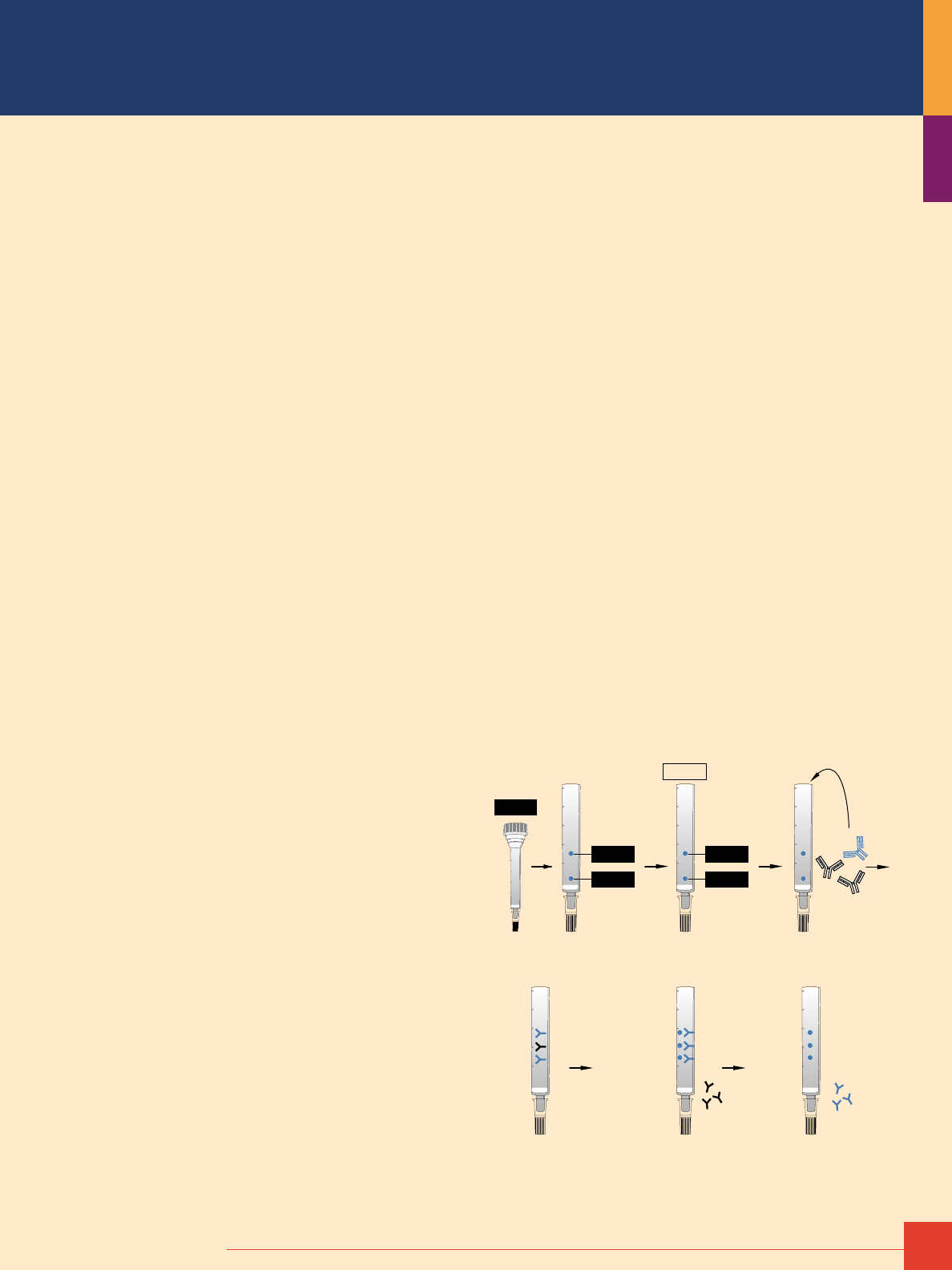
1. Immobilize the antigen to
an appropriate support.
4. Bind anti-antigen
antibodies.
5. Wash off unbound
antibodies.
6. Elute anti-antigen
antibodies.
2. Quench the unreacted
sites and wash.
3. Add the antibody solution.
5ml
4ml
3ml
2ml
1ml
5ml
4ml
3ml
2ml
1ml
5ml
4ml
3ml
2ml
1ml
5ml
4ml
3ml
2ml
1ml
5ml
4ml
5ml
4ml
5ml
4ml
Antigen
Antigen
Antigen
Antigen
Antigen
TBS
Typical antibody purification using an immobilized antigen column.

4
For more information, or to download product instructions, visit www.thermoscientific.com/pierce
Affinity Purification Supports
Affinity purification involves the separation of molecules in solution
(mobile phase) based on differences in binding interaction with a
ligand that is immobilized to a stationary material (solid phase). The
solid phase in affinity purification is a support or matrix material
that a biospecific ligand may be covalently attached. Typically, the
material to be used as an affinity matrix is insoluble in the system
in which the target molecule is found. Usually, but not always,
the insoluble matrix is a solid. Hundreds of substances have been
described and employed as affinity matrices.
Useful affinity supports are those with a high surface area to
volume ratio, chemical groups that are easily modified for covalent
attachment of ligands, minimal nonspecific binding properties,
good flow characteristics, and mechanical and chemical stability.
When choosing an affinity support or matrix for any separation,
the most important question to answer is whether a reliable
commercial source exists for the desired matrix material in the
quantities required. Fortunately, we offer a wide range of practical
and efficient matrices in volumes ranging from 1mL to much larger
bulk quantities.
Porous Beaded Resins
Porous beaded supports generally provide the most useful proper-
ties for affinity purification of proteins. We offer affinity purifica-
tion products in two main porous gel support formats: crosslinked
beaded agarose and Thermo Scientific UltraLink Biosupport.
The various features of these two supports are listed in the
accompanying table. Agarose is good for routine applications but
crushes easily, making it suitable for gravity-flow column or small-
scale batch procedures using low-speed centrifugation. UltraLink
Biosupport is incompressible and can be used in high-pressure
applications with a peristaltic pump or other liquid chromatogra-
phy system. In addition, UltraLink
®
Supports display extremely low
nonspecific binding characteristics because they are polyacryl-
amide-based. Both supports perform well in typical gravity-flow
and spin column purification and immunoprecipitation procedures.
Magnetic Particles
When a matrix is required for affinity purification of cells within a
population, we recommend Thermo Scientific MagnaBind Beads.
Magnetic affinity separation is a convenient method for isolating
antibodies, antigens, lectins, enzymes, nucleic acids and cells
while retaining biological activity. Samples containing the mole-
cule of interest are incubated with beads that are derivatized with
an antibody or other binding partner. A rare earth magnet is used
to pull the MagnaBind
™
Beads out of solution and onto a surface.
The buffer can be carefully removed, containing any non-bound
molecules or cells.
MagnaBind Beads consist of a silanized surface over an iron
oxide core (see Table 2). The silanized surface has been deriva-
tized to contain active groups, such as carboxylic acids or primary
amines, or specific affinity molecules such as streptavidin; Protein
A; Protein G; or goat anti-mouse, anti-rabbit or anti-rat IgG. Due
to the nature of the MagnaBind Beads, strong elution conditions
are not recommended with these products. See page 80 for a
complete listing of MagnaBind Supports.
Introduction to Protein Purification
To order, call 800-874-3723 or 815-968-0747. Outside the United States, contact your local branch office or distributor.
5
Table 2. Characteristics of underivatized Thermo Scientific MagnaBind Beads.
Composition
Silanized iron oxide
Magnetization
25–35EMU/g
Type of Magnetization
Superparamagnetic (no magnetic memory)
Surface Area
>100m
2
/g
Settling Rate
4% in 30 minutes
Effective Density
2.5g/mL
Number of Beads
1 x 10
8
beads/mg
pH Stability
Aqueous solution, above pH 4.0
Concentration
5mg/mL
Note: To establish a microbe-free preparation, MagnaBind Beads can be washed with
antibiotic medium or g-irradiated.
Microplates
Polystyrene microplates are another type of matrix commonly
used for immobilization of proteins. Proteins passively adsorb
to the polystyrene surface through hydrophobic interactions.
Generally, this adsorption of proteins onto the polystyrene surface
occurs best in carbonate/bicarbonate buffer at an alkaline pH
(9.0–9.5). In addition, polystyrene surfaces can be derivatized with
certain chemistries that will allow peptides and other nonprotein
molecules to adhere to the surface to perform affinity assays in
the wells of the plates.
We offer precoated plates to allow researchers an easy-to-use,
consistent method for affinity purification or identification of
specific molecules of interest. The plates offered include those
specific for fusion proteins (6xHis, GST and GFP), antibodies
(Protein A, Protein G, Protein A/G, Protein L, goat anti-mouse and
goat anti-rabbit IgG), biotin (streptavidin and Thermo Scientific
NeutrAvidin Protein) and those with reactive chemistries (maleic
anhydride and maleimide) to allow binding of nonprotein samples
that do not adsorb to the plastic microplate well surface. Only
selected microplate products are featured in this handbook. For
a complete selection of precoated plates, visit www.thermosci-
entific.com/pierce.
There are a variety of activated supports that allow a researcher
to purify proteins and other biological molecules of interest either
alone or when present in complexes with their binding partners.
Many of these supports are discussed on the following pages.
Physical properties of porous gel supports.
Support
4% Agarose
(crosslinked beaded agarose)
6% Agarose
(crosslinked beaded agarose)
Thermo Scientific UltraLink Biosupport
(co-polymer of crosslinked bis-acrylamide and azlactone)
Bead
45–165μm 45–165μm 50–80μm
Exclusion Limit
20,000,000 daltons 4,000,000 daltons 2,000,000 daltons (1,000 Å pore size)
Durability
Crushes under pressure Crushes under pressure Sturdy (>100 psi, 6.9 bar)*
Types of Chromatography
Gravity and small spin columns Gravity and small spin columns FPLC Systems, medium pressure, gravity flow
Coupling Capacity
Medium Medium High
pH range
3–11 3–11 3–11
Form
Preswollen Preswollen Dry or Preswollen
* Note: The indicated maximum pressure of 100 psi refers to the maximum pressure
drop across the gel bed that the support can withstand. It does not necessarily
refer to the indicated system pressure shown on a liquid chromatography apparatus
because the system pressure may not actually be measuring the pressure drop
across the column. Typical system pressures are usually much higher due to
pumping through small I.D. tubing, auto-samplers, detectors, etc. When packed into
a 3mm ID x 14cm height glass column, these exclusive supports have been run to
approximately 650 psi (system pressure) with no visual compression of the gel or
adverse effects on chromatography. These columns can be run at linear flow rates
or 85–3,000cm/hour with excellent separation characteristics.
6
For more information, or to download product instructions, visit www.thermoscientific.com/pierce
Protein Extraction, Binding and Elution Buffers
Protein purification is preceeded by expression of the target
protein in a host organism. In in vitro protein expression, the
simple introduction of DNA and a bacteriophage RNA polymerase
to a cell free lysate initiates protein production, with no extrac-
tion reagents required before purification. For in vivo protein
expression, such as in E. coli or tissue culture cells, expression
can be driven from constituitive promoters or it can be induced
chemically during bacterial growth or induced genetically
through transient DNA transfection into tissue culture cells.
For in vivo protein expression, the total protein content of a cell
culture or tissue sample must be extracted from the cell's mem-
branes and organelles before a chromatography resin can be
used for purification. A list of Thermo Scientific Pierce Protein
Extraction Reagents for different cell and tissue type is listed
on page 7. Additionally, protease and phosphatase inhibitor
cocktails that can be used to preserve protein integrity during
extraction (see page 8).
Most affinity purification procedures involving protein:ligand inter-
actions use binding buffers, such as phosphate buffered saline
(PBS), at physiologic pH and ionic strength. This is especially true
when antibody:antigen or native protein:protein interactions are
the basis for the affinity purification. Once the binding interaction
occurs, the support is washed with additional buffer to remove
unbound components of the sample.
Nonspecific (e.g., simple ionic) binding interactions can be mini-
mized by moderate adjustments to salt concentration or by adding
low levels of detergent in the binding and/or wash buffer. Finally,
elution buffer is added to break the binding interaction and release
the target molecule, which is then collected in its purified form.
Elution buffer can dissociate binding partners by extremes of pH
(low or high), high salt (ionic strength), the use of detergents or
chaotropic agents that denature one or both of the molecules,
removal of a binding factor, or competition with a counter ligand.
In most cases, subsequent dialysis or desalting is required to
exchange the purified protein from elution buffer into a more
suitable buffer for storage or downstream analysis. For more
information on dialysis or desalting, download or request our
a free high-performance dialysis technical handbook.
The most widely used elution buffer for affinity purification of
proteins is 0.1M glycine•HCl, pH 2.5–3.0. This buffer effectively
dissociates most protein:protein and antibody:antigen binding
interactions without permanently affecting protein structure.
However, some antibodies and proteins are damaged by low pH,
so eluted protein fraction(s) should be neutralized immediately
by collecting the fractions in tubes containing 1/10th volume of
alkaline buffer such as 1M Tris•HCl, pH 8.5. Other elution buffers
for affinity purification of proteins are listed in the accompanying
table. In addition, we offer several preformulated binding and
elution buffers designed for affinity purification involving antibodies.
Common elution systems for protein affinity purification.
Condition Buffer
pH 100mm glycine•HCl, pH 2.5–3.0
100mm citric acid, pH 3.0
50–100mm triethylamine or triethanolamine, pH 11.5
150mm ammonium hydroxide, pH 10.5
Ionic strength and/or
chaotropic effects
3.5–4.0M magnesium chloride, pH 7.0 in 10mm Tris
5M lithium chloride in 10mm phosphate buffer, pH 7.2
2.5M sodium iodide, pH 7.5
0.2–3.0 sodium thiocyanate
Denaturing 2–6M guanidine•HCl
2–8M urea
1% deoxycholate
1 % SDS
Organic 10% dioxane
50% ethylene glycol, pH 8–11.5 (also chaotropic)
Competitor >0.1M counter ligand or analog
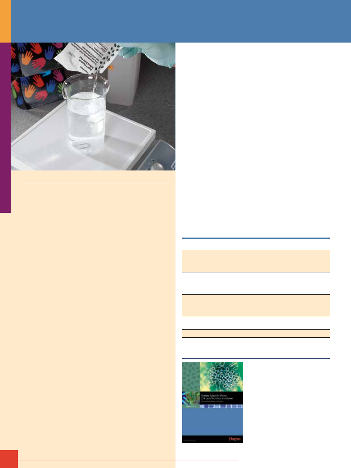
Protein Purification Accessories
Cell Lysis Technical Handbook
This 50-page handbook provides
protocols and technical and product
information to help maximize results
for protein/gene expression studies.
The handbook provides helpful hints
and troubleshooting for cell lysis,
protein purification, cell fractionation,
protease inhibitors and protein
refolding. (# 1601756)
To order, call 800-874-3723 or 815-968-0747. Outside the United States, contact your local branch office or distributor.
7
Thermo Scientific Pierce Protein Extraction Reagents.
Name Description
Organisms/Samples
B-PER
®
- Bacterial Protein
Extraction Reagent
Product # 90084
Efficient, gentle lysis and extraction of soluble proteins from E. coli and other
bacterial cells. Uses mild nonionic detergents to disrupt cells and solubilizing
proteins without denaturation, eliminating the need for harsh mechanical
procedures like sonication.
Gram(-) bacteria, S. aureus, H. pylori, E. coli strains
BL21(D3)>JM109>DH5a>M15, Archaebacteria,
nematodes and Acinetobacter sp.
B-PER II
Product # 78260
Similar to B-PER, but optimized for low cell density, or for proteins with low
expression levels.
Gram(-) bacteria, S. aureus, H. pylori, E. coli strains
BL21(D3)>JM109>DH5a>M15, Archaebacteria,
nematodes and Acinetobacter sp.
B-PER PBS
Product # 78266
Similar to B-PER, but in Phosphate Buffer. This amine free formulation is ideal
for amine-reactive labeling and/or crosslinking applications.
Gram(-) bacteria, S. aureus, H. pylori, E. coli strains
BL21(D3)>JM109>DH5a>M15, Archaebacteria,
nematodes and Acinetobacter sp.
B-PER with Enzymes
Product # 90078
Similar to B-PER, but kit contains DNAse I and lysozyme, which
improve cell membrane and DNA digestion for increased yields,
and increases the recovery of large molecular weight proteins
and insoluble proteins from inclusion bodies.
Gram(-) bacteria, S. aureus, H. pylori, E. coli strains
BL21(D3)>JM109>DH5a>M15, Archaebacteria,
nematodes and Acinetobacter sp.
B-PER Direct with Enzymes
Product # 90080
Similar to B-PER with Enzymes, but bacteria can be lysed directly in cell
culture media; ideal for screening 96-well microplate samples.
Gram(-) bacteria, S. aureus, H. pylori, E. coli strains
BL21(D3)>JM109>DH5a>M15, Archaebacteria,
nematodes and Acinetobacter sp.
Y-PER
®
- Yeast Protein
Extraction Reagent
Product # 78990
Easy-to-use solution gently disrupts the tough yeast cell wall in less than
20 minutes at room temperature, using a mild detergent. No mechanical
disruption needed; yields more than twice as much protein as glass
bead methods.
S. cerevisiae, Schizo-saccharomyces pombe,
C. albicans, B. subtilis, E. coli, P. pastoris, Strep.
avidinii and Acinetobacter sp.
Y-PER Plus
Product # 78998
More stringent than Y-PER, but entire formulation (including detergent)
are dialyzable.
Yeast (S. cerevisiae) and Acinetobacter sp.
M-PER
®
- Mammalian
Protein Extraction Reagent
Product # 78501
Highly efficient total protein extraction from cultured mammalian cells;
extracts proteins in nondenatured state, enabling protein to be directly
immunoprecipitated; amine-free and fully dialyzable; adhered cells can be
directly lysed in plate or after scraping and washing in suspension.
Cultured mammalian cells, COS-7, NIH 3T3, Hepa 1-6, 293,
CHO, MDA, MB231 and FM2
P-PER - Plant Protein
Extraction Reagent
Product # 89803
Contains organic lysing reagent and two aqueous reagents, which, in
conjuction with mild mechanical agitation, effectively extract high quality
protein extracts from plant leaves, stem, root, seed and flower cells without
liquid nitrogen or harsh mechanical aids, such as mortar and pestle.
Multiple plant organs (leaf, stem, root, seed and flowers);
multiple plant species (Arabidopsis, tobacco, maize,
soybeans, peas, spinach, rice and other
plant tissues); and fresh, frozen and dehydrated
plant tissues
T-PER
®
- Tissue Protein
Extraction Reagent
Product # 78510
Simple, easy to use reagent for extracting total protein from tissue in 1:20
(w/v) of tissue to T-PER, using centrifugation to pellet cell/tissue debris. Mild
detergent is dialyzable.
Heart, liver, kidney and brain.
I-PER - Insect Cell Protein
Extraction Reagent
Product # 89802
Optimized mild nonioinic detergent formulation provides maximum extraction
of soluble proteins from insect cells; better yield than sonication; can be used
for both suspended or adherent insect cells
Baculovirus-infected insect cells grown in
suspension or monolayer culture.
NE-PER
®
- Nuclear and
Cystoplasmic Extraction Kit
Product # 78833
Obtain functional concentrated nuclear extracts and cytoplasmic fractions
from mammalian cells and tissues in less than two hours, eliminating the need
for freeze/thaw cycles, Dounce homogenization, lengthy centrifugation times
and cold-room work.
Tissue: calf liver. Tissue: mouse heart, kidney, lung and
liver; Cultured cells: epithelial (HeLa), fibroid (COS-7),
kidney (NIH 3T3), liver (Hepa 1) and brain (C6).
Mem-PER
®
- Eukaryotic
Membrane Protein
Extraction Kit
Product # 89826
Efficient, gentle reagents that solubilize and isolate membrane proteins from
mammalian and yeast cells, as well as soft and hard tissues, in less than an
hour. Minimal cross contamination (less than 10%) of hydrophilic proteins into
the hydrophobic (membrane protein) fraction
Cultured cells: brain (C6), epithelial (HeLa), fibroblasts
(NIH 3T3) and yeast (S. cerevisiae).
Subcellular Protein
Fractionation Kit
Product # 78840
Includes a combination of reagents for stepwise lysis of mammalian cells into
functional cytoplasmic, membrane, soluble nuclear, chromatin-bound, and
cytoskeletal protein fractions in a single kit; includes a stabilized nuclease
and protease inhibitors. Extracts from each compartment have less than 15%
contamination between fractions, with sufficient purity for studying protein
localization and redistribution.
Cultured mammalian cells.
Mitochondrial Isolation
Kit for Cultured Cells
Product # 89874
Isolate intact mitochondria from cultured mammalian cells in approximately 40
minutes, with an optional Dounce homogenization protocol for increased yield.
Mammalian cells.
Mitochondrial Isolation
Kit for Tissues Product # 89801
Isolate intact mitochondria from soft or hard tissue in less than 60 minutes,
with an optional Dounce homogenization protocol for increased yield.
Heart, liver, kidney and brain.
Lysosome Enrichment
Kit for Tissues and Cells
Product # 89839
Uses density gradient centrifugation to separate lysozyme from
contaminating celluar structures in both mammalian cells and soft
and hard tissue.
Tissues and cultured cells.
Peroxisome Enrichment
Kit for Tissues Product # 89840
Uses density gradient centrifugation to separate peroxisome from
contaminating celluar structures in both soft and hard tissue.
Heart, liver, kidney and brain.
Nuclei Enrichment
Kit for Tissue Product # 89841
Uses density gradient centrifugation to separate nuclei from
contaminating celluar structures in both soft and hard tissue
Heart, liver, kidney and brain.
Pierce
®
RIPA Buffer
Product # 89900
Extracts cystoplasmic, membrane, and nuclear proteins from cultured
mammalian cells; can be used for both plated cells and cells pelleted from
suspension cultures. Protease and phosphatase inhibitors are compatible
with this formulation.
Cultured mammalian cells and cytoplasmic, membrane
and nuclear proteins.
Pierce IP Lysis Buffer
Product # 87787
Gently extracts cytoplasmic, membrane and nuclear proteins while
maintaining protein complexes for immunoprecipitation (IP), Pulldowns, and
co-IP; does not liberate DNA which can cause high viscosity.
Cultured mammalian cells.
For more details on these products, request the Cell Lysis Technical Handbook (#1601756) at www.thermoscientific.com/pierce
8
For more information, or to download product instructions, visit www.www.thermoscientific.com/pierce
Protein Purification Accessories
Protease and Phosphatase Inhibitors
for Protein Purification
Thermo Scientific Halt Protease and Phosphase Inhibitors
Our broad-spectrum protease and phosphatase inhibitor cocktails
and individual protease inhibitors accommodate specific and
general needs in cell lysis and protein extraction methods.
Highlights
• Halt Protease Inhibitor Cocktails target serine, cysteine and
aspartic acid proteases and aminopeptidases. Metalloproteases
are inhibited by the optional addition of EDTA. Individual protease
inhibitors targeting separate classes of
proteases are also available for custom cocktail development.
• Halt
™
Phosphatase Inhibitor Cocktails contain chemical com-
pounds that target serine/threonine and tyrosine phosphatases.
• The Halt Protease and Phosphatase Inhibitor Cocktail prevents
protein degradation and preserves phosphorylation simultaneously,
providing protection that is similar to the individual cocktails.
For a complete listing of the Halt Protease and Phosphatase
Inhibitors at www.thermoscientific.com.
Buffers for Protein Purification
Thermo Scientific BupH Pack Dry Blend Buffers
BupH
®
Packs are pre-blended and pre-measured dry mixtures of
commonly needed buffers that are easy to prepare; simply empty
contents of foil envelope pack into a beaker, add ultrapure water,
and stir to dissolve. The packs eliminate weighing time and tedious
pH adjustments. BupH Pack Dry Blend Buffers are offered for use in
common laboratory techniques and to support other Pierce Protein
Research Products.
Highlights:
• Convenient – dissolve contents of one envelope in 500ml of water
and the buffer is ready to use
• Save time and trouble – no weighing, no pH adjustment, no need
to stock individual components and no need to make and store
large volumes of stock solution in advance of daily needs
• Long shelf life – stocking and storage as dry packs eliminates
concerns about long-term stability of stock solutions
• Eliminate variables – our quality control ensures that every pack
will yield the same, consistent buffer
Ordering Information
Product #
Description
Pkg. Size
Applications
Formulation of each pack
after reconstitution
U.S.
Price
28384 Borate Buffer
40 packs
Protein modification procedures that
require amine-free buffer at alkaline pH
500mL of 50mM borate, pH 8.5
$132
28382 Carbonate-Bicarbonate Buffer
40 packs
Microplate protein and antibody coating
for ELISA or RIA
500mL of 0.2M carbonate-bicarbonate, pH 9.4
$125
28388 Citrate-Carbonate Buffer
10 packs
Protein immobilization to UltraLink
®
Biosupport (Product # 53110)
100mL of 0.6M sodium citrate,
0.1M sodium carbonate, pH 9
$ 64
28386 Citrate-MOPS Buffer
10 packs
Protein immobilization to UltraLink
Biosupport (Product # 53110)
100mL of 0.6M sodium citrate,
0.1M MOPS, pH 7.5
$ 58
28390 MES Buffered Saline
10 packs
Crosslinking using carbodiimide
(EDC, Product # 22980)
500mL of 0.1M MES, 0.9% NaCl, pH 4.7
$112
28372 Phosphate Buffered Saline (PBS)
40 packs
Crosslinking and biotinylation requiring
amine-free buffer
500mL of 0.1M sodium phosphate,
0.15M NaCl, pH 7.2
$135
28374 Modified Dulbecco’s PBS
40 packs
Wash buffers and antibody diluents
for ELISA, Western blotting and other
immunoassays
500mL of 8mM sodium phosphate,
2mM potassium phosphate, 0.14M NaCl,
10mM potassium chloride, pH 7.4
$ 96
28379
28376
Tris Buffered Saline
10 packs
40 packs
Wash buffers and antibody diluents
for ELISA, Western blotting and other
immunoassays
500mL of 25mM Tris,
0.15M NaCl, pH 7.2
$ 61
$134
28378 Tris-Glycine-SDS Buffer
40 packs
Running buffer for Tris-Glycine gel
electrophoresis
500mL of 25mM Tris, 192mM glycine,
0.1% SDS, pH 8.3
$120
28398 Tris-HEPES-SDS Running Buffer 10
10 packs
Running buffer for Tris-HEPES electrophoresis
with Precise
™
Precast Gels
500mL of 100mM Tris, 100mM HEPES,
3mM SDS, pH 8±0.25
$ 36
28380 Tris-Glycine Transfer Buffer
40 packs
Running buffer for gel to membrane
electrophoretic transfer
500mL of 25mM Tris, 192mM glycine,
pH 8 (use 20% methanol to dissolve)
$120
Need a larger quantity?
Call our Bulk and Custom
department at 1-800-874-3723
or +1 815-968-0747 ext. 300 for
pricing and information. Or, visit
www.thermoscientific.com/protein-custom
To order, call 800-874-3723 or 815-968-0747. Outside the United States, contact your local branch office or distributor.
9
Thermo Scientific Concentrated Liquid Buffers
Save counter space and time.
Pierce 10X and 20X Concentrated Buffers are ready to use without
having to reconstitute with ultrapure water and, in the case of
Tris-Glycine Buffer, methanol. Buffers are designed for use in
dialysis, cross-linking, enzyme assays, ELISAs, immunohistochem-
istry, protein plate-coating, biotinylation and other applications.
Keep our concentrated buffers close at hand to cover your lab’s
research needs.
Highlights:
• Easy to use – no packets to open and no powder to dissolve
• Increased accuracy – eliminates the possibility of powder
remaining in a packet
• Saves time – 10X or 20X concentration eliminates time spent
waiting for powder to dissolve
• Saves space – storage as concentrated stock minimizes bench
space needed for large volume solutions
PBS
Tris-Glycine-SDS
Tris-Hepes-SDS
Ordering Information
Product #
Description
Pkg. Size
Applications
1X Formulation
U.S.
Price
28341 20X Borate Buffer
500mL
Ideal for protein modification procedures
requiring amine free buffer at alkaline PH
500mL of 50mM borate, pH 8.5
$68
28344 20X Modified Dulbecco's
PBS Buffer
500mL
Used for wash buffers and antibody diluents
in applications such as ELISA, Western
blotting and other immunoassays
8mM Sodium Phosphate,
2mM Potassium Phosphate,
0.14M NaCl, 100mM KCl, pH 7.4
$68
28346 20X Modified Dulbecco's
PBS Tween-20 Buffer
500mL
A wash buffer for ELISA, Western and
other Immunoassays as well as a blocking
buffer for plate based assays
8mM Sodium Phosphate,
2mM Potassium Phosphate,
0.14M NaCl, 100mM KCl,
0.05% Tween-20, pH 7.4
$68
28348 20X Phosphate Buffered Saline
500mL
Its ionic strength makes it ideal for
crosslinking and biotinylation requiring
amine free buffer
0.01M Sodium Phosphate,
0.15M NaCl, pH 7.5
$68
28352 20X PBS Tween-20 Buffer
500mL
A wash buffer for ELISA, Western and other
Immunoassays as well as a blocking buffer
for plate based assays
0.01M Sodium Phosphate,
0.15M NaCl, 0.05% Tween-20, pH 7.5
$68
28354 20X TAE Buffer
500mL
Historically the most common buffer used
for agarose gel electrophoresis in the
analyses of nucleic acids
0.04M Tris, 0.04M Acetate,
0.001M EDTA, pH 8.2-8.4
$68
28355 10X TBE Buffer
1L
Often used for agarose gel electrophoresis
in the analysis of nucleic acids
0.089M Tris, 0.089 M Borate,
0.002M EDTA, pH 8.2-8.4
$65
28358 20X TBS Buffer
500mL
Used for wash buffers and antibody diluents
in applications such as ELISA, Western
blotting and other immunoassays
25mM Tris, 0.15M NaCl, pH 7.2
$68
28360 20X TBS Tween-20 Buffer
500mL
A wash buffer for ELISA, Western and other
Immunoassays as well as a blocking buffer
for plate based assays
25mM Tris, 0.15M NaCl,
0.05% Tween-20, pH 7.5
$68
28362 20X Tris/HEPES/SDS Buffer
1L
A running buffer for Tris-HEPES
electrophoresis with Precise Precast Gels
0.025 M Tris, 0.192 M Glycine,
0.1% SDS, pH 8.5
$45
28363 10X Tris-Glycine Buffer
1L
Great for electrophoretic transfer from
gel to membrane
0.025M Tris, 0.192M Glycine, pH 8.5
$45
28368 20X Tris/HEPES/SDS Buffer
500mL
A running buffer for Tris-HEPES
electrophoresis with Precise Precast Gels
0.1M Tris, 0.1M HEPES,
3mM SDS, pH 8+0.25
$68
10
For more information, or to download product instructions, visit www.www.thermoscientific.com/pierce
Spin Cups and Columns
Thermo Scientific Pierce Spin Columns are convenient tools for
manipulating small volumes of affinity supports (5–500μL) for
protein purification. Add the affinity resin and sample to one of
the columns, use a microcentrifuge to efficiently wash away
contaminants and elute your purified sample without losing any
resin in the process. Spin columns allow you to affinity-purify
more protein in less time!
Highlights:
• Efficient washing of samples means fewer washes are needed
to remove contaminating proteins
• Efficient elution of samples means more antigen and
co-precipitated proteins are recovered
• No resin loss means more consistent IP and co-IP results
• No need to decant supernatant from IP or co-IP pellet
• Spin protocols drastically reduce the time required for IPs and co-IPs
• Low protein-binding polypropylene column construction
minimizes nonspecific binding
Spin Cups – Paper Filter
Paper filter with collection tubes.
Highlights:
• Paper filters are resistant to clogging
from cellular debris
• Column Volume: 600μL
• Resin Volume: 20–300μL
• Filter Type: Paper, ~10μm pore size
• Cap Type: Collection tube cap fits onto inserted spin cup
Spin Cups – Cellulose Acetate Filter
Cellulose acetate filter with collection tubes.
Highlights:
• Used in our IP and Co-IP Kits
• Column Volume: 800μL
• Resin Volume: 20–400μL
• Filter Type: Cellulose acetate, 0.45μm pore size
• Cap Type: Collection tube cap fits onto inserted spin cup
Spin Columns – Screw Cap
Screw cap with Luer-Lok
®
Adaptors.
Highlights:
• Luer-Lok Adaptors allow these columns
to be used for syringe-based purifications
• Column Volume: 900μL
• Resin Volume: 20–400μL
• Filter Type: Polyethylene, ~10μm pore size
• Small & large frit options for different sample sizes
• Cap Type: O-ring screw top caps; press-in bottom plugs
Spin Columns – Snap Cap
Snap cap with collection tubes.
Highlights:
• Used in the Cell Surface Protein Isolation
and Glycoprotein Isolation Kits
• Column Volume: 1,000μL
• Resin Volume: 20–500μL
• Filter Type: Polyethylene, ~30μm pore size
• Cap Type: Snap cap on column (no cap on collection tube);
press-on bottom caps
Micro-Spin Columns
Highlights:
• Column Volume: 400μL
• Resin Volume: 5–100μL
• Filter Type: Polyethylene, ~30μm pore size
• Cap Type: O-ring screw top caps; press-on
bottom caps
Ordering Information
Product #
Description
Pkg. Size
U.S.
Price
69700 Spin Cups – Paper Filter
Includes cups and collection tubes
50/pkg
$109
69715 Microcentrifuge Tubes
Collection Tubes for Product # 69700
72/pkg
$ 32
69702 Spin Cups – Cellulose Acetate Filter
Includes cups and collection tubes
50/pkg
$108
69720 Microcentrifuge Tubes
Collection Tubes for Product # 69702
72/pkg
$ 32
69705 Spin Columns – Screw Cap
with Luer-Lok Adaptors
Includes: Spin Columns, Screw Caps and Column Plugs
Luer-Lok Adaptors
Large Frits (6.8mm diameter 10μm pore size)
Small Frits (2.7mm diameter 10μm pore size)
Large and Small Frit Tools
Kit
25 each
5 each
25 each
25 each
1 each
$ 99
69725 Spin Columns – Snap Cap
with Collection Tubes
Includes: Spin Columns and Bottom Caps
Collection Tubes
Kit
50 each
100 each
$109
89879 Micro-Spin Columns
50/pkg
$ 55
Protein Purification Accessories
To order, call 800-874-3723 or 815-968-0747. Outside the United States, contact your local branch office or distributor.
11
Disposable Plastic Columns
Automatic “stop-flow” action provided by porous polyethylene
discs prevents column beds from drying out.
Highlights:
• Supplied complete with porous polyethylene discs, stoppers, end caps
• Compatible with most types of aqueous buffer eluents commonly
used in chromatography
• Can be pre-packed and stored until needed
Ordering Information
Product #
Description
Pkg. Size
U.S.
Price
29920 Disposable Polystyrene Columns
Ideal for packing 0.5–2.0mL bed volumes.
100/pkg
$144
29922 Disposable Polypropylene Columns
Ideal for packing 1–5mL bed volumes.
100/pkg
$174
29924 Disposable Polypropylene Columns
Ideal for packing 2–10mL bed volumes.
100/pkg
$190
29923 Disposable Polypropylene Funnels
Buffer reservoirs that fit Product #’s 29920,
29922 and 29924.
50/pkg
$109
29925 Disposable Column Trial Pack
Includes accessories plus two each of Product #’s
29920, 29922 and 29924 and one of Product # 29923.
Trial Pack
$ 63
Centrifuge Columns
Efficiently handle a wide variety of resin volumes for affinity
purification! Thermo Scientific Pierce Centrifuge Columns are
convenient tools for handling 40μL–10mL of an affinity purification
support. Add the affinity resin to one of the polypropylene columns,
remove the twist-off bottom and allow the resin to pack itself.
Then add your sample and allow it to bind to the support. Use a
centrifuge to efficiently wash away any contaminants and elute
your purified protein.
Pierce Centrifuge Columns allow you to use a spin-column format
in addition to traditional gravity flow to reduce the time required for
column washing and elution. This accelerates sample processing
time and makes multiple-sample processing possible. Centrifuge
columns allow you to affinity-purify more protein in less time!
Centrifuge columns are made from low protein-binding
polypropylene for compatibility with protein purification, and
they fit into standard centrifuge tubes for use in any centrifuge.
Applications for Centrifuge Columns:
• Affinity purification/affinity chromatography
• Immunodepletion
• Spin desalting
0.8mL Centrifuge Columns
Highlights:
• Total Volume: 800μL
• Resin Volume: 40–400μL
• Filter Type: Polyethylene, ~30μm pore size
• Receiver Tube: Fits standard microcentrifuge tubes
(e.g., Product # 69720)
• Cap Type: O-ring screw-top cap
• Twist-off bottom
2mL Centrifuge Columns
Highlights:
• Total Volume: 5mL (2mL resin bed, 3mL reservoir)
• Resin Volume: 2mL
• Filter Type: Polyethylene, ~30μm pore size
• Receiver Tube: Fits standard 15mL conical
centrifuge tubes
• Cap Type: Screw-top cap
• Twist-off bottom and press-on cap to reseal
5mL Centrifuge Columns
Highlights:
• Total Volume: 8mL (5mL resin bed, 3mL reservoir)
• Resin Volume: 5mL
• Filter Type: Polyethylene, ~30μm pore size
• Receiver Tube: Fits standard 15mL conical
centrifuge tubes
• Cap Type: Screw-top cap
• Twist-off bottom and press-on cap to reseal
10mL Centrifuge Columns
Highlights:
• Total Volume: 22mL (10mL resin bed, 12mL reservoir)
• Resin Volume: 10mL
• Filter Type: Polyethylene, ~30μm pore size
• Receiver Tube: Fits standard 50mL conical
centrifuge tubes
• Cap Type: Screw-top cap
• Twist-off bottom and press-on cap to reseal
Ordering Information
Product #
Description
Pkg. Size
U.S.
Price
89868 Centrifuge Columns, 0.8mL capacity
Includes: Pierce Centrifuge Columns
Screw Caps
Kit
50 each
50 each
$ 62
89869 Centrifuge Columns, 0.8mL capacity
Includes: Pierce Centrifuge Columns
Screw Caps
Kit
4 x 50 each
4 x 50 each
$192
89896 Centrifuge Columns, 2mL capacity
Includes: Pierce Centrifuge Columns
Screw Caps and Tips
Kit
25 each
25 each
$ 39
89897 Centrifuge Columns, 5mL capacity
Includes: Pierce Centrifuge Columns
Screw Caps and Tips
Kit
25 each
25 each
$ 44
89898 Centrifuge Columns, 10mL capacity
Includes: Pierce Centrifuge Columns
Screw Caps and Tips
Kit
25 each
25 each
$ 49
69707 Column Extender
Fits 89896, 89897 and 89898. Increases column
capacity by 35mL
10 each
$ 28
5ml
4ml
3ml
2ml
1ml
10ml
9ml
8ml
7ml
6ml
5ml
4ml
3ml
2ml
1ml

12
For more information, or to download product instructions, visit www.thermoscientific.com/pierce
Fusion Protein Purification
Cultures of E. coli or Picchia are common vehicles for protein
expression. They are low cost and low maintenance platforms
which can be easily scaled up to deliver protein at milligram to
gram yields. These microoganisms are also very easy to trans-
form with a DNA vector containing the gene of interest. Typically,
researchers use common expression vectors which possess the
proper promoter elements for expression and inclusion of an
affinity tag sequence. The affinity tag sequence is cloned in frame
with the DNA sequence of the target protein, and will flank either
the N- or C-terminus. The two most common affinity tags are
polyhistidine (6xHis) and glutathione-S-trasferase (GST).
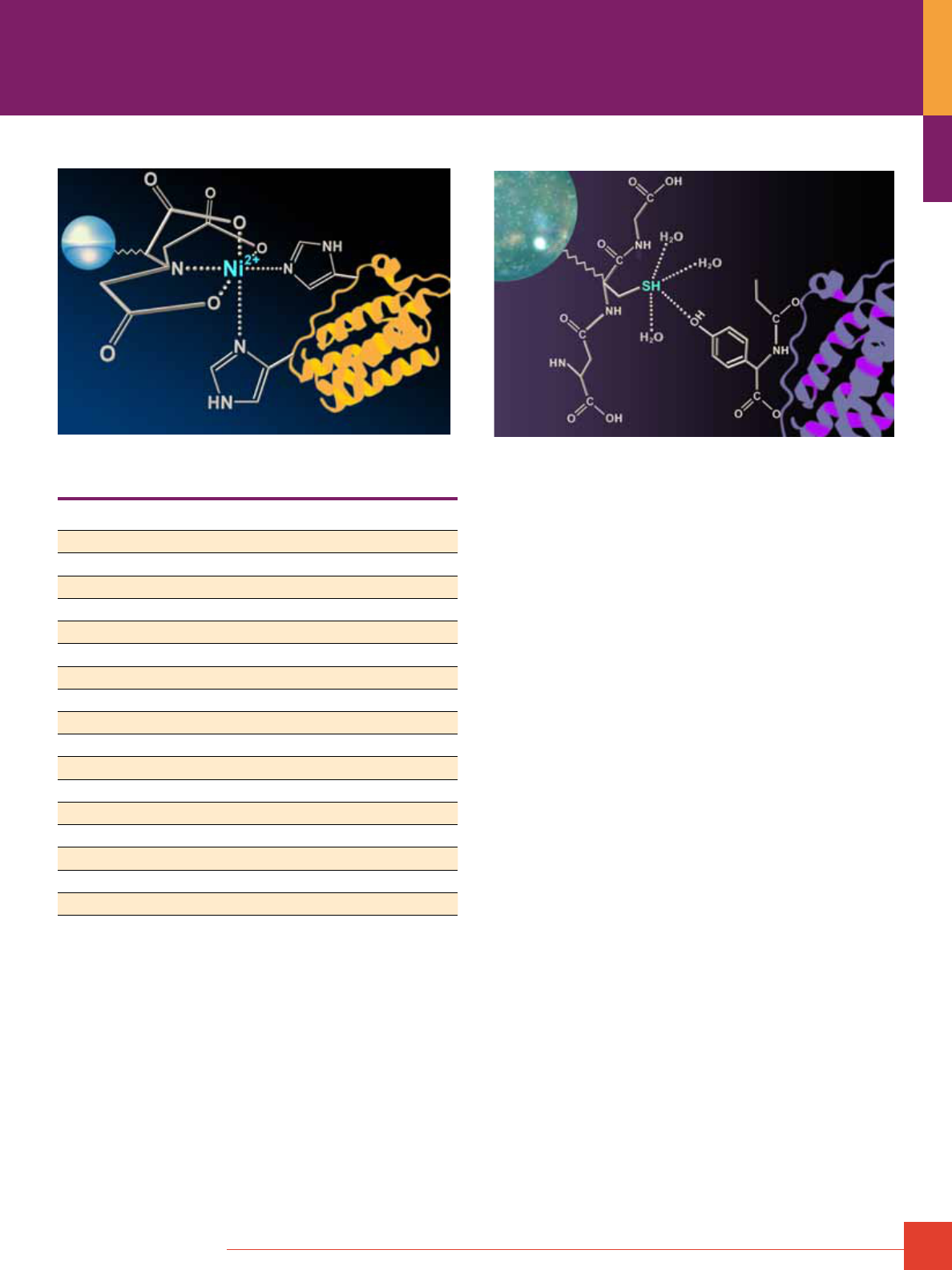
Polyhistidine Purification
Cobalt-histidine binding.
The polyhistidine tag is a sequence of five to nine histidine amino
acids attached to the terminus of a target protein. The polyhistidine
tag is purified using immobilized metal affinity chromatography
(IMAC). For histidine tag purification, either nickel or cobalt is
immobilized onto a solid chromatography resin. While the two
metals can be used interchangeably, typically nickel has a higher
binding capacity whereas cobalt binds less non-specific protein to
deliver a purer final protein.
IMAC resins work by charge interactions with the nitrogen atoms
on the histidine amino acid side chain to bind and immobilize the
histidine-tagged protein from a cell lysate. The incorporation of
multiple histidine residues as an affinity tag is designed to improve
this charge association. Because IMAC affinity for histidine resi-
dues is not dependent on the secondary structure of the protein,
IMAC purification can be performed under denaturing conditions.
IMAC purification is, however, sensitive to pH and the presence of
chelators and reducing agents. See Table 1 for a list of common
interfering agents and their concentration tolerance for both nickel
and cobalt resin.
Fusion Protein Purification
To order, call 800-874-3723 or 815-968-0747. Outside the United States, contact your local branch office or distributor.
13
Nickel-histidine binding.
Table 1. Common interfering agents for Ni-NTA and cobalt resins.
Reagent Tolerance for Ni-NTA resin Tolerance for Cobalt resin
ß-Mercaptoethanol 10mM 10mM
CHAPS 1% 1%
Ethanol 30% 30%
Ethylene glycol 30% 30%
HEPES 50mM 50mM
Glycerol 20% 20%
Guanidinium 6M 6M
Imididazole 500mM 200mM at pH 7.0-8.0 for elution
KCl 500mM 500mM
MES 20mM 20mM
MOPS 50mM 50mM
NaCl 1.0M 1.0M
NP-40 1% 1%
SDS 1% 1%
TRIS 50mM 50mM
Triton
®
-X-100 <1% <1%
Urea 8M 8M
Once immobilized, imidazole is used to disrupt the charge
attractions between the immobilized metal affinity chromatography
resin and the histidine-tagged protein. The eluted histidine-tagged
protein can be easily cleaned-up using a desalting column or
dialysis cassette to remove imidazole. Often trace amounts of
imidazole are included in the protein binding and wash steps
to compete away binding of endogenous proteins with multiple
histidines.
Glutathione-S-transferase Purification
GST binding.
GST is a 26kDa endogenous enzyme found in both prokaryotes
and eukaryotes. In the cell GST detoxifies reactive oxygen species
such as free radicals and peroxides. GST performs this task by
neutralizing reactive oxygen species with the antioxidant glutathione
(GSH). GSH is an endogenous tripeptide (Glu-Cys-Gly) containing
a cysteine residue, whose sulfhydryl side chain functions as a
reducing agent.
GST makes an ideal affinity tag because of its strong binding to
reduced glutathione. GST possess numerous tyrosine residues in
the in the GSH binding pocket, and one of these tyrosines hydrogen
bonds to the substrate glutathione forming a stable complex.
In vivo, GST would transfer glutathione to a reactive oxygen
species and neutralize it.
As a research tool, scientists have taken advantage of the strong
interaction between GST and GSH to selectively extract recom-
binant proteins. In this strategy, glutathione is immobilized onto
a solid support (typically an agarose bead) and the amino acid
sequence for the enzyme GST is cloned into either the C- or
N-terminus of the target gene (commonly using the pGEX
®
-series
of expression vectors). After binding of the GST-tagged fusion
protein to the immobilized glutathione agarose, excess reduced
glutathione is introduced (typically between 10-50mM) to compete
off the target protein. Again, simple dialysis or desalting columns
can be used to clean up the final GST-tagged protein. For reasons
that have not been fully characterized in the literature, the struc-
ture of the GST fusion tag often degrades upon denaturation and
reduction for protein gel electrophoresis (e.g., SDS-PAGE). As a
result, electrophoresed samples often appear as a ladder of lower
MW bands below the full-sized fusion protein.
In contrast to polyhistidine purification, GST purification requires
the target protein maintain native tertiary structure. Additionally,
the GST tag is quite large (26kDa) compared to the six histidines
which comprise a typical polyhistidine tag. To circumvent the
problems associated with a 26kDa appendage, selective proteases
are used to cleave the GST tag from the GST-fusion protein. These
proteases include Factor Xa (Product # 32520) or thrombin.
14
For more information, or to download product instructions, visit www.thermoscientific.com/pierce
His-Tagged Protein Purification Resin
A cost-effective Ni-NTA resin is now available.
The expression and purification of
recombinant proteins is central to protein
regulation, structure and function stud-
ies. The majority of recombinant proteins
are expressed as fusions with short
affinity tags with the most popular being
the polyhistidine (6xHis) tag. The method
used to purify recombinant His-tagged
proteins is immobilized metal affinity
chromatography (IMAC), consisting of
chelating resins charged with either
nickel or cobalt ions that coordinate
with the histidine side chains. The new
Thermo Scientific HisPur Ni-NTA Resin
complements the HisPur
™
Cobalt Resin. Ni-NTA resins are the most
common IMAC resin choice for 6xHis-tagged protein purifications
because of the four metal-binding sites on the chelate, which
enables high-protein binding and low-metal ion leaching.
Highlights:
• High capacity – bind up to 60mg of 6xHis-tagged protein per
milliliter of resin
• Versatile – purify proteins using native or denaturing conditions
• Compatible – use with Thermo Scientific Cell Lysis Reagents
and a variety of buffer additives
• Flexible – available in multiple formats, including bulk resin, spin
columns and chromatography cartridges
• Cost-effective – reuse the same batch of resin at least five times
• Easy to use – pre-formulated buffers available for kit formats
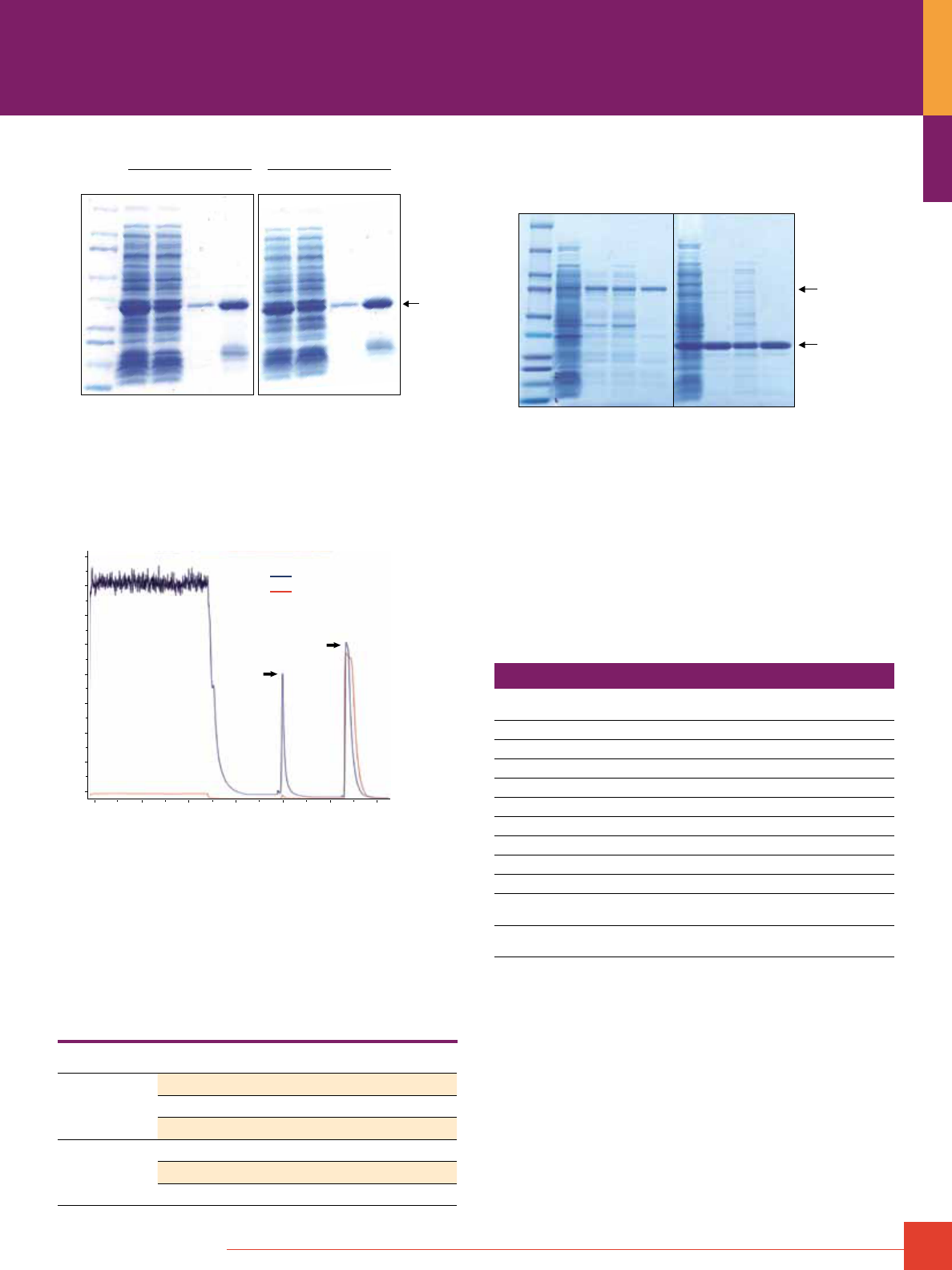
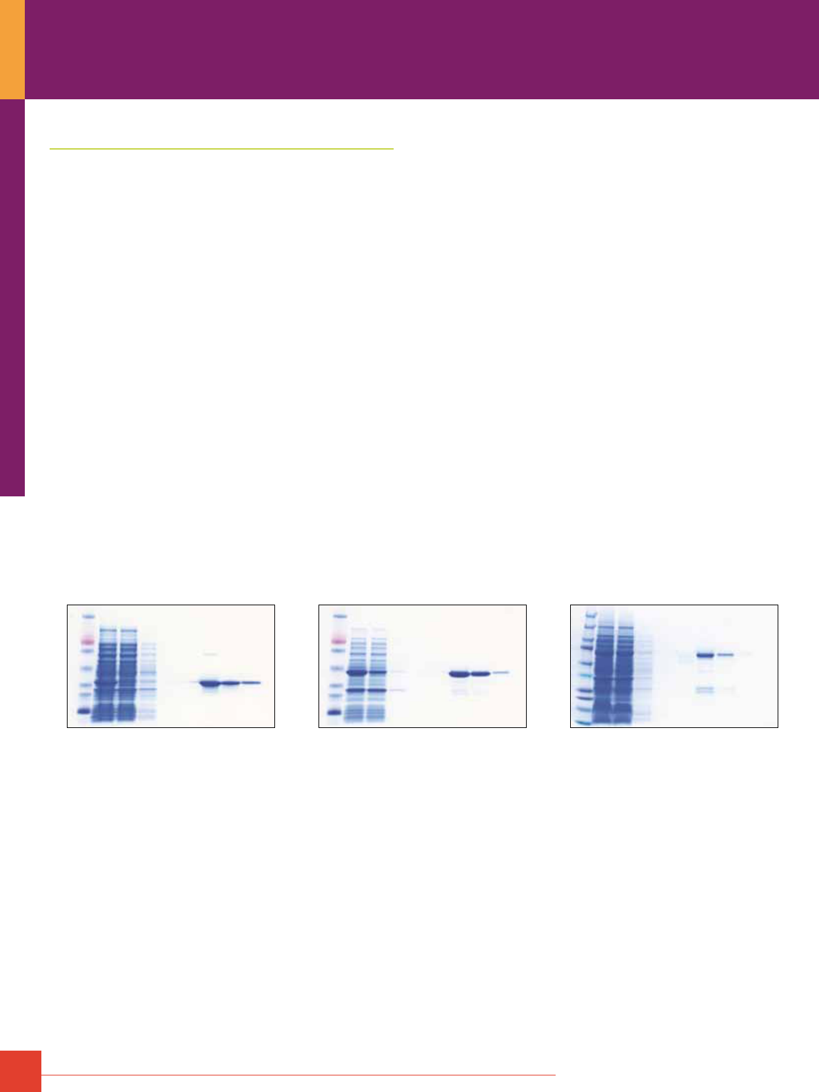
His-tagged green fluorescent protein (GFP) was recovered in
similar or greater purity and yield compared to other commercially
available nickel-chelated resins (Figure 1). HisPur Ni-NTA Resin
is also cost-effective because it can be used at least five times
without loss in performance (Figure 2). The resin is amenable to
chromatography-cartridge format, resulting in a sharp elution
peak evident in the chromatogram (Figure 3). When HisPur Ni-NTA
Resin is compared to HisPur Cobalt Resin (Product # 90090) and
Ni-IDA resin using the batch-bind method, the results depend on
protein expression level (Table 2 and Figure 4). When purifying a
low-expression protein, such as 6xHis-AIF2, there is a dramatic
difference in purity. HisPur Cobalt Resin yields the purest protein
followed by HisPur Ni-NTA Resin with Ni-IDA being the least pure.
When purifying 6xHis-GFP, a high expresser, there was minimal
difference between HisPur Ni-NTA and Cobalt Resins. Similar
results were obtained using spin purification (data not shown).
M
10
15
25
20
37
50
75
100
150
250
250kDa
L
HisPur Ni-NTA
Supplier Q
Supplier C
Ni-IDA
6xHis-GFP
Figure 1. Thermo Scientific HisPur Ni-NTA resin is comparable to or
performs better than other suppliers’ nickel resins. Bacterial lysate
(12mg total protein) containing over-expressed 6xHis-green fluorescent
protein (GFP) was applied to HisPur Ni-NTA Resin (200μL) and purified by
the batch-bind method as described. The same amount of total protein
was applied to Supplier Q, Supplier C and Ni-IDA resins per the manu-
facturers’ instructions. Gel lanes were normalized to equivalent volume.
M = molecular-weight marker and L = lysate load.
Fusion Protein Purification
To order, call 800-874-3723 or 815-968-0747. Outside the United States, contact your local branch office or distributor.
15
M
First Use
L FT W E
Fifth Use
L FT W E
10
15
25
20
37
50
75
100
150
kDa
6xHis-
Protein L
Figure 2. Thermo Scientific HisPur Ni-NTA Resin can be used at least five
times without losing performance. Bacterial lysate (20mg total protein) con-
taining over-expressed 6xHis-Protein L was applied to HisPur Ni-NTA Resin
(200μL) and purified by the batch-bind method as described. Before each
reuse, the resin was washed with 20mm MES buffer, 0.1 M NaCl; pH 5 (1mL)
and followed by a water wash (1mL). Gels lanes were normalized to equivalent
volume. M = molecular-weight marker, L = lysate load, FT = flow-through and
E = elution.
280 nm
485 nm
0
500
1000
1500
2000
2500
3000
3500
mAU
Flow-through
Wash
0 20 40 60 80 100 120mL
6xHis-GFP
Figure 3. Thermo Scientific HisPur Ni-NTA Chromatography Cartridges
provide an automatable solution for obtaining high-purity proteins. Bacterial
lysate (140mg total protein) containing over-expressed 6xHis-GFP was diluted
with equilibration buffer and applied to a HisPur Ni-NTA Chromatography
Cartridge at a flow rate of 1mL/min. The cartridge was washed with PBS,
60mm imidazole until the baseline absorbance was reached. 6xHis-GFP was
eluted with PBS, 300mm imidazole. Elution was monitored at 280nm (blue line)
and 485nm (red line).
Table 2. Elution fractions were analyzed for protein content using the Thermo
Scientific Coomassie Plus (Bradford) Protein Assay Kit (Product # 23236).
Purity was determined by analyzing the stained SDS-PAGE gel (Figure 4)
with densitometry software.
Sample Resin Total yield (mg) Purity
6xHis-AIF2
HisPur Ni-NTA 0.5 32%
Ni-IDA 0.5 25%
HisPur Cobalt 0.4 49%
6xHis-GFP
HisPur Ni-NTA 0.8 90%
Ni-IDA 0.6 52%
HisPur Cobalt 0.7 91%
M
10
15
25
20
37
50
75
100
150
250
kDa
L
HisPur Ni-NTA
Ni-IDA
HisPur Cobalt
L
HisPur Ni-NTA
Ni-IDA
HisPur Cobalt
6xHis-GFP
6xHis-AIF2
Figure 4. Thermo Scientific HisPur Cobalt Resin produces the most pure
results. Bacterial lysate containing over-expressed 6xHis-AIF2 (6mg total
protein) or 6xHis-GFP (4mg total protein) was applied to HisPur Ni-NTA Resin
(200μL) and purified by the batch-bind method as described. The same amount
of total protein was applied to Ni-IDA and HisPur Cobalt Resins and purified
as described in the instructions. Gels lanes were normalized to equivalent
volume. M = molecular-weight marker, L = lysate load.
The HisPur Ni-NTA Resin offers a cost-effective alternative to
other commercially available nickel-IMAC resins, without compro-
mising yield, purity or performance. The HisPur Ni-NTA and Cobalt
Resins are available in multiple formats to accommodate a variety
of applications and purification volumes.
Ordering Information
Product # Description Pkg. Size
U.S.
Price
88221 HisPur Ni-NTA Resin
10mL
$ 85
88222 HisPur Ni-NTA Resin
100mL
$ 579
88223 HisPur Ni-NTA Resin
500mL
$2275
88224 HisPur Ni-NTA Spin Columns, 0.2mL
25 columns
$ 155
88225 HisPur Ni-NTA Spin Columns, 1mL
5 columns
$ 99
88226 HisPur Ni-NTA Spin Columns, 3mL
5 columns
$ 150
88227 HisPur Ni-NTA Purification Kit
§
, 0.2mL
25 columns
$ 269
88228 HisPur Ni-NTA Purification Kit
§
, 1mL
5 columns
$ 253
88229 HisPur Ni-NTA Purification Kit
§
, 3mL
5 columns
$ 269
90098 HisPur Ni-NTA Chromatography
Cartridges, 1mL
5 cartridges
$ 155
90099 HisPur Ni-NTA Chromatography
Cartridges, 5mL
2 cartridges
$ 200
§ For complete ordering information and kit components, please visit
www.thermoscientific.com/pierce
16
For more information, or to download product instructions, visit www.thermoscientific.com/pierce
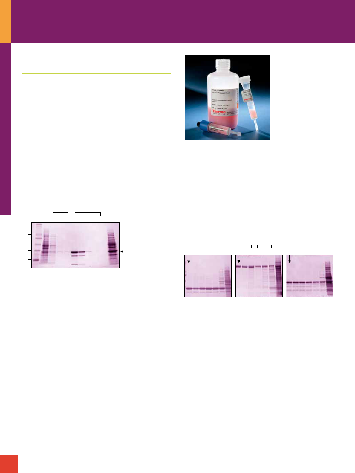
Cobalt Resin, Spin Columns
and Chromatography Cartridges
Specific, fast and gentle purification of His-tagged proteins.
The preferred method for purifying recombinant His-tagged
proteins is immobilized metal affinity chromatography (IMAC).
Traditionally, chelating chromatography resins are charged with
either nickel or cobalt ions that coordinate with the histidine side
chains in the 6xHis-tag. HisPur Cobalt Resin is a tetradentate
chelating resin charged with cobalt that binds His-tagged proteins
with high specificity and releases them under lower imidazole
concentrations than required with nickel resins (see Figure 5).
HisPur Cobalt Resin can be used to obtain high-purity proteins with
no metal contamination.
The HisPur Chromatography Cartridges are convenient, reliable
and ready-to-use pre-packed devices for the isolation of proteins
in solution and purification of His-tagged fusion proteins. The
HisPur Cartridges are compatible with automated LC instrumen-
tation, such as the ÄKTA
®
and BioLogic Systems, and adapt to
manual syringe processing.
Wash Elution
16.5
25
32
47
110
210
kDa
M F 1 2 3 1 2 3 4 5 L
6xHis-GFP
Thermo Scientific HisPur Cobalt Resin is specific for His-tagged proteins and
allows mild, efficient elutions. Bacterial lysate (1.0mg total
protein) was applied to a 200μL bed volume of HisPur Cobalt Resin in a spin
column. Gel lanes were normalized to equivalent volume. M = Molecular
Weight Markers (Product # 26691), L = lysate load and F = flow-through.
Highlights:
• High purity – obtain pure protein without optimizing imidazole
washing conditions
• Specificity – cobalt:chelate binding core binds fewer contaminants,
resulting in lower background than nickel (see Table 3)
• Low metal leaching – no metal contamination in eluted sample
• Versatility – purify proteins under native or denaturing conditions;
compatible with cell lysis reagents and a variety of buffer additives
• Flexibility – available as bulk resin or predispensed columns
compatible with both spin and gravity-flow formats
• Cost effective – reuse or discard
• Superior – performs better than other commercially available
IMAC Resins (see Figure 5)
Co
2
+
1
GFP β-gal Protein L
2 3 4 5 6 L 1 2 3 4 5 6 L 1 2 3 4 5 6 L
Ni
2
+
Ni
2
+
Co
2
+
Ni
2
+
Co
2
+
Figure 5. Thermo Scientific HisPur Cobalt Resin outperforms other IMAC
resins. Comparable yield and higher purity obtained with HisPur Cobalt Resin
(lane 1) compared to other IMAC resins (lanes 2 to 6). Cell lysates containing
over-expressed recombinant 6xHis-tagged protein were prepared in B-PER
Bacterial Protein Extraction Reagent (Product # 78243) and Protease Inhibitors
(Product # 78410). Protein concentrations were determined by Coomassie
Plus Protein Assay (Product # 23238). E. coli lysates containing over-expressed
His-tagged GFP, β-galactosidase or Protein L were applied to 0.2mL bed
volumes of each IMAC resin in spin column format. Binding, wash and elution
buffers were prepared and used per each manufacturers’ instructions. The first
elution fraction for each IMAC resin was analyzed by SDS-PAGE and protein
purity determined by densitometry. Gel lanes were normalized to equivalent
volume. Lanes: 1= HisPur Cobalt Resin, 2= supplier C cobalt resin, 3= supplier
S cobalt resin, 4= supplier G nickel resin, 5= supplier Q nickel resin, 6= Ni-IDA
and L= lysate load.
Fusion Protein Purification
To order, call 800-874-3723 or 815-968-0747. Outside the United States, contact your local branch office or distributor.
17
Table 3. Thermo Scientific HisPur Cobalt Resin yields more His-tagged protein and higher purity than other Co
2+
and Ni
2+
IMAC Resins.
His-GFP
His-β-Gal
His-Protein L
Yield (μg)* Purity (%)** Yield (μg)* Purity (%)** Yield (μg)* Purity (%)**
Thermo Scientific HisPur Cobalt Resin 298 87 78 93 42 77
Supplier C Cobalt Resin 206 78 26 90 35 76
Supplier S Cobalt Resin 211 85 27 65 38 77
Supplier G Nickel Resin 239 84 42 83 29 68
Supplier Q Nickel Resin 242 85 24 48 30 72
Ni
2+
-IDA Resin 70 37 6 16 17 46
* Recovered from a 5mg total protein load (total protein yield x purity). ** Purity of the first elution fraction.
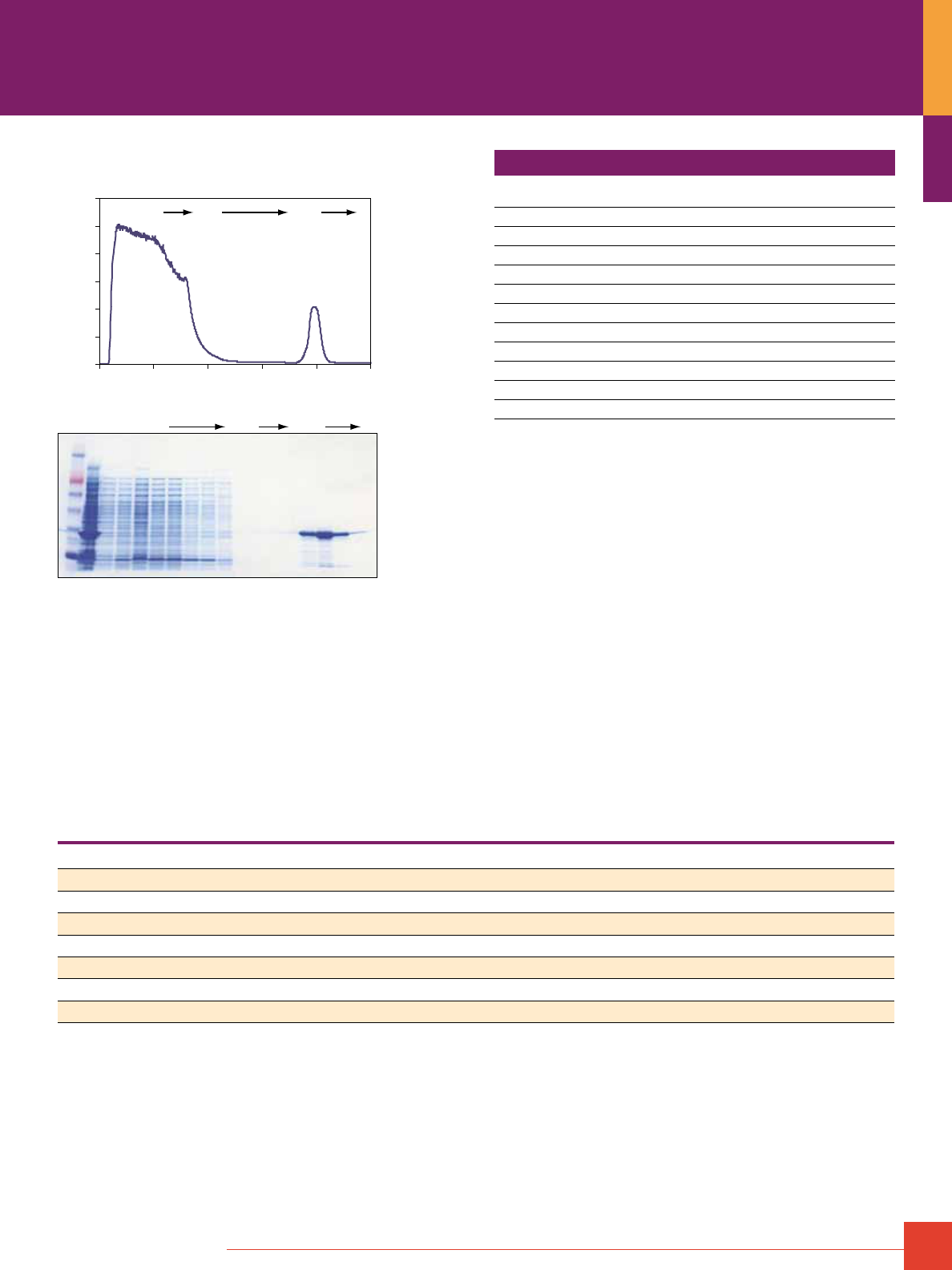
HisPur Cobalt Cartridge Performance Data:
0
1000
2000
3000
4000
5000
6000
0 5 10 15 20 25
Chromatography Progress (mL)
Flow-through Wash Elution
Absorbance 280nm (mAU)
Flow-through (FT)M S Wash Elution
Electrophoresed Fractions
Chromatography Profile
Purification of 6xHis-GFP from E. coli lysate using a Thermo Scientific HisPur
Cobalt Cartridge. His-tagged green fluorescent protein (GFP) was extracted
from E. coli using Thermo Scientific B-PER Bacterial Protein Extraction
Reagent in Phosphate Buffer (Product # 78266) containing Halt Protease
Inhibitor Cocktail, EDTA-Free (Product # 78415). The lysate was diluted 1:1
with equilibration/wash buffer (50mM sodium phosphate, 300mM sodium
chloride, 10mM imidazole, pH 7.4) and applied to a Thermo Scientific HisPur
Cobalt Chromatography Cartridge at a flow rate of 0.3mL/min. The cartridge
was washed with equilibration/wash buffer until the baseline absorbance
at A
280
was reached. His-tagged GFP was eluted (50mM sodium phosphate,
300mM sodium chloride, 150mM imidazole; pH 7.4) and selected fractions
were analyzed by SDS-PAGE and Thermo Scientific GelCode Blue Stain
Reagent (Product # 24592). M = MW Marker; S = non-fractionated lysate;
FT = flow-through.
Ordering Information
Product #
Description
Pkg. Size
U.S.
Price
89967 HisPur Cobalt Spin Columns, 0.2mL
25 columns
$ 160
89968 HisPur Cobalt Spin Columns, 1mL
5 columns
$ 105
89969 HisPur Cobalt Spin Columns, 3mL
5 columns
$ 155
89964 HisPur Cobalt Resin
10mL bottle
$ 88
89965 HisPur Cobalt Resin
100mL bottle
$ 600
89966 HisPur Cobalt Resin
500mL bottle
$2347
90090 HisPur Purification Kit, 0.2mL
25 columns
$ 277
90091 HisPur Purification Kit, 1mL
5 columns
$ 262
90092 HisPur Purification Kit, 3mL
5 columns
$ 277
90093 HisPur Chromatography Cartridges
5 x 1mL
$ 160
90094 HisPur Chromatography Cartridges
2 x 5mL
$ 267
18
For more information, or to download product instructions, visit www.thermoscientific.com/pierce
Fusion Protein Purification
High-quality purification of GST-fusion proteins
Multiple formats available to meet your purification needs
Purification of glutathione S-transferase (GST) fusion proteins
using glutathione agarose beads provides one-step, high-purity
affinity purification. The Thermo Scientific Pierce Glutathione
Agarose is composed of 6% crosslinked beaded agarose with
glutathione (GSH) immobilized by its central sulfhydryl. GST-fusion
proteins are purified with high yield because of the 12-atom GSH
linker that
minimizes steric hindrance. The Pierce Glutathione Agarose is
available in resin slurry packages, spin columns, complete
purification kits and FPLC-ready chromatography cartridges.
Highlights:
• High capacity – binds at least 40mg of pure GST per milliliter
of resin
• Cost-effective – resin is economically priced and can be reused
at least five times without reduction in performance
• High yield and purity – each milliliter of resin can purify ≥ 10mg
of GST-tagged protein from lysates with > 90% purity
• Compatible – validated and effective with Thermo Scientific
Cell Lysis Reagents
• Versatile – purify lysates or pulldown protein-protein interactions
• Flexible – available in multiple formats
Glutathione S-transferase and polyhistidine are the most
commonly used protein tags for affinity purification. GST
produces cleaner purifications than histidine tags and stabilizes
overexpressed fusion proteins. GST binds specifically to reduced
GSH in near-neutral, nondenaturing conditions. The bound
GST-tagged protein is easily dissociated (eluted) from GSH resin
by competitive displacement with a buffer containing free, reduced
GSH. Alternatively, if the fusion protein is designed with a protease
cleavage site, it may be released (cleaved) from the column at the
GST tag junction using thrombin, HRV 3C protease or Factor Xa.
The Pierce Glutathione Agarose is highly effective for purifying a
variety of GST-fusion proteins (Figure 6). The purity and yield of
GST-fusion protein recovery is comparable to or better than
glutathione resins from other suppliers (Figure 7). The resin is
composed of 6% crosslinked beaded agarose, making it a
structurally stable and resilient affinity support that enables
repeated uses. The Pierce Glutathione Agarose is also amenable
to chromatography cartridge format, producing baseline
resolution and a sharp elution peak (Figure 8) with no visible
resin bed compression.
GST-Pak1
kDa M L FT W1 W2 W3 E1 E2 E3
210
110
80
47
32
25
16.5
GST-ERK
M L FT W1 W2 W3 E1 E2 E3kDa
210
110
80
47
32
25
16.5
GST-Bid
M L FT W1 W2 W3 E1 E2 E3kDa
210
110
80
47
32
25
16.5
Figure 6. The Thermo Scientific Pierce Glutathione Agarose effectively purifies different GST-fusion proteins. Three GST-fusion proteins were expressed in E.
coli and extracted with Thermo Scientific B-PER Reagent with Enzymes (Product # 90078). GST-Pak1 and GST-Bid were purified using the spin format, and GST-
ERK was purified by gravity flow. GST-Pak1 and GST-Bid, 2.4mg and 11mg of total protein, respectively, were purified with a 0.2mL Pierce Glutathione Spin Column
(Product # 16106) per kit instructions. For GST-ERK, 11.2mg of total lysate protein in buffer (50mM Tris, 150mM NaCl; pH 8.0) was applied to a 0.5mL bed of Pierce
Glutathione Agarose (Product #16100) in a drip column and eluted with 125mM Tris, 150mM NaCl, 10mM glutathione at pH 8.0. Chromatography fractions were sep-
arated by SDS-PAGE and stained with Thermo Scientific GelCode Blue Stain Reagent (Product # 24590). M = Molecular weight markers; L = Lysate load; FT = Flow-
through; W = Wash; E = Elution.
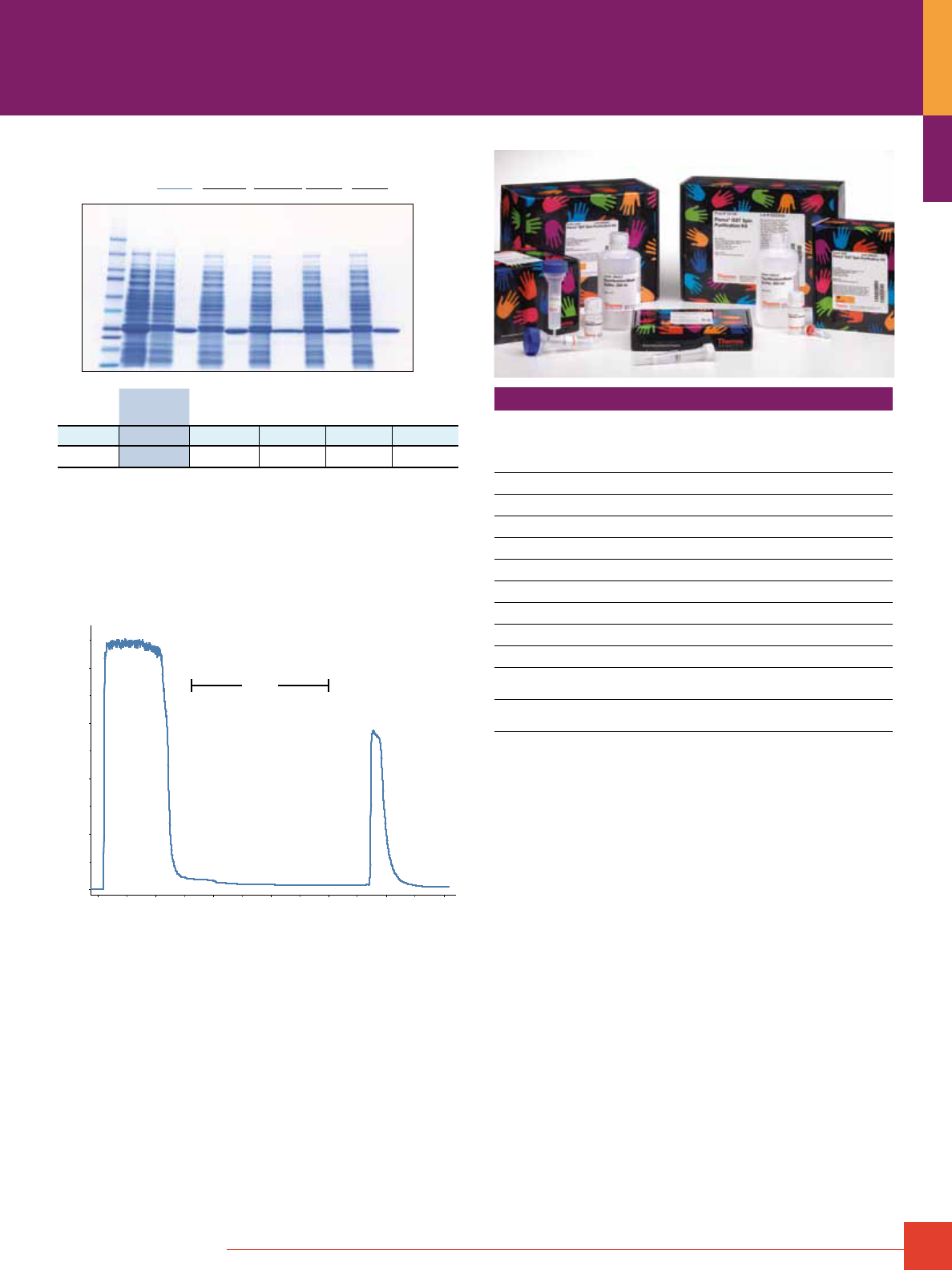
To order, call 800-874-3723 or 815-968-0747. Outside the United States, contact your local branch office or distributor.
19
kDa
250
150
100
75
50
27
25
20
15
10
M L FT
Thermo
Scientific
GE
Healthcare Qiagen Clontech Sigma
E FT E FT E FT E FT E
Vendor
Thermo
Scientific
GE
Healthcare Qiagen Clontech Sigma
Yield
537μg 562μg 285μg 299μg 410μg
Purity
93% 93% 90% 91% 94%
Figure 7. Thermo Scientific Pierce Glutathione Agarose delivers high yield
and high-purity GST-fusion proteins. E. coli lysate (14.4mg total protein) con-
taining an overexpressed GST-fusion protein was incubated with 50μL GSH
resin from various suppliers and purified per manufacturers’ instructions. The
amount of GST eluted from the resin (yield) was quantified by Thermo Scientific
Coomassie Plus Protein Assay. Purity was assessed by densitometry of the
stained gel lanes. M= Molecular weight markers; L= Lysate load; FT= Flow-
through; E= Elution.
0
0.0 10.0 20.0 30.0 40.0 50.0 ml
mAU
4000
Flow-
through
Wash Elution
3000
2000
1000
Figure 8. The glutathione chromatography cartridges provide an automatable
solution for GST-fusion protein purification. Clarified E. coli lysate (50mg)
containing overexpressed GST-Pak1 (34kDa) was applied to a 1mL glutathione
chromatography cartridge in buffer (50mM Tris, 150mM sodium chloride;
pH 8.0) at a flow rate of 0.5mL/minute using an ÄKTApurifier
®
System. The
cartridge was washed with the same buffer at 1mL/minute. GST-Pak1 was
eluted (50mM Tris, 150mM sodium chloride, 10mM glutathione; pH 8.0) at
1mL/minute.
Ordering Information
See catalog or www.thermoscientific.com/pierce for a complete description
of products and kits.
Product # Description Pkg. Size
U.S.
Price
16100 Pierce Glutathione Agarose
10mL
$ 180
16101 Pierce Glutathione Agarose
100mL
$ 975
16102 Pierce Glutathione Agarose
500mL
$3000
16103 Pierce Glutathione Spin Columns, 0.2mL
25 columns
$ 175
16104 Pierce Glutathione Spin Columns, 1mL
5 columns
$ 175
16105 Pierce Glutathione Spin Columns, 3mL
5 columns
$ 315
16106 Pierce GST Spin Purification Kit, 0.2mL
25-column kit
$ 275
16107 Pierce GST Spin Purification Kit, 1mL
5-column kit
$ 275
16108 Pierce GST Spin Purification Kit, 3mL
5-column kit
$ 375
16109 Pierce Glutathione Chromatography
Cartridges, 1mL
5 cartridges
$ 230
16110 Pierce Glutathione Chromatography
Cartridges, 5mL
2 cartridges
$ 400

20
For more information, or to download product instructions, visit www.thermoscientific.com/pierce
GST- and PolyHis-Tagged Pull-Down Assay Kits
Prepare to discover a new protein:protein interaction with your
GST- or polyHis-tagged bait protein.
Identifying and characterizing the interactions of a given protein has
emerged as the most valuable information that can be developed
in the post-genomic era. Thermo Scientific Pierce Pull-Down Kits
contain the necessary components to capture interacting proteins
using the popular pull-down method. The only item you provide is an
appropriately tagged fusion protein as the “bait.” Our Pull-Down Kits
are designed to teach the method to the first-time user and to shorten
the time to a result for those experienced in this method.
Step 3. Bind “prey” protein to immobilized
“bait” protein.
Step 4. Wash away unbound protein. Step 5. Elute protein:protein interaction complex.
Step 2. Wash away unbound protein.Step 1. Immobilize the fusion-tagged “bait”
from the lysate.
Step 6. Analyze protein:protein interaction
complex on SDS-PAGE.
=
Fusion Tag (GST or polyHis)
Bait
Prey
Marker Purified
Protein:Protein
Interaction
Agarose
Resin
Control
Purified
Bait
Displaced Interacting Complex
P re y
P ro te in
Bait Protein
Prey
Protein
Bait Protein
Prey Protein-
Containing Lysate
Bait Protein
Bait Protein
Bait Protein-
Containing Lysate
Affinity Ligand
(Glutathione or
Co
2+
Chelate)
Agarose
Bead
Prey
Protein
Bait Protein
Prey
Protein
Bait Protein
Prey
Protein
Glutathione
or
Imidazole
Spin
Spin
Highlights:
• Provides a complete, affordable and easy-to-use strategy
for discovery of protein:protein interactions
• Uses common laboratory equipment (e.g., microcentrifuges
and mini-gels)
• Adapts to single- or multiple-sample demands
• Supplied complete with cell lysis buffer
• Flexible protocol aids in the capture of weak or transient
interactions
• Efficient recovery of interacting complexes
Applications:
• Discover a new protein:protein interaction from a cell lysate
• Confirm a putative interaction from a cell lysate or with a
previously purified protein
• Extract protein:protein interaction information from in vitro
transcription/translation lysates
Generalized scheme for use of a Thermo Scientific Pierce Pull-Down Protein:Protein Interaction Kit using a GST-tagged or
PolyHis-tagged protein as the “bait.”
Fusion Protein Purification
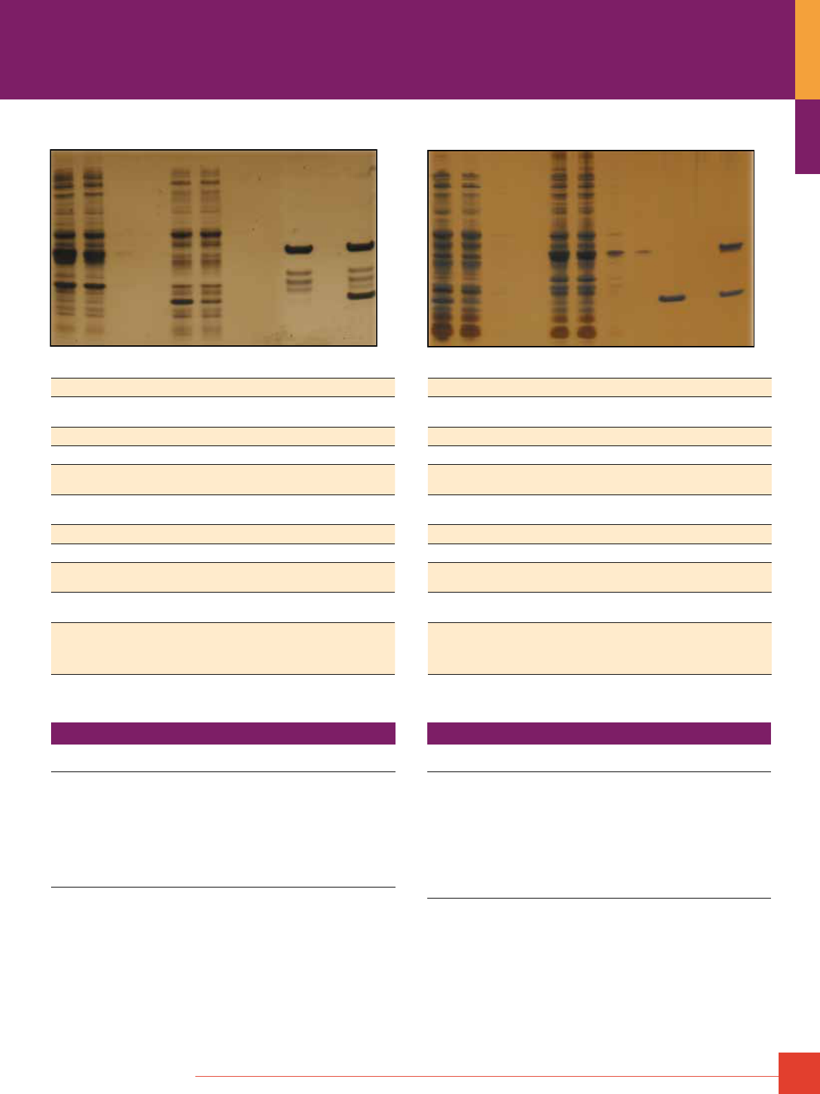
To order, call 800-874-3723 or 815-968-0747. Outside the United States, contact your local branch office or distributor.
21
Ordering Information
Product #
Description
Pkg. Size
U.S.
Price
21516 Pierce Pull-Down GST Protein Interaction Kit
Sufficient materials for performing 25 pull-down
assays using a GST-tagged protein as the bait.
Includes: Immobilized Glutathione
Lysis Buffer
Glutathione
BupH Tris Buffered Saline
Spin Columns Accessory Pack
Collection Tubes and Caps
Accessory Pack
Kit
750μL settled gel
250mL
1g
1 pack (500μL)
27 columns
100 tubes
$320
Acknowledgment
We gratefully acknowledge Dr. Yigong Shi of Princeton University for supplying the
GST-BIR2- and 9xHis Smac/DIABlO-expressing clones. Dr. Shi’s laboratory is engaged in
research aimed at understanding the structural and molecular mechanisms involved in
tumorigenesis and apoptosis.
Ordering Information
Product #
Description
Pkg. Size
U.S.
Price
21277 Pierce Pull-Down PolyHis
Protein Interaction Kit
Sufficient materials for performing 25 pull-down
assays using a polyhistidine-tagged protein as the bait.
Includes: Immobilized Glutathione
Lysis Buffer
4 M Imidazole Stock Solution
BupH Tris Buffered Saline
Spin Columns Accessory Pack
Collection Tubes and Caps
Accessory Pack
Kit
750μL settled gel
250mL
5mL
1 pack (500μL)
27 columns
100 tubes
$320
References
1. Chai, J., et al. (2000). Nature 406, 855-862.
2. Kaelin, W.G., et al. (1991). Cell 64, 521-532.
3. Sambrook, J. and Russell, D.W. (2001). Molecular Cloning: A Laboratory Manual
3rd Edition. Chapter 18: Protein Interaction Technologies, Protocol #3: Detection of
Protein-Protein Interactions using the GST Fusion Protein Pull-Down Technique.
Cold Spring Harbor Laboratory Press.
4. Soutoglou, E., et al. (2000). Mol. Cell 5, 745-751.
Lane # A. GST-Tag Pull-Down B. PolyHis-Tag Pull-Down
1
Lysate from E. coli expressing GST-tagged BIR2 (bait protein). Lysate from E. coli expressing 9xHis-tagged wild-type Smac (bait protein).
2
Flow-through from the lysate in Lane 1 bound to an immobilized
reduced glutathione support for 1 hour at 4°C.
Flow-through from the lysate in Lane 1 bound to an immobilized cobalt chelate support
for 1 hour at 4°C.
3
Wash #1 of the support. Wash #1 of the support.
4
Wash #2 of the support. (Washes 3-5 not shown.) Wash #2 of the support. (Washes 3-5 not shown.)
5
Lysate from E. coli expressing 9xHis-tagged wild-type Smac
(prey protein).
Lysate from E. coli expressing GST-tagged BIR2 (prey protein).
6
Flow-through from the lysate in Lane 5 is allowed to interact
with the immobilized bait for 1 hour at 4°C.
Flow-through from the lysate in Lane 5 is allowed to interact with the immobilized bait
for 1 hour at 4°C.
7
Wash #1 of the support. Wash #1 of the support.
8
Wash #2 of the support. (Washes 3-5 not shown.) Wash #2 of the support. (Washes 3-5 not shown.)
9
Bait control. Bait treated as described in Lanes 1-8 and
subsequently eluted. No prey added – just binding buffer.
Bait control. Bait treated as described in Lanes 1-8 and subsequently eluted. No prey
added – just binding buffer.
10
Prey control. Prey treated as described in Lanes 1-8 and
subsequently eluted. No bait added – just binding buffer.
Prey control. Prey treated as described in Lanes 1-8 and subsequently eluted. No bait
added – just binding buffer.
11
Elution of bait:prey complex (prepared in Lanes 1-8) from the
support with 100mm reduced glutathione. Western blotting
confirms that the minor bands observed in Lanes 9 and 11
are degradation products of GST-tagged BIR2.
Elution of bait:prey complex (prepared in Lanes 1-8) from the support with 250mm imidazole.
Validation of the Thermo Scientific Pierce Pull-Down Protein Interaction Kits using a known interacting pair.
1 2 3 4 5 6 7 8 9 10 11
1 2 3 4 5 6 7 8 9 10 11
A. B.
22
For more information, or to download product instructions, visit www.thermoscientific.com/pierce
Covalent Immobilization of Ligands
Affinity chromatography uses the specific interactions between two
molecules for the purification of a target molecule. In practice, a
ligand having affinity for a target molecule is covalently attached to
an insoluble support and functions as bait for capturing the target
from complex solutions.
The affinity ligand can be virtually any molecule that can specifically
bind the target without displaying significant nonspecific binding
toward other molecules in the solution. Ligands that have been
used for affinity separations include small organic compounds that
are able to dock into binding sites on proteins, inorganic metals that
form coordination complexes with certain amino acids in proteins,
hydrophobic molecules that can bind nonpolar pockets in biomole-
cules, proteins with specific binding regions that are able to interact
with other proteins, and antibodies, which can be designed to target
any biomolecule through their antigen-binding sites.
The concept of using immobilized affinity ligands to target
biomolecules has extended beyond chromatographic applications.
Affinity ligands are now coupled to latex beads, nanoparticles,
macro-beads, membranes, microplates, array surfaces, dipsticks
and a host of other devices that facilitate the capture of specific
biomolecules. The application of affinity targeting includes
purification, scavenging (or removal of contaminants), catalysis
(or modification of target molecules) and a broad range of
analytical uses to quantify a target molecule in a sample solution.
Designing custom affinity supports that are able to target unique
biomolecules requires methods to covalently link a ligand to an
insoluble matrix. Regardless of the intended application, the
chemical reactions that make ligand attachment possible are
well characterized and facilitate the attachment of biomolecules
through their common chemical groups. The types of functionalities
generally used for attachment include easily reactive components
such as primary amines, sulfhydryls, aldehydes, carboxylic acids,
hydroxyls, phenolic groups and histidinyl residues. Usually, the
solid-phase matrix first is activated with a compound that is
reactive toward one or more of these functional groups. The
activated complex then can form a covalent linkage between
the ligand and the support, resulting in ligand immobilization.
The type of linkage that is formed between the matrix and the
immobilized ligand affects the performance of the affinity support
in a number of ways. A linkage that allows the coupled ligand to
leach from the matrix will result in contamination of the purified
protein and shorten the useful life of the affinity support. A linkage
that introduces a charged functional group into the support can
cause nonspecific binding by promoting ion-exchange effects.
A linkage that alters the structure of the matrix can change the
flow and binding characteristics of the support. Cyanogen
bromide (CNBr)-activated supports are informative as an example
of these principles. This popular immobilization method results
in a linkage that:
1. Has a constant leakage of ligand from the matrix that
becomes a contaminant in the purified preparation.
2. Includes a charged isourea group in the linkage,
resulting in nonspecific binding.
3. Causes extensive crosslinking of the matrix, reducing the
ability of large molecules to penetrate into the interior of
the resin.
We offer a number of activated affinity supports that are designed
to couple ligands of every type via stable, uncharged covalent
linkages that avoid introducing undesirable properties into the
supports. The activation chemistry and protocols have been
optimized to ensure excellent coupling yields with minimal effort
under a variety of conditions. Each activated support comes with
instructions for use and literature references as examples. The
associated kits contain all the coupling buffers, wash buffers and
columns necessary to perform the ligand immobilization and
produce a support ready to perform an affinity separation.
Covalent Coupling of Affinity Ligands
to Chromatography Supports
22
To order, call 800-874-3723 or 815-968-0747. Outside the United States, contact your local branch office or distributor.
23
Coupling Affinity Ligands through Amine Groups
The most common functional target for immobilizing protein
molecules is the amine group, which is present on the vast
majority of proteins because of the abundance of lysine side chain
e-amines and N-terminal a-amines. Thermo Scientific AminoLink
Coupling Resin and AminoLink Plus Coupling Resin are prepared
from crosslinked agarose supports, and they are designed to
create a stable linkage between amine groups and the support
material. AminoLink
®
Resins are activated to contain numerous
aldehyde groups, which can be used to immobilize amine-containing
ligands by reductive amination.
The immobilization reaction using reductive amination involves
the formation of an initial Schiff base between the aldehyde and
amine groups, which then is reduced to a secondary amine by
the addition of sodium cyanoborohydride. The cyanoborohydride
reducing agent used during the coupling process is mild enough
not to cleave disulfides in most proteins, and it will not reduce the
aldehyde reactants – only the Schiff base intermediates. It is best
to avoid stronger reducing agents such as sodium borohydride
because of the potential for disulfide reduction of the protein and
reduction of the aldehydes on the support to hydroxyls, effectively
quenching the reaction. Depending on the type and amount of
ligand present, a coupling reaction using reductive amination can
achieve immobilization yields of greater than 85%.
H
H
2
N–R
NaCNBH
3
O
N
H
R
Aldehyde Group on
Thermo Scientific
AminoLink Coupling Resin
Affinity Ligand Coupled
via Secondary Amine Bond
Another amine-reactive strategy that can be used for immobilization
is the azlactone ring present in UltraLink Biosupport. A primary
amine will react with an azlactone group in a ring-opening
process that produces an amide bond at the end of a five-atom
spacer. The group is spontaneously reactive with amines,
requiring no additives or catalysts to drive the coupling process.
The UltraLink Biosupport is supplied dry to ensure stability of the
azlactone groups until use. Adding a quantity of the support to a
sample containing a protein or other amine-containing molecule
causes immobilization to occur within about one hour. For protein
immobilization at high yield, it is recommended that the coupling
buffer contain a lyotropic salt, which functions to drive the protein
molecules toward the bead surface. This brings the hydrophilic
amines close enough to the azlactone rings to react quickly. The
simple nature of coupling affinity ligands to the UltraLink
Biosupport along with its inherently low nonspecific binding makes
it one of the best choices for immobilization.
O
N
O
CH
3
CH
3
N
H
O
H
N
O
R
Azlactone Group on
Thermo Scientific
UltraLink Biosupport
Affinity Ligand Coupled
via Amide Bond
H
2
N—R
Thermo Scientific UltraLink Biosupport binding capacity for various proteins.
Capacity Protein Coupling Buffer
35.0mg/mL Myoglobin 0.1M CHES, 1.0M sodium citrate, pH 9.0
21.5mg/mL Penicillin Acylase 0.1M sodium phosphate, 1.1M sodium
sulfate, pH 7.4
20.9mg/mL a-chymotrypsin 0.1M borate, 1.5 sodium sulfate, pH 9.0
35.5mg/mL BSA 0.1M borate, 1.5 sodium sulfate, pH 9.0
29.8mg/mL Lysozyme 0.1M borate, 1.0 sodium sulfate, pH 9.0
21.0mg/mL Human IgG 0.1M borate, 1.5 sodium sulfate, pH 9.0
A third option for immobilizing amine-containing affinity ligands
is the use of carbonyl diimidazole (CDI) to activate hydroxyls on
agarose supports to form reactive imidazole carbamates. This
reactive group is formed on the support in organic solvent and
stored as a suspension in acetone to prevent hydrolysis. Reaction
of the support in an aqueous coupling buffer with primary
amine-containing ligands causes loss of the imidazole groups
and formation of carbamate linkages. The coupling process
occurs at basic pH (8.5–10), but it is a slower reaction with
proteins than reductive amination or azlactone coupling. Thermo
Scientific Pierce CDI Supports are available with the CDI-activated
group, and they are particularly adept at immobilizing peptides and
small organic molecules. The reaction also can be done in
organic solvent to permit coupling of water-insoluble ligands.
Reactive Imidazole Carbamate
on Thermo Scientific Pierce
CDI Supports
Affinity Ligand Coupled
via Carbamate Bond
O
N
H
O
R
N
O
O
N
H
2
N—R
NHS-Activated Agarose resin uses safe, reliable NHS-ester
chemistry and does not require hazardous chemicals for
immobilization. Other amine-reactive supports, such as periodate-
oxidized resins, use toxic sodium cyanoborohydride to stabilize
the reaction linkage to primary amines and take 4 to 6 hours
to complete. Traditional methods such as cyanogen bromide-
activated supports also couple amines; however, this chemistry
results in nonspecific binding and constant slow leakage of the
coupled ligand. The Pierce NHS-Activated Agarose uses the
stable NHS-ester chemistry that allows immobilization reactions
to be completed in less than 1 hour.
The NHS-Activated Agarose coupling reaction is performed in an
amine-free buffer at pH 7-9. Protein coupling efficiency is typically
greater than 80%, regardless of the ligand’s molecular weight or pI.
Once a ligand is immobilized, the prepared resin can be used for
multiple affinity purification procedures. The crosslinked beaded
agarose has fast linear flow potential, making it useful for gravity-
flow and low- to medium-pressure applications.
24
For more information, or to download product instructions, visit www.thermoscientific.com/pierce
NHS-Activated Agarose resin is available in an anhydrous acetone
slurry and as dry powder. The unique dry form does not require
storage in or removal and disposal of the acetone solvent. In
addition, the dry resin is ideal for coupling reactions with dilute
samples because it concentrates the sample as the resin swells,
reducing the volume of the starting material and resulting in highly
efficient ligand immobilization.
O
N
H
O
O
O
O
O
N
NH
2
–R
O
N
H
O
O
R
H
N
Coupling Affinity Ligands through Sulfhydryl Groups
It is often advantageous to immobilize affinity ligands through
functional groups other than just amines. In particular, the thiol
group can be used to direct coupling reactions away from active
centers or binding sites on certain protein molecules. Because
amines occur at many positions on a protein’s surface, it is usually
difficult to predict where an amine-targeted coupling reaction will
occur. However, if sulfhydryl groups that typically are present in
fewer numbers are targeted for immobilization, then coupling can
be done at discrete sites in a protein or peptide. Thiol groups
(sulfhydryls) may be indigenous within a protein molecule or they
can be added through the reduction of disulfides or through the
use of various thiolation reagents. Sulfhydryls also can be added
to peptide affinity ligands at the time of peptide synthesis by
adding a cysteine residue at one end of the molecule. This ensures
that every peptide molecule will be oriented on the support in the
same way after immobilization.
Thermo Scientific SulfoLink Coupling Resin is designed to
efficiently react with thiol-containing molecules and immobilize
them through a thioether linkage. The support contains an iodo-
acetyl group at the end of a long spacer arm, which reacts with
sulfhydryls through displacement of the iodine. Optimal conditions
for the reaction are an aqueous environment at slightly basic pH,
wherein amines are not very reactive toward the iodoacetyl
function, but thiols are highly reactive due to their increased
nucleophilicity. The thioether bond that is formed provides a
stable linkage to any sulfhydryl-containing molecule.
HS
R
Reactive Iodoacetyl Group
on Thermo Scientific
SulfoLink Supports
Affinity Ligand
Coupled via
Thioether Bond
H
N
O
S
R
H
N
O
I
Coupling Affinity Ligands through Carbonyl Groups
Most biological molecules do not contain carbonyl ketones or
aldehydes in their native state. However, it might be useful to
create such groups on proteins to form a site for immobilization that
directs covalent coupling away from active centers or binding sites.
Glycoconjugates, such as glycoproteins or glycolipids, usually
contain sugar residues that have hydroxyls on adjacent carbon
atoms, which can be periodate-oxidized to create aldehydes.
Controlled oxidation using 1mm sodium meta-periodate at 0°C will
selectively oxidize sialic acid groups to form an aldehyde functionality
on each sugar. Using higher concentrations of periodate (10mm)
at room temperature will result in oxidation of other sugar diols to
create additional formyl groups. Aldehydes on the carbohydrate
portion of glycoconjugates can be covalently linked with
affinity supports through an immobilized hydrazide, hydrazine or
amine group by Schiff base formation or reductive amination.
Thermo Scientific CarboLink Coupling Resin contains long spacer
arms that terminate in hydrazide groups. Reaction of the hydrazides
with aldehydes forms hydrazone linkages, which are a form of
Schiff base displaying better stability than those formed between
an amine and an aldehyde. The CarboLink
™
Resin can be used to
immobilize glycoproteins, such as antibodies, after periodate
oxidation of the carbohydrate. Coupling antibodies in this manner
specifically targets the heavy chains in the Fc portion of the
molecule. Since this is away from the antigen-binding sites at the
end of the Fv regions, immobilization using this route often results
in the best retention of antigen-binding activity.
N
H
O
NH
2
N
H
O
N
R
Reactive Hydrazide Group
on Thermo Scientific
CarboLink Supports
Affinity Ligand
Coupled via
Hydrazone Bond
R H
O
Covalent Coupling of Affinity Ligands
to Chromatography Supports
To order, call 800-874-3723 or 815-968-0747. Outside the United States, contact your local branch office or distributor.
25
The CarboLink Resin also can be used to couple carbohydrates and
sugars through their reducing ends. Aldehyde- or ketone-containing
sugars will react with the immobilized hydrazide groups without
oxidation of other sugar hydroxyls. However, this reaction may be
dramatically slower than coupling with oxidized sugars because
these native aldehydes or ketones are usually tied up in acetal or
ketal ring structures. These rings can open in aqueous solution to
reveal the aldehyde or ketone, but the open structure is present
only a small percentage of the time. Thus, the reducing ends of
sugars have decreased reactivity toward an immobilized hydrazide,
sometimes requiring days of reaction time to obtain acceptable
immobilization yields.
Although the hydrazone bond created between the immobilized
hydrazide and an aldehyde is much more stable than amine-aldehyde
Schiff bases, to obtain a leach-resistant linkage it is recommended
that the Schiff base be reduced with sodium cyanoborohydride.
This is especially true if a ligand is coupled that has only a single
point of attachment to the support. Reduction of the hydrazone
creates a stable bond that will perform well in affinity
chromatography applications.
Affinity Ligand
Coupled via
Hydrazone Bond
Stable Ligand
Linkage
N
H
O
N
R
N
H
O
H
R
N
NaCNBH
3
Coupling Affinity Ligands through Carboxyl Groups
The carboxyl group is a frequent constituent of many biological
molecules. Particularly, proteins and peptides typically contain
numerous carboxylic acids due to the presence of glutamic acid,
aspartic acid and the C-terminal a-carboxylate group. Carboxylic
acids may be used to immobilize biological molecules through the
use of a carbodiimide-mediated reaction. Although no activated
support contains a reactive group that is spontaneously reactive
with carboxylates, chromatography supports containing amines
(or hydrazides) may be used to form amide bonds with carboxyl-
ates. Molecules containing carboxylates may be activated to react
with an immobilized amine (or hydrazide) through reaction with the
water-soluble carbodiimide EDC.
EDC reacts with carboxylates to form an intermediate ester that is
reactive with nucleophiles such as primary amines. The reaction
takes place efficiently between about pH 4.5 and pH 7.5, and it is
complete within two to four hours, depending on the temperature.
The intermediate ester is subject to hydrolysis; therefore, it is
beneficial if the amine-containing ligand to be immobilized is
included in the reaction medium upon addition of EDC, so it can
react immediately with the ester as it forms.
Thermo Scientific CarboxyLink Coupling Resin or the UltraLink
DADPA Resin may be used to immobilize carboxylate-containing
ligands by EDC. CarboxyLink
®
Resin contains a nine atom spacer
arm and UltraLink DADPA contains a 12-atom spacer arm to mini-
mize steric hindrance.
R
OH
O
Carboxylate-Containing
Affinity Ligand
+
EDC
Immobilized DADPA on
Thermo Scientific CarboxyLink Resin
Immobilized Ligand via
Amide Bond Formation
H
N
H
N
H
N
O
R
H
N
H
N
NH
2
26
For more information, or to download product instructions, visit www.thermoscientific.com/pierce
Products for Immobilizing Ligands through
Primary Amines
Thermo Scientific Pierce NHS Ester-Activated Agarose
Pierce NHS-Activated Agarose Resin, available as a slurry or
dry powder, allows for the simple and efficient immobilization of
proteins to a beaded-agarose support. The activated agarose
contains N-hydroxysuccinimide (NHS) ester with a spacer arm of
at least 10 atoms in length that reacts with primary amines form-
ing stable amide linkages. The NHS-Activated Agarose coupling
reaction is performed in an amine-free buffer at pH 7-9, with typi-
cal coupling efficiencies of more than 85%. The prepared resin
can be used for multiple affinity purification procedures. The
crosslinked beaded agarose has fast linear flow potential, making
it useful for gravity-flow and low- to medium-pressure applications.
O
N
H
O
O
O
O
O
N
O
N
H
H
O
O
N
Agarose
Bead
Agarose
Bead
Protein
+
NH
2
Protein
Thermo Scientific NHS Ester-Activated Agarose Support
immobilization chemistry.
Target: –NH
2
Support: 6% agarose
Binding capacity: >25mg protein/mL resin
Time: 30 minutes
Ordering Information
Product #
Description
Pkg. Size
U.S.
Price
26196
Pierce NHS-Activated Agarose, Dry
Swell volume: 6-7.5mL/g of dry resin
1g
$ 65
26197
Pierce NHS-Activated Agarose, Dry
Swell volume: 6-7.5mL/g of dry resin
5g
$200
26198
Pierce NHS-Activated Agarose
Spin Columns, 0.2mL
25 spin columns containing 33mg
of NHS-Activated Agarose
Swell volume: 6- 7.5uL/mg of dry resin
25 columns
$125
26199
Pierce NHS-Activated Agarose
Spin Columns, 2mL
5 spin columns containing 330mg of
NHS-Activated Agarose
Swell volume: 6- 7.5uL/mg of dry resin
5 columns
$125
26200
Pierce NHS-Activated Agarose Slurry
25mL of settled resin in anhydrous acetone
25mL
$195
Covalent Coupling of Affinity Ligands
to Chromatography Supports
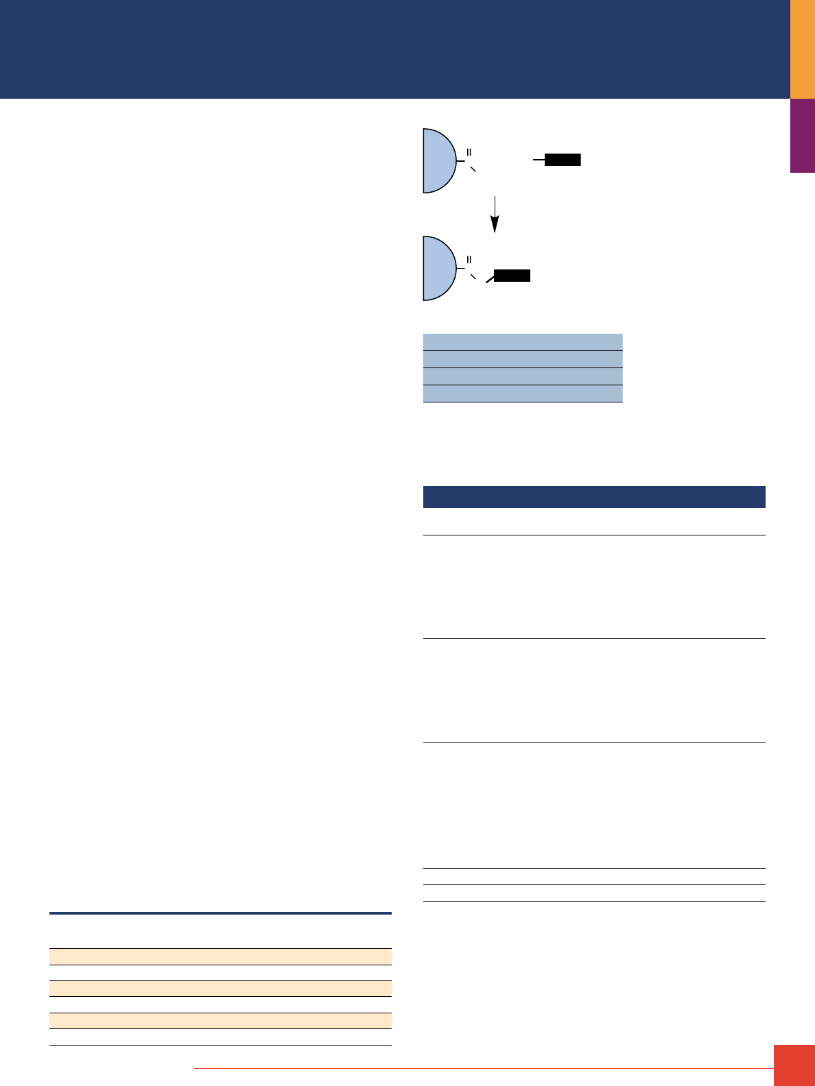
To order, call 800-874-3723 or 815-968-0747. Outside the United States, contact your local branch office or distributor.
27
Thermo Scientific AminoLink Plus Immobilization Kits
and Coupling Resin
The simplest and surest method for making an affinity purification
resin with antibodies or other proteins.
AminoLink Plus Coupling Resin and Immobilization Kits use activated
beaded agarose and a robust coupling chemistry to immobilize
proteins and other ligands through primary amines (–NH
2
) to the
resin. Once an antibody or other ligand is immobilized, the prepared
affinity resin can be used for a variety of purification methods
involving batch or column chromatography. The resin and linkage
are stable in binding and elution conditions typically used in affinity
chromatography, enabling prepared resin to be used for at least
10 rounds of affinity purification.
The AminoLink Plus Coupling Reaction involves spontaneous
formation of Schiff base bonds between aldehydes (on the support)
and amines (on the ligand) and their subsequent stabilization by
incubation with a mild reductant (sodium cyanoborohydride; see
more detailed reaction scheme on next page). The entire coupling
reaction, called reductive amination, occurs in four to six hours
in simple non-amine buffers such as PBS. Coupling efficiency with
antibodies and typical proteins is generally greater than 85%,
resulting in 1 to 20mg of immobilized protein per milliliter of
agarose resin.
Highlights:
• AminoLink Plus Coupling Resin – aldehyde-activated crosslinked
4% beaded agarose
• Ideal for antibodies and other proteins – immobilize molecules
via primary amines (–NH
2
)
• Flexible coupling conditions – efficient (>85%) coupling over
a wide range of pH (4–10) and buffer conditions (PBS or other
non-amine buffer with or without organic solvent); regular
(PBS, pH 7.2) and enhanced (borate, pH 10) coupling
protocols provided
• Stable, permanent immobilization – coupling reaction results
in stable, leak-resistant secondary amine bond between resin
and ligand
• Better than immobilization to CNBr-activated agarose – bond is
more stable and uncharged, resulting in less nonspecific binding
in affinity purification procedures
• Versatile and reusable – prepared affinity resin is adaptable to
column and batch affinity techniques and the resin is reusable
for typical applications based on protein binding interactions
• Convenient kits and product sizes – choose one- or five-column
kit containing complete sets of buffers, reagents and versatile
spin/drip columns, or select bulk resin. Bulk quantities are
available for manufacturing applications
Efficient immobilization of antibodies and other proteins. Percent coupling
efficiency of different proteins to 2mL of Thermo Scientific AminoLink Plus
Coupling Resin using the pH 7.2 coupling protocol.
Protein
Protein Applied
(mg/mL)
Protein Coupled
(mg/mL)
Percent Coupled
Protein G 4.6 4.0 83
Mouse IgG 4.7 4.5 96
Rat IgG 4.7 4.4 93
Goat IgG 0.9 0.8 84
Human IgG 4.8 4.6 97
Human IgM 0.9 0.8 93
Agarose
Bead
C
O
H
+
NH
2
Protein
Agarose
Bead
C
O
N
H
Protein
Thermo Scientific AminoLink Support immobilization chemistry.
Target: –NH
2
Support: Highly crosslinked 4% agarose
Binding capacity: 20mg protein/mL resin
Time: 4 hours
References
Beall, A., et al. (1999). J. Biol. Chem. 274(16), 11344–11351.
Nakasato, Y.R., et al. (1999). Clin. Chem. 45, 2150–2157.
Allan, B.B., et al. (2000). Science 289, 444–448.
Lu, R., et al. (2000). J. Neurochem. 74, 320–326
Ordering Information
Product #
Description
Pkg. Size
U.S.
Price
44894 AminoLink Plus Immobilization Kit
Sufficient reagents for preparing five affinity columns.
Includes: AminoLink Column
Phosphate Buffered Saline (PBS)
Citrate-Carbonate Buffer
Quenching Buffer
Wash Solution
Sodium Cyanoborohydride Solution (5 M)
Column Accessories
Kit
5 x 2mL
1 pack
1 pack
50mL
240mL
0.5mL
$385
20394 AminoLink Plus Immobilization Trial Kit
Sufficient reagents for preparing one affinity column.
Includes: AminoLink Column
Phosphate Buffered Saline (PBS)
Citrate-Carbonate Buffer
Quenching Buffer
Wash Solution
Sodium Cyanoborohydride Solution (5 M)
Column Accessories
Kit
1 x 2mL
1 pack
1 pack
15mL
50mL
0.5mL
$105
20475 AminoLink Plus Micro Immobilization Kit
Sufficient reagents for 10 coupling reactions
using 25–100µg of protein and 20 affinity purifications.
Includes: AminoLink Plus Spin Columns,
each containing 400μL of 25% slurry
Phosphate Buffered Saline
Quenching Buffer
Sodium Cyanoborohydride Solution
Wash Solution
Elution Buffer
Microcentrifuge Collection Tubes
Kit
10 each
1 pack
60mL
0.5mL
25mL
50mL
200 each
$301
20501 AminoLink Plus Coupling Resin
10mL
$105
20505 AminoLink Plus Coupling Resin
50mL
$360
28
For more information, or to download product instructions, visit www.thermoscientific.com/pierce
Thermo Scientific AminoLink Immobilization Kits
and Coupling Resin
Links with primary amines (lysine residues and N-terminus)
on proteins, peptides, antigens or antibodies.
AminoLink Coupling Resin is crosslinked 4% beaded agarose that
has been activated with aldehyde groups. Proteins and other
molecules with primary amines can be covalently attached
(immobilized) to AminoLink Resin to make chromatography columns
for use in affinity purification. The aldehyde groups form stable
secondary amine bonds with primary amines such as exist in the
side chain of lysine (K) residues, which are generally abundant
and readily accessible in proteins. Once a protein is immobilized,
the prepared affinity resin can be used for a variety of batch and
column affinity purification methods involving binding interactions
with the immobilized protein. The resin and linkage are stable
in most binding and elution conditions typically used in affinity
chromatography, enabling prepared resin to be used for multiple
rounds of affinity purification procedures.
Highlights:
• AminoLink Coupling Resin – aldehyde-activated crosslinked
4% beaded agarose
• Ideal for antibodies and other proteins – immobilize molecules
via primary amines (–NH
2
)
• Flexible coupling conditions – efficient (>85%) coupling over a
wide range of pH (4–10) and buffer conditions (PBS or other
non-amine buffer with or without organic solvent)
• Stable, permanent immobilization – coupling reaction results in
stable, leak-resistant secondary amine bond between resin
and ligand
• Better than immobilization to CNBr-activated agarose – bond is
more stable and uncharged, resulting in less nonspecific binding
in affinity purification procedures
• Versatile and reusable – prepared affinity resin is adaptable to
column and batch affinity techniques and the resin is reusable
for typical applications based on protein binding interactions
Agarose
Bead
C
O
H
+
NH
2
Protein
Agarose
Bead
C
O
N
H
Protein
Thermo Scientific AminoLink Support detailed immobilization chemistry.
Target: –NH
2
Support: 4% agarose
Binding capacity: 1-20mg protein/mL resin
Time: 4 hours
Efficient immobilization at a variety of pH values. The effect of coupling
buffer pH on percent coupling efficiency of 9.58mg of human IgG to 2mL of
Thermo Scientific AminoLink Resin using the standard coupling protocol.
pH Coupling Efficiency of 9.58mg Human IgG
4 91.8%
5 92.7%
6 89.1%
7 87.3%
8* 85.3%
9* 94.9%
10* 98.4%
* Schiff-base formation occurs readily at high pH, but reduction to stable secondary
amine bond requires subsequent incubation with sodium cyanoborohydride
(AminoLink Reductant) at pH 7.2.
References
Cheadle, C., et al. (1994). J. Biol. Chem. 269(39), 24034–24039.
Cofano, F., et al. (1990). J. Biol. Chem. 265(7), 4064–4071.
DeSilva, B.S. and Wilson, G.S. (1995). J. Immunol. Method 188, 9–19.
Rivero-Lezcano, O.M., et al. (1994). J. Biol. Chem. 269(26), 17363–17366.
Czermak, B.J., et al. (1999). J. Immunol. 162, 2321–2325.
Assad, F.F., et al. (2001). J. Cell Biol. 152, 531–543.
Zuk, P.A. and Elferink, L.A. (2000). J. Biol. Chem. 275(35), 26754–26764.6.
Ordering Information
Product #
Description
Pkg. Size
U.S.
Price
20381 AminoLink Coupling Resin
10mL
$105
20382 AminoLink Coupling Resin
50mL
$380
44890 AminoLink Immobilization Kit
Sufficient reagents to prepare five affinity columns.
Each column can be used for up to
10 affinity purifications.
Includes: AminoLink Columns
AminoLink Coupling Buffer
Quenching Buffer
Wash Solution, Sodium Cyanoborohydride
Solution (5 M)
Column Accessories
Kit
5 x 2mL
250mL
50mL
240mL
0.5mL
$399
20384 AminoLink Immobilization Trial Kit
Includes: AminoLink Column Reagents and Buffers
Trial Kit
1 x 2mL
$106
44892 AminoLink Reductant
(Sodium cyanoborohydride)
2 x 1g
$ 39
Covalent Coupling of Affinity Ligands
to Chromatography Supports
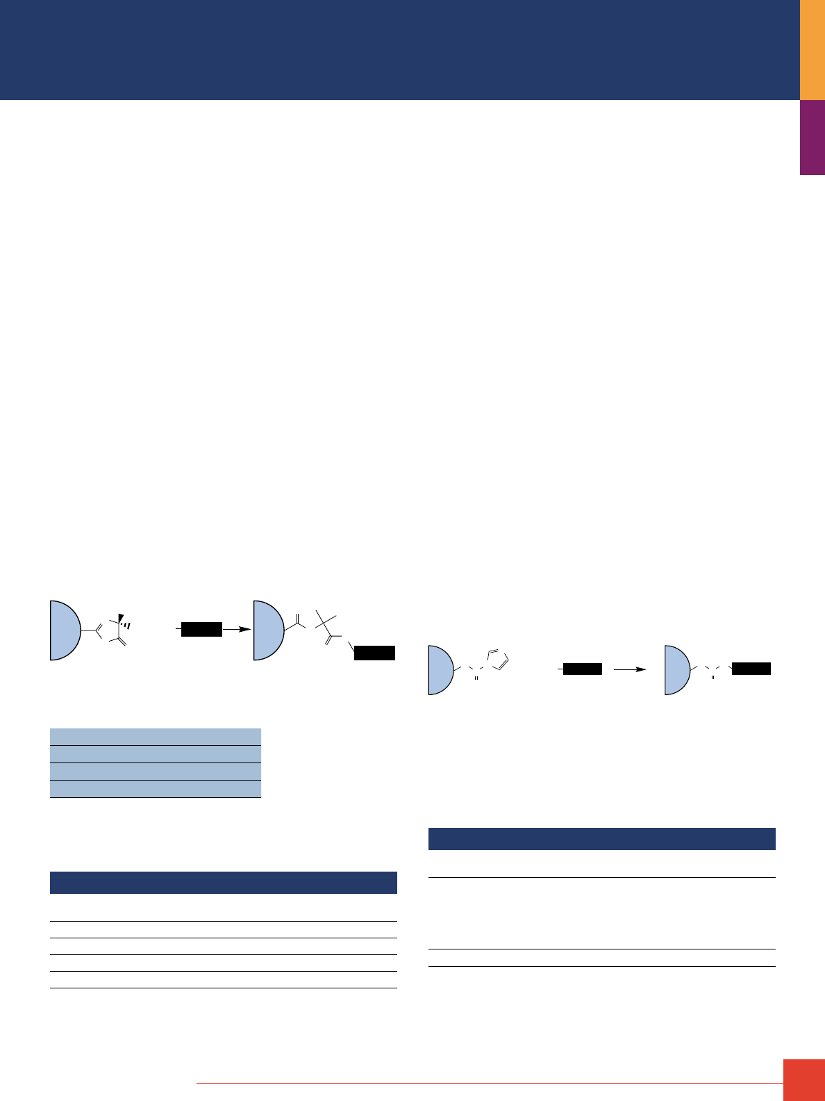
To order, call 800-874-3723 or 815-968-0747. Outside the United States, contact your local branch office or distributor.
29
Thermo Scientific UltraLink Biosupport
A durable, polyacrylamide resin, activated for efficient coupling
of proteins.
UltraLink Biosupport is a durable, porous resin that is activated to
enable efficient and direct covalent immobilization of proteins and
other biomolecules through their primary amines for use in affinity
purification procedures.
Highlights:
• High coupling efficiency and capacity – immobilizes proteins
with very high efficiency and coupling capacity in 1 hour
• Specific and leak-proof coupling chemistry – reacts specifically
with primary amines (–NH
2
), resulting in amide bonds that are
stable for use in many affinity purification procedures; coupling
reaction has no leaving group to contaminate samples
• Easy to use – no pre-swelling or secondary reagents required;
simply weigh the needed amount of dry support and add the
ligand solution to initiate coupling reaction
• Flexible coupling conditions – perform immobilization reaction
in any of a variety of non-amine buffers and pH levels; coupling
is most efficient in buffers containing a lyotropic salt such as
sodium citrate; coupling compatible with or without organic solvent
• Excellent reusability – prepared affinity resin can be used with
typical binding and elution procedures for more than 100 cycles
of affinity purification without significant loss of binding capacity
• Durable, high-performance resin – porous beads have a 60μm
diameter, can withstand 100 psi (6.9 bar) and allow for linear
flow-through rates of 3,000cm/hour
H
2
N
+
Amine
Ligand
Protein
UtraLink Biosupport
(Azlactone Ring on Bead)
Amide Bond Formation
with Ring Opening
NH
O
N
H
O
O
N
O
CH
3
CH
3
Resin
Bead
Resin
Bead
Protein
Thermo Scientific UltraLink Biosupport immobilization chemistry.
Target: –NH
2
Support: Polyacrylamide/azlactone copolymer
Binding capacity: >18mg protein/mL resin
Time: 4 hours
References
1. Ju, T., et al. (2002). J. Biol. Chem. 277, 169–177.
2. Ju, T., et al. (2002). J. Biol. Chem. 277, 178–186.
3. Kornfeld, R., et al. (1998). J. Biol. Chem. 273, 23202–23210.
4. Liu, L.A. and Engvall, E. (1999). J. Biol. Chem. 274, 38171–38176.
Ordering Information
Product #
Description
Pkg. Size
U.S.
Price
53110 UltraLink Biosupport (8-10mL)
1.25g
$155
53111 UltraLink Biosupport (50mL)
6.25g
$539
28388 BupH Citrate-Carbonate Buffer Packs
10 packs
$ 64
28386 BupH Citrate-MOPS Buffer Packs
10 packs
$ 58
Thermo Scientific Pierce CDI Supports
Carbonyldiimidazole-activated resins for ligand immobilization.
Highlights:
• Reliable coupling chemistry – immobilization occurs through the
reaction of N-nucleophiles with 1,1’-carbonyldiimidazole groups
of the resin to form a stable, uncharged N-alkylcarbamate linkages
• Easy-to-use – no secondary reagents needed; just wash equilibrate
resin in alkaline coupling buffer and add ligand; reaction proceeds
spontaneously
• Stable activation – half-life of hydrolysis is much longer than
hydroxysuccinimide ester activations, making immobilization
reactions simpler to prepare and control; simplifies filtration and
washing before adding a ligand or protein
• Well-defined coupling conditions – reaction is most efficient with
primary amines at pH 9-11
CDI-Activated Crosslinked 6% Beaded Agarose:
• Beaded agarose – the most popular resin for routine affinity
purification methods
• Highly activated – at least 50μmol 1,1’-carbonyldiimidazole (CDI)
groups per milliliter of resin
• Convenient form – supplied as stabilized, 50% slurry in acetone
CDI-Activated Trisacryl
®
GF-2000:
• Trisacryl resin – rigid, polyacrylamide matrix allows for high flow rates
• Hydrophilic matrix without charge effects – provides for low
nonspecific binding
• Highly activated – at least 50μmol 1,1’ -carbonyldiimidazole (CDI)
groups per milliliter of resin
• Convenient form – supplied as stabilized, 50% slurry in acetone
Resin
Bead
O
C
N
N
O
O
C
H
N
O
H
2
N
Imidazole
Carbamate
Amine
Ligand
Carbamate
Linkage
Resin
Bead
Protein
Protein
+
CDI immobilization chemistry.
References
1. Shenoy, S.K., et al. (2001). Science 294, 1307–1313.
2. Richardson, R.T., et al. (2000). J. Biol. Chem. 275, 30378–30386.
3. Tanaka, M, et al. (2005). PLoS Biology 3, 764–776.
Ordering Information
Product #
Description
Pkg. Size
U.S.
Price
20259 Pierce CDI (6X) Support
1,1’-Carbonyldiimidazole activated crosslinked
6% beaded agarose
Supplied: stabilized in acetone slurry agarose
hydrated particle size: 45–165μm
Activation level: >50μmol/mL of resin
10mL
$ 85
20377 Pierce CDI Trisacryl Support
50mL
$442
30
For more information, or to download product instructions, visit www.thermoscientific.com/pierce
Products for Immobilizing Ligands
through Sulfhydryl Groups
Thermo Scientific SulfoLink Immobilization Kits
and Coupling Resin
Covalent immobilization of sulfhydryl-containing peptides or
proteins for affinity purification.
SulfoLink Coupling Resin is porous, crosslinked 6% beaded agarose
that has been activated with iodoacetyl groups. When incubated
with a solution of peptide or protein that contains reduced cysteine
residues, the iodoacetyl groups react specifically and efficiently
with the exposed sulfhydryls (–SH) to form covalent and irrevers-
ible thioether bonds that permanently attach the peptide or protein
to the resin. The result is a custom-made affinity resin for purifica-
tion of antibodies, antigens and other molecules of interest.
Highlights:
• Specific conjugation through sulfhydryl (–SH) groups – the
iodoacetyl groups react specifically with sulhydryls to form
irreversible thioether bonds
• Separate kits optimized for peptides or proteins – kits include
optimized reagents for preparing peptide or protein samples for
efficient immobilization
• Fast – spin columns increase protocol speed; prepare and
couple samples in 2 hours (peptides) to 3.5 hours (proteins)
• Flexible coupling conditions – use pH 7.5–9.0 aqueous buffers,
organic solvent (e.g., 20% DMSO) or denaturant (guanidine•HCl),
as needed for protein or peptide solubility during coupling reaction
• Easy-to-follow instructions – streamLined protocols for sample
preparation, immobilization, and affinity purification
• High capacity – immobilize 1–2mg peptide or 2–20mg protein
per 2–mL column of SulfoLink Coupling Resin
HS
+
H
N
I
O
H
N
S
O
Thermo Scientific
SulfoLink Coupling Resin
Thioether
Bond
12-atom
Spacer
Iodoacetyl
Group
+
HI
Immobilized Peptide
Reduced Sulfhydryl
Molecule
Agarose
Bead
Peptide
Peptide
Agarose
Bead
Thermo Scientific SulfoLink Support immobilization chemistry.
Target: –SH
Support: 6% agarose or UltraLink
®
Resin
Binding capacity: 20mg protein/mL resin
Time: 2-3.5 hours
Peptide A Calcitonin Peptide
0
25
50
75
100
+TCEP
+TCEP
-TCEP
-TCEP
Percent
Immobilized
Improved retention of peptides on Thermo Scientific SulfoLink Resin with TCEP.
TCEP effectively reduces peptides to maximize immobilization efficiency. Two
peptides (Peptide A and human calcitonin peptide) were incubated with 25mm
TCEP for 30 minutes and immobilized onto SulfoLink Resin via their reduced
sulfhydryl groups. Peptide A’s cysteine had oxidized during long-term storage and
the calcitonin peptide contained an internal disulfide bond. Each peptide also con-
tained an amine-terminal fluorescent probe by which the binding of the peptide
could be monitored during the immobilization and wash steps.
References
Grunwald, R. and Meissner, G. (1995). J. Biol. Chem. 270(19), 11338–11347.
Seubert, P., et al. (1993). Nature 361, 260–263.
Sukegawa, J., et al. (1995). J. Biol. Chem. 270(26), 15702–15706.
Wisniewski, J.R., et al. (1994). J. Biol. Chem. 269(46), 29261–29264.
Sakaguchi, K., et al. (2000). J. Biol. Chem. 275, 9278–9283.
Tan, M., et al. (2000). Proc. Natl. Acad. Sci. USA 97, 109–114.
Quill, T.A., et al. (2001). Proc. Natl. Acad. Sci. USA 98, 12527–12531.
Tokumaru, H., et al. (2001). Cell 104, 421–432.
Assad, F.F., et al. (2001). J. Cell Biol. 152, 531–543.
Ordering Information
Product #
Description
Pkg. Size
U.S.
Price
44995 SulfoLink Immobilization Kit for Proteins
Sufficient reagents to prepare five affinity columns.
Kit contains: SulfoLink Columns
SulfoLink Sample Preparation Buffer
SulfoLink Coupling Buffer
Wash Solution
2-Mercaptoethylamine•HCl
L-Cysteine•HCl
Zeba Desalt Spin Columns
Phosphate Buffered Saline
Columns Accessories
Kit
5 x 2mL
7.5mL
500mL
120mL
5 x 6mg
100mg
5 x 5mL
1 Pack
$ 399
44999 SulfoLink Immobilization Kit for Peptides
Sufficient reagents to prepare five affinity columns.
Kit contains: SulfoLink Columns
SulfoLink Coupling Buffer
Wash Solution
Bond-Breaker TCEP Solution
L-Cysteine•HCl
Phosphate Buffered Saline
Column Accessories
Kit
5 x 2mL
120mL
120mL
0.5mL
100mg
1 Pack
$ 399
20325 SulfoLink Immobilization Trial Kit
Sufficient reagents to prepare 1 affinity column with a
peptide or protein.
Kit contains: SulfoLink Column
SulfoLink Sample Preparation Buffer
SulfoLink Coupling Buffer
Wash Solution
2-Mercaptoethylamine•HCl
Bond-Breaker TCEP Solution
Cysteine•HCl
Zeba Desalt Spin Column
Phosphate Buffered Saline
Columns Accessories
Kit
1 x 2mL
7.5mL
120mL
25mL
6mg
0.5mL
100mg
5mL
1 Pack
$ 170
20401 SulfoLink Coupling Resin
10mL
$ 135
20402 SulfoLink Coupling Resin
50mL
$ 474
20404 SulfoLink Coupling Resin
250mL
$1900
Covalent Coupling of Affinity Ligands
to Chromatography Supports
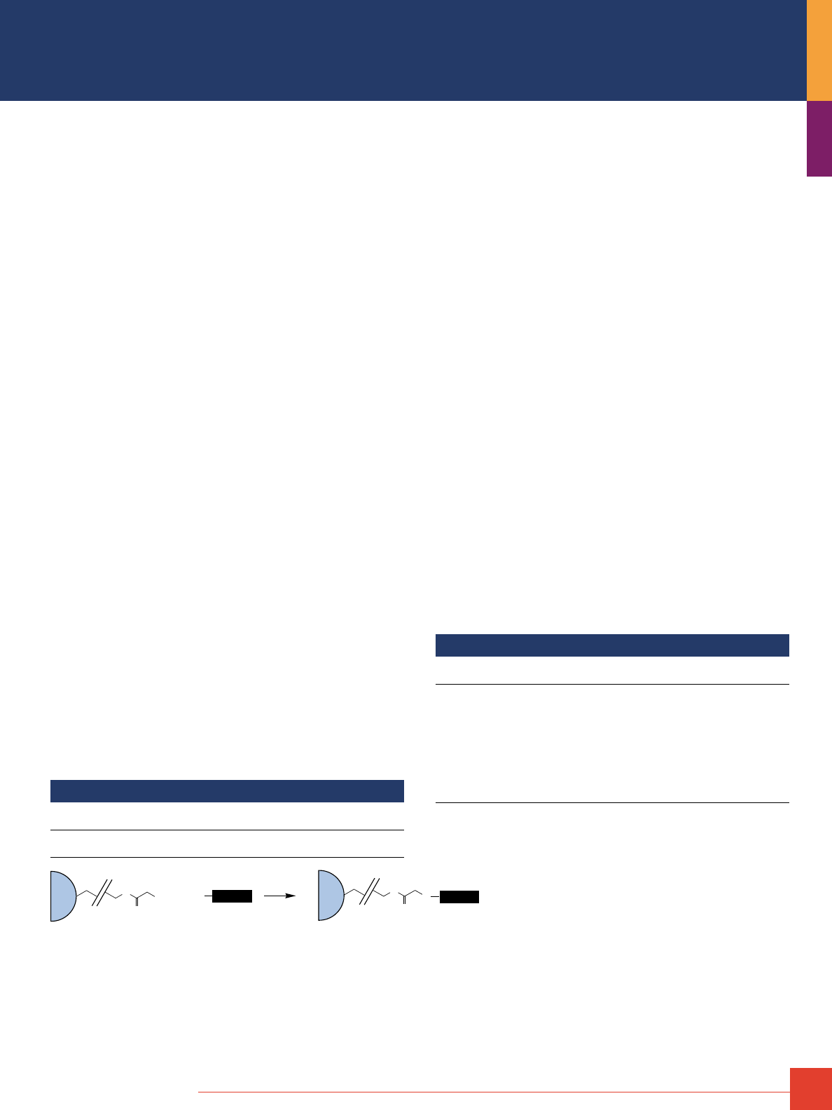
To order, call 800-874-3723 or 815-968-0747. Outside the United States, contact your local branch office or distributor.
31
Thermo Scientific UltraLink Iodoacetyl Resin
Polyacrylamide resin for coupling sulfhydryl-containing ligands.
UltraLink Iodoacetyl Resin is a durable, porous resin that is activated
to enable efficient and direct covalent immoblization of peptides
and other ligands through their sulfhydryl groups (–SH) for use in
affinity purification procedures. The beaded resin is a hydrophilic
copolymer of polyacrylamide and azlactone having a rigid poly-
meric structure with high surface area and pore volume. Each
azlactone group has been modified to form a 15-atom spacer arm
that terminates in an iodoacetyl group, which is capable of react-
ing with sulfhydryl groups (e.g., side chain of reduced cysteine
residues) to covalently immobilize peptide or other sulfhydryl-
containing ligands. The bead structure and efficiently coupling
chemistry of Iodoacetyl-Activated UltraLink Support results in high
protein binding capacity, high linear flow rates, low nonspecific
binding and overall superior performance in affinity chromatography.
UltraLink Resins are ideal for medium pressure applications such
as FPLC.
Highlights:
• High coupling efficiency and capacity – immobilizes sulfhydryl-
containing proteins or other ligands with high efficiency and
coupling capacity in 1 hour
•
Specific and leak-proof coupling chemistry – reacts specifically
with sulfhydryl groups (reduced thiols), resulting in thioether
bonds that are stable for use in many affinity purification procedures
•
Simple coupling conditions – perform immobilization reaction in
any of a variety of buffers; coupling is most efficient and specific
at pH 8.0–8.5; coupling compatible with or without organic solvent
•
Excellent reusability – prepared affinity resin can be used with
typical binding and elution procedures for many cycles of affnity
purification without significant loss of binding capacity
•
Durable, high-performance resin – porous beads have a 60μm
diameter, can withstand 100 psi (6.9 bar) and allow for linear
flow-through rates of 3,000cm/hour
References
1. Ju, T., et al. (2002). J. Biol. Chem. 277, 169–177.
2. Hill, K., et al. (2000). J. Biol. Chem. 275(6), 3741–3744.
3. Liu, L.A. and Engvall, E. (1999). J. Biol. Chem. 274, 38171–38176.
4. Bicknell, A.B., et al. (2001). Cell 105, 903–912.
Ordering Information
Product #
Description
Pkg. Size
U.S.
Price
53155
UltraLink Iodoacetyl Resin
Support: UltraLink Biosupport
10mL
$188
HS
+
H
N
I
O
H
N
S
O
Iodoacetyl-Activated
Thermo Scientific UltraLink Resin
Thioether
Bond
15-atom
Spacer
Iodoacetyl
Group
+
HI
Immobilized Peptide
Reduced Sulfhydryl
Molecule
Thermo Scientific
UltraLink Bead
Thermo Scientific
UltraLink Bead
Peptide
Peptide
Thermo Scientific UtraLink Iodoacetyl immobilization chemistry.
Thermo Scientific UltraLink Iodoacetyl Micro Peptide Coupling Kit
Easily prepare a small-scale affinity column with sulfhydryl-
containing peptides.
The Micro Peptide Coupling Kit is a microcentrifuge spin
column kit for immobilizing small amounts (25–250μg) of sulfhydryl-
containing peptides (e.g., cysteine-terminated peptides) onto a
beaded porous resin to create a small, reusable affinity column.
The coupling and affinity purification procedures are optimized
for small sample volumes (200–300μL). Wash and elution steps are
achieved rapidly and efficiently with the convenient microcentrifuge
spin columns. Each kit contains sufficient reagents for 10 coupling
reactions and 20 affinity purifications. The kit is ideal for immobilizing
peptide antigens that containing a terminal cysteine residue for
use in purifying specific antibodies from small serum, ascites or
culture supernatant samples.
Highlights:
• Optimized for small scale – ideal for coupling small amounts
(25–250μg) of sulfhydryl-containing peptide or protein for
purification of specific antibodies from crude serum
•
High-performance affinity resin – uses durable, polyacrylamide-
based UltraLink Iodoacetyl Resin for specific reaction to
sulfhydryl groups
•
Efficient coupling chemistry – immobilization efficiency >85%
(as measured with 1 hour reaction using insulin, calcitonin and
osteocalcin peptides)
•
Fast and easy to use – perform wash and elution steps using
a microcentrifuge
•
Reusable – use prepared peptide resin several times with no
significant loss of capacity
Ordering Information
Product #
Description
Pkg. Size
U.S.
Price
20485
Micro Peptide Coupling Kit
Sufficient for coupling 10 sulfhydryl-containing peptides
or proteins and perform 20 affinity purifications.
Kit contents: UltraLink Iodoacetyl Spin Columns, 0.1mL
Coupling Buffer
L-Cysteine-HCl
Wash Solution
PBS Pack (makes 500mL)
IgG Elution Buffer
Collection Tubes
Kit
10 columns
100mL
100mg
25mL
1 pack
50mL
200 tubes
$357
32
For more information, or to download product instructions, visit www.thermoscientific.com/pierce
Thermo Scientific Disulfide Reducing Agents
Reduce disulfide bonds to produce sulfhydryl groups for
immobilization on SulfoLink or UltraLink Resins.
Free sulfhydryls are required for immobilization onto sulfhydryl-
reactive affinity supports. Cysteines in proteins and peptides
usually exist as cystines (disulfide bridges) and must be reduced
to expose sulfhydryls for coupling. Reduction can be accomplished
with free or immobilized reducing agents. Free reducing agents
are efficient in reducing all disulfides in proteins, including those
buried in the tertiary structure, but they must be removed from the
reduced sample with a desalting column before coupling to the
support. Immobilized reducing agents enable reduction of disulfides
and simple removal of the reduced sample from the reducing
agent. This is especially helpful when reducing peptides whose
small size prevents them from being effectively desalted.
P
HO
O
OH
O
OH
O
Cl
–
TCEP•HCl
MW 286.65
H
+
HS
NH
3
Cl
–
2-Mercaptoethylamine•HCI
MW 113.61
SH
HS
OH
OH
DTT
MW 154.25
+
Ordering Information
Product #
Description
Pkg. Size
U.S.
Price
20408 2-Mercaptoethylamine•HCl
6 x 6mg
$135
20290 DTT, Cleland’s Reagent
(Dithiothreitol)
5g
$105
20291 Dithiothreitol (DTT) in No-Weigh™ Format
7.7mg DTT/tube. Makes 100μL of 0.5 M DTT.
48 tubes
$125
20490 TCEP•HCl
(Tris[2-carboxyethyl]phosphine hydrochloride)
1g
$ 50
20491 TCEP•HCl
10g
$299
77720 Bond-Breaker TCEP Solution, Neutral pH
5mL
$108
77712 Immobilized TCEP Disulfide Reducing Gel
5mL
$118
Thermo Scientific Ellman’s Reagent and Sulfhydryl Addition Kit
Measure and add free sulfhydryls to ensure success of
cysteine-targeted immobilization.
Ellman’s Reagent, also called DTNB, is a versatile water-soluble
compound for quantifying free sulfhydryl groups in solution. A
solution of this compound produces a measurable yellow-colored
product when it reacts with sulfhydryls (–SH groups). By testing
an unknown sample, such as a peptide having a terminal cysteine
residue, compared to a standard curve made with known amounts
of free, reduced cysteine (Product # 44889), availability of reduced
sulfhydryls in the sample can be determined.
The Sulfhydryl Addition Kit provides the essential reagents and
procedure for creating new sulfhydryl groups on a protein or
other molecule that contains available primary amines (–NH
2
).
The kit uses SATA reagent, which forms covalent bonds to primary
amines. The result is addition of a stable (capped) sulfhydryl
group, which can later be exposed by gentle treatment with
hydroxylamine, making the molecule ready for conjugation to
SulfoLink Coupling Resin, UltraLink Iodoacetyl Resin or other
sulfhydryl-reactive immobilization method.
Ordering Information
Product #
Description
Pkg. Size
U.S.
Price
23460 Sulfhydryl Addition Kit
Adds free sulfhydryl groups to proteins.
Includes: SATA
Hydroxylamine•HCl
10X Conjugation Buffer Stock
Phosphate Buffered Saline Pack
Dimethylformamide (DMF)
Dextran Desalting Column
Column Extender
Ellman’s Reagent (DTNB)
Cysteine•HCl H
2
O
Kit
2mg
5mg
20mL
1 pack
1mL
1 x 5mL
1
2mg
20mg
$223
22582 Ellman’s Reagent
(5,5’-Dithio-bis-[2-nitrobenzoic acid])
5g
$ 74
44889 Cysteine•HCl
5g
$ 45
Covalent Coupling of Affinity Ligands
to Chromatography Supports
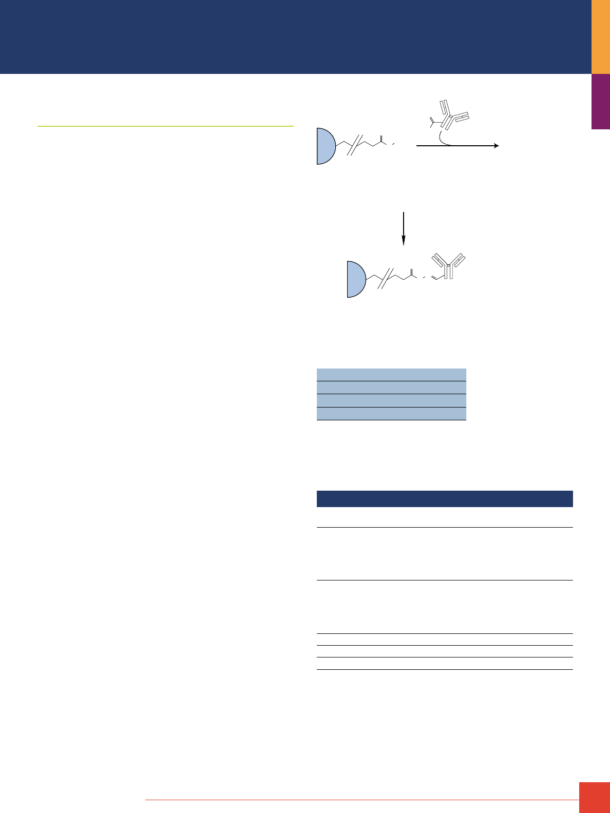
To order, call 800-874-3723 or 815-968-0747. Outside the United States, contact your local branch office or distributor.
33
Products for Immobilizing Ligands
through Carbonyl Groups
Thermo Scientific CarboLink Immobilization Kit and
Coupling Resins
Immobilize polyclonal antibodies and other glycoproteins through
carbohydrate groups.
CarboLink Coupling Resin and Kits provide for covalent immobiliza-
tion of glycoproteins and other carbohydrate-containing molecules
to beaded agarose (or polyacrylamide UltraLink Support) for use
in affinity purification procedures. Carbohydrate moieties in glyco-
proteins contain common sugars whose cis-diol groups are easily
oxidized with sodium meta-periodate (included in the CarboLink
Kit) to yield aldehydes. When incubated with the CarboLink Resin,
these aldehyde groups react spontaneously with the hydrazide
group of the activated resin to form stable, covalent bonds. The
immobilization strategy is especially useful for glycoproteins,
such as polyclonal antibodies, because it allows attachment of the
molecule at domains that will not interfere with binding sites that
are critical for the intended affinity purification. Once a molecule
is coupled, the prepared affinity resin can be used multiple times in
typical protein affinity purification procedures.
Highlights:
• CarboLink Coupling Resin – hydrazide-activated crosslinked
6% beaded agarose (or hydrazide-activated UltraLink Support,
a beaded, polyacrylamide resin)
• Efficient immobilization – couple 1–5mg of oxidized polyclonal
antibody or other glycoprotein per milliliter of resin (CarboLink
Resin contains greater than 14μmol hydrazide groups per milliliter)
• Stable linkage – resonance structure of the hydrazone bonds
are sufficiently stable to allow multiple rounds of affinity
purification with one batch of prepared resin; no stabilizing
reductant required
• Flexible and gentle coupling conditions – immobilization reaction
completed in simple buffers (PBS or other non-amine buffer with
or without organic solvent) at near-neutral pH
• Ideal for polyclonal antibodies – immobilizes IgG through
carbohydrates in the Fc region, so both antigen binding sites are
free to interact with the antigen in the mobile phase
• Effective for any molecule with oxidizable sugars – first step is
oxidation of the sugar groups, which allows the cis-diols of the
IgG to be transformed into reactive aldehyde moieties; these
aldehydes then combine with hydrazide groups on the matrix to
form stable, leak-resistant linkages
• Convenient kits and product sizes – choose one- or five-column
kit containing complete sets of buffers, oxidizing reagent and
versatile spin/drip columns containing the beaded agarose resin;
or choose the polyacrylamide-based UltraLink Support. Bulk
quantities are available for manufacturing applications
Agarose
Bead
Agarose
Bead
N
H
NH
2
O
N
H
N
O
Thermo Scientific
CarboLink Resin
23-atom
Spacer
Hydrazide
Group
Antibody with Carbohydrate
Groups Oxidized to Form
Aldehyde Groups
Immobilized Glycoprotein
Oxidized Glycoprotein
Hydrazone
Bond
H
O
Thermo Scientific CarboLink Support immobilization chemistry.
Target: –CHO
Support: 6% agarose
Binding capacity: 5mg protein/mL resin
Time: 8 hours
References
1. Kumar, P.G., et al. (2001). J. Biol. Chem. 276, 41357–41364.
2. Strakova, Z., et al. (1997). Mol. Pharmacol. 51, 217–224.
3. Brown, M.A., et al. (2000). J. Biol. Chem. 275, 19795–19802.
4. Butko, P., et al. (1999). J. Immunol. 163, 2761–2768.
5. Sequra, M., et al. (1999). Infect. Immun. 67(9), 4646
Ordering Information
Product #
Description
Pkg. Size
U.S.
Price
20355 CarboLink Immobilization Trial Kit
Kit contains: CarboLink Column
CarboLink Coupling Buffer
CarboLink Wash Solution
Sodium meta periodate
Zeba Desalting Column
Kit
1 x 2mL
60mL
15mL
1 x 5mg
1 x 5mL
$155
44910 CarboLink Immobilization Kit
Kit contains: CarboLink Columns
CarboLink Coupling Buffer
CarboLink Wash Solution
Sodium meta periodate
Zeba Desalting Columns
Kit
5 x 2mL
250mL
100mL
5 x 5mg
5 x 5mL
$454
20391 CarboLink Coupling Resin
10mL
$113
53149 UltraLink Hydrazide Resin
10mL
$186
20504 Sodium meta-Periodate
25g
$ 29
34
For more information, or to download product instructions, visit www.thermoscientific.com/pierce
Products for Immobilizing Ligands
through Carboxyl Groups
Thermo Scientific CarboxyLink Immobilization Kits
and Coupling Resins
Immobilize peptides via carboxyl groups to create an affinity column.
CarboxyLink Coupling Resin and Kits provide for covalent immobil-
ization of peptides or other carboxyl-containing (–COOH) molecules
to a porous, beaded resin for use in affinity purification procedures.
CarboxyLink Resin is crosslinked beaded agarose (or polyacryl-
amide UltraLink Support) that has been activated with diamino-
dipropylamine (DADPA) to contain long spacer arms, each with
a primary amine at the end. When incubated with the resin and
the carbodiimide crosslinker EDC (included in the CarboxyLink
Immobilization Kit), carboxyl-containing molecules become per-
manently attached to the support by stable amide bonds. Once
a molecule is coupled, the prepared affinity column can be used
multiple times in typical protein affinity purification procedures.
CarboxyLink Coupling Resins can also be used to immobilize other
kinds of molecules using alternative amine-reactive crosslinking
chemistries.
Highlights:
• CarboxyLink Coupling Resin – DADPA-activated crosslinked
4% beaded agarose (or DADPA-activated UltraLink Support,
a beaded polyacrylamide resin)
• Efficient immobilization – couple 1–2mg of peptide per milliliter
of resin (CarboxyLink Agarose Resin activated with greater than
16μmol amine milliliter of resin; DADPA on UltraLink Support
activated with greater than 40μmol amine milliliter of resin)
• Stable linkage – immoblization results in covalent attachment of
carboxyl groups by amide bonds, allowing for multiple rounds of
affinity purification with one batch of prepared resin
• Flexible and gentle coupling conditions – immobilization reaction
completed in simple MES or other non-amine and non-carboxyl,
near-neutral buffer, with or without organic solvent.
• Ideal for unmodified peptides – immobilizes peptides with
high capacity and various orientations without steric
hindrance, allowing for effective use in affinity purification of
specific antibodies
• Convenient kits and product sizes – choose five-column kits with
complete sets of buffers, crosslinker and versatile spin/drip
columns containing either type of resin (agarose or
polyacrylamide) or choose stand-alone resin for other uses
H
N
H
N
NH
2
Immobilized DADPA
(Thermo Scientific CarboxyLink Resin)
H
N
H
N
H
N
+
O
HO
O
EDC Crosslinker
Carboxyl Ligand
(e.g., peptide C-terminus)
Covalently Immobilized Ligand
(attached by amide bond and long spacer arm)
Agarose
Bead
Peptide
Peptide
Agarose
Bead
Thermo Scientific CarboxyLink Support immobilization chemistry.
Target: –COOH
Support: 4% agarose or UltraLink
®
Resin
Binding capacity: 5mg protein/mL resin
Time: 4 hours
Reference
Yoo, B.C., et al. (2002). J. Biol. Chem. 277, 15325–15332.
Ordering Information
Product #
Description
Pkg. Size
U.S.
Price
44899 CarboxyLink Immobilization Kit
Kit contains: CarboxyLink Columns (DADPA agarose)
EDC Crosslinker
Coupling Buffer (MES-buffered Saline)
Wash Solution (1 M NaCl)
Column Accessories
Kit
5 x 2mL
5 x 60mg
500mL
120mL
$393
20266 CarboxyLink Coupling Resin
Support: Crosslinked 4% beaded agarose
Loading: 16–20μmol available amino groups/mL of resin
25mL
$145
53154 CarboxyLink Immobilization Kit
with UltraLink Support
Kit contains: DADPA UltraLink Columns
EDC Crosslinker
Coupling Buffer (MES-buffered Saline)
Wash Solution (1 M NaCl)
Column Accessories
Kit
5 x 2mL
5 x 60mg
500mL
120mL
$432
22980 EDC
1-Ethyl-3-[3-dimethylaminopropyl]carbodiimide
hydrochloride
5g
$ 83
28390 BupH MES Buffered Saline Packs
10 pack
$112
Covalent Coupling of Affinity Ligands
to Chromatography Supports

To order, call 800-874-3723 or 815-968-0747. Outside the United States, contact your local branch office or distributor.
35
Antibody immobilization: choosing the best Thermo Scientific Support.
AminoLink Plus
Coupling Resin
UltraLink
Biosupport
CarboLink
Coupling Resins
SulfoLink
Coupling Resins
Crosslink IP Kits
See page 41
Monoclonal Antibodies
Advantages:
• Good choice when
only small amounts of
antibody are available
• Couple over a broad
pH range
• Good coupling efficiency
Disadvantages:
• Reduction of Schiff’s base
with sodium cyanoborohy-
dride may adversely affect
monoclonals
• Some antibodies may
be coupled through
antigen-binding site
Advantages:
• Good choice if antibody
can withstand 1.0M
sodium citrate or sulfate
• High capacity
• Fast, efficient coupling
• Good, for large-scale or
fast-flow applications
Disadvantages:
• Some antibodies may be
coupled through antigen-
binding site
• Some antibodies may pre-
cipitate in high-salt buffer
Advantages:
• Correctly orients antibody
• Antibody must be able
to withstand oxidation
conditions
• Good for antibodies with
low avidity for antigen
Disadvantages:
• Not all monoclonals have
carbohydrate accessible
for coupling
• Conditions necessary for
coupling may adversely
affect some monoclonals
Advantages:
• Good choice for
antibodies that have
extremely high avidity
for their antigen
• Allows for gentle elution
conditions
Disadvantages:
• Must first reduce antibody
prior to coupling
• Not good for antibodies
with low affinity for
their antigens
Advantages:
• Allows for correct
orientation of antibodies
• Gentle coupling conditions
• Protein A/G will bind
most antibodies
Disadvantages:
• If purifying antigen from
serum, antibodies may
bind to Protein A/G and
co-purify with antigen
• Crosslinking results in
some loss of antibody
activity
Polyclonal Antibodies
Advantages:
• Excellent coupling
efficiency
• Good antigen recovery
Disadvantages:
• Some antibodies may
be coupled through
antigen-binding site
Advantages:
• Good choice for
large-scale or fast-flow
applications
• High capacity
• Fast, efficient coupling
Disadvantages:
• Some antibodies may
be coupled through
antigen-binding site
• Some antibodies may
precipitate in high-salt
buffer
Advantages:
• Correctly orients antibody
• Antibody must be able
to withstand oxidation
conditions
• Good for antibodies with
low avidity for antigen
Disadvantages:
• Conditions necessary for
coupling may adversely
affect some antibodies
Advantages:
• Good choice for anti-
bodies that have avidity
for their antigen
• Allows for gentle elution
conditions
Disadvantages:
• Must first reduce antibody
prior to coupling
• Not good for antibodies
with low affinity for their
antigens
Advantages:
• Allows for correct
orientation of antibodies
• Gentle couple conditions
• Protein A/G will bind
most antibodies
Disadvantages:
• If purifying antigen from
serum, antibodies may
bind to Protein A/G and
co-purify with antigen
• Crosslinking results in
some loss of antibody
activity
High-Activity Antibodies
Advantages:
• Immobilization of reduced
antibody allows for
gentler elution conditions
Low-Activity Antibodies
Advantages:
• Correctly orients antibody
Disadvantages:
• Conditions necessary for
coupling may adversely
affect some monoclonals
Advantages:
• Allows for correct
orientation of antibodies
• Gentle couple conditions
• Protein A/G will bind
most antibodies
Disadvantages:
• If purifying antigen from
serum, antibodies may
bind to Protein A/G and
co-purify with antigen
• Crosslinking results in
some loss of antibody
activity
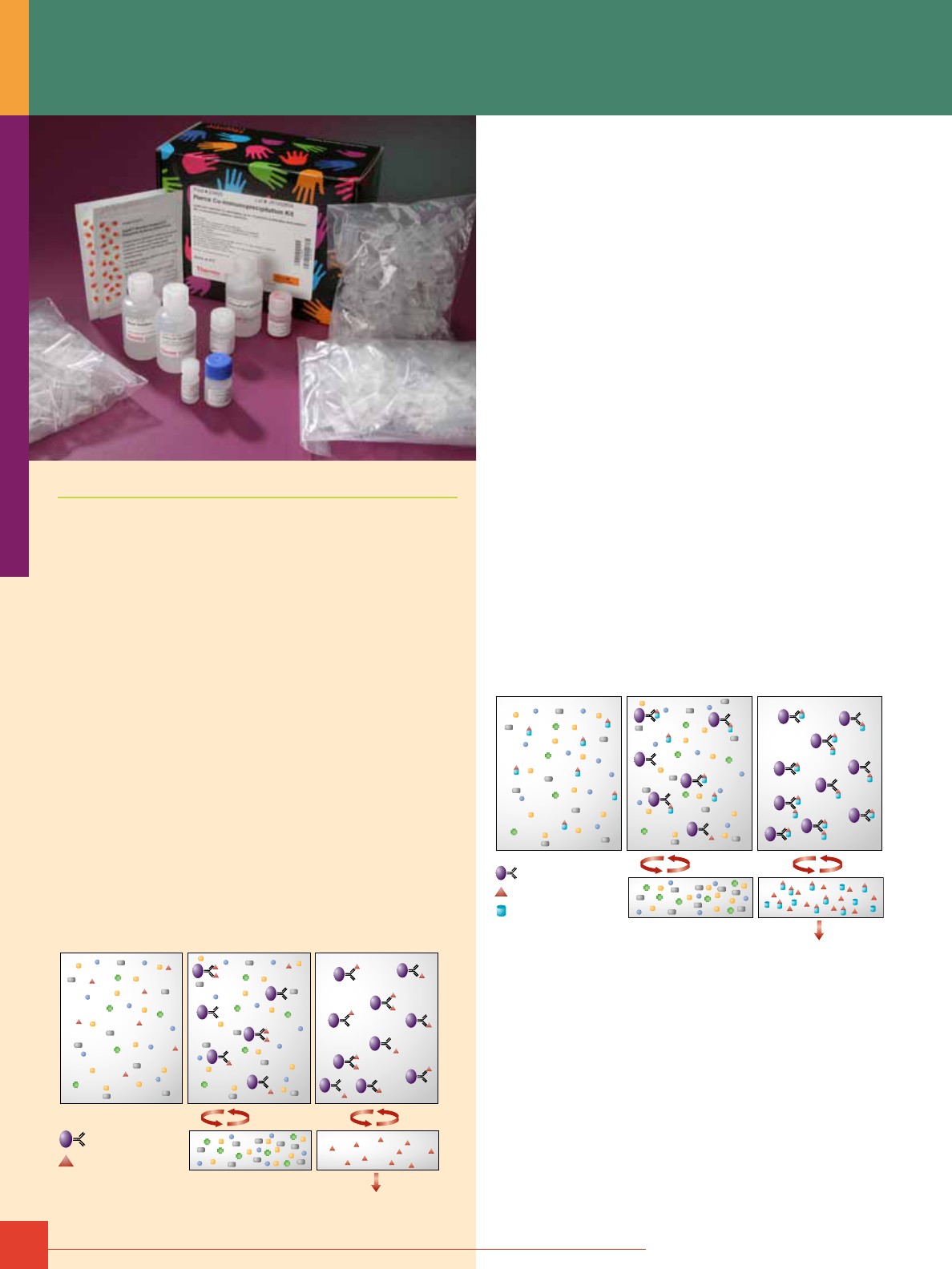
36
For more information, or to download product instructions, visit www.thermoscientific.com/pierce
Immunoprecipitation
The topic of co-immunoprecipitation (co-IP) is best preceded by
a discussion of immunoprecipitation (IP) to help frame an under-
standing of the principles involved.
IP is one of the most widely used methods for antigen detection
and purification. An important characteristic of IP reactions is
their potential to deliver not only the target protein, but also other
macromolecules that interact with the target.
The IP Principle
The principle of an IP is very simple (Figure 1). An antibody
(monoclonal or polyclonal) against a specific target antigen is
allowed to form an immune complex with that target in a sample,
such as a cell lysate. The immune complex is then captured on
a solid support to which either Protein A or Protein G has been
immobilized (Protein A or Protein G binds to the antibody, which
is bound to its antigen). The process of capturing this complex
from the solution is referred to as precipitation. Any proteins not
“precipitated” by the immobilized Protein A or Protein G support
are washed away. Finally, components of the bound immune
complex (both antigen and antibody) are eluted from the support
and analyzed by SDS-PAGE (gel electrophoresis), often followed
by Western blot detection to verify the identity of the antigen.
Coupled Antibody
Spin and wash
Cell Lysate or Protein Mixture
Incubation with
Antibody-coupled Resin
Antigen
Elute
Analyze
Figure 1. Summary of a traditional immunoprecipitation procedure.
Traditional immunoprecipitation involves the following steps:
1. Form the antigen-antibody complex (immune complex) by
incubating specific antibody with the antigen-containing sample
for 1 hour to several hours.
2. Capture the immune complex to Protein A or Protein G agarose
beads by incubation for 0.5-2 hours.
3. Remove any non-bound protein (non-immune complex sample
components) from the precipitated complex by washing beads
with additional sample buffer.
4. Boil beads in reducing SDS-PAGE sample loading buffer.
5. Recover eluted sample in loading buffer and analyze by
SDS-PAGE.
6. Perform Western blot analysis, probing with antigen-
specific antibody.
Co-IP vs. IP
Co-IP is a popular technique for protein interaction discovery.
Co-IP is conducted in essentially the same manner as IP (Figure 2).
However, in a co-IP the target antigen precipitated by the antibody
“co-precipitates” a binding partner/protein complex from a lysate;
i.e., the interacting protein is bound to the target antigen, which
becomes bound by the antibody that becomes captured on the
Protein A or Protein G resin. The assumption usually made when
associated proteins are co-precipitated is that these proteins are
related to the function of the target antigen at the cellular level. This
is only an assumption, however, that is subject to further verification.
Coupled Antibody
Spin and wash
Cell Lysate or Protein Mixture
Incubation with
Antibody-coupled Resin
Antigen
Protein Interacting
with Antigen
Elute
Analyze
Figure 2. Summary of a traditional co-immunoprecipitation procedure.
IP/Co-IP

To order, call 800-874-3723 or 815-968-0747. Outside the United States, contact your local branch office or distributor.
37
Traditional Methods vs.
Thermo Scientific Pierce Innovations for Co-IP
Problems with Traditional Co-IP Methods
The traditional co-IP protocol has certain deficiencies relating to
the fundamental format of the assay, the antibody and associated
chemistry. One of the most commonly encountered problems
with the traditional IP and co-IP approach is interference from
antibody bands in gel analysis. In those cases in which several
proteins may be co-precipitated with the target, presence of the
co-eluted antibody heavy and light chains (25 and 50 kDa bands
in reducing SDS-polyacrylamide gel) in the preparation can
obscure the results. The ideal situation would be to conduct
the co-IP without contamination of the eluted antigen with
antibody. With this potential interference eliminated, only the
co-precipitated proteins will be present and detected on a gel.
This and other shortcomings of the traditional protocol and our
solutions are summarized in Table 1.
Table 1. Comparison of traditional Co-IP and Thermo Scientific Pierce
Co-IP Products.
Traditional Co-IP Problems Thermo Scientific Pierce Product Solutions
Batch processing of the precipitated
complex in a single tube: results in
inefficient washing of non-bound proteins
from the support and in resin loss due to
decanting wash buffer from tube via a
pipette, which lowers protein yields
Spin cup or spin tube processing:
dedicated IP and co-IP kits that
contain spin-cup or spin tube devices
that increase washing efficiency offer
more effective elution of antigen and
associated protein and eliminate resin
loss, yielding significantly higher protein
recoveries and more consistent results
Antibody fragment interference:
co-elution of antibody fragments with
antigen often results in bands interfering
with detection of any co-precipitated
proteins on SDS-PAGE
Antibody immobilization: chemistries
designed to immobilize the antibody to
the support, thereby allowing elution
of only the target and any associated
proteins in a co-IP complex
Eliminate detection of antibody used
for immunoprecipitation: contaminating
antibody fragments become denatured
during SDS-PAGE. In contrast, the primary
antibody used during Western blotting is
still native. Use CleanBlot HRP Detection
Reagent (Product # 21230) to only detect
native antibody and not contaminants.
Antibody sacrificed: as a consequence
of harsh elution conditions, the target
antibody is destroyed; antibody loss by
way of the protocol can be costly
Antibody re-used: immobilization
chemistry and mild elution conditions
for the target and associated proteins
allow the immobilized antibody to be
re-equilibrated and recycled several times
in the co-IP protocol
Approaches to Co-IP Free of Antibody Interference
Three approaches have been incorporated into several products
targeted to IP and co-IP applications.
Activated Support for Antibody Immobilization — Direct Strategy
In this approach, an antibody is immobilized directly through its
surface amine groups (contributed primarily by the side chain
epsilon-amino group of lysine) to a high-capacity aldehyde-
activated beaded agarose support (Thermo Scientific AminoLink
Plus Coupling Resin). The support forms a Schiff’s base with these
available amines that is reduced to form stable secondary amine
bonds during the immobilization process. The wide range of coupling
conditions that can be used with this support make it ideal for
maintaining biological binding activity critical to the successful
execution of a co-IP experiment. When using the Direct Strategy,
the antibody source should be free of carrier protein, which can also
be immobilized. Products such as Thermo Scientific Melon Gel IgG
Purification Kits (Product # 45206) can easily clean-up an antibody
to remove carrier proteins. The direct immobilization of antibodies is
not species dependent, which allows for the use on non-traditional
antibody sources that don’t bind globulin binding proteins (Protein A
or Protein G). Thermo Scientific Pierce Co-Immunoprecipitation Kits
(Product # 26149) and Thermo Scientific Pierce Direct IP Kit (Product
# 26148) use this direct immobilization approach. For co-IP applica-
tions, the flexibility, simplicity and d urability of the direct method
as an antibody-coupling strategy makes it the method of choice for
delivering results free from antibody interference.
Antibody Orientation and Immobilization — Indirect Strategy
This strategy takes advantage of the binding characteristics of the
traditional Protein A or Protein G agarose combined with chemical
crosslinking to covalently link the antibody to the support. Protein
A and Protein G bind IgG class antibodies through the Fc region
that is characterized primarily by dimerized heavy chain modified
by carbohydrate. Fc region binding naturally orients the antigen-
binding domains of the antibody (Fab) away from the support,
making them available for binding to their respective target antigen.
The Indirect Strategy is compatible with antibody samples that
contain carrier proteins, provided the carrier proteins are washed
away prior to crosslinking. To ensure that the antibody remains on
the support during the requisite antigen binding, wash and elution
steps of the protocol, this bound and oriented antibody is chemi-
cally crosslinked to the Protein A or Protein G with the bifunctional
reagent disuccinimidyl suberate (DSS). Thermo Scientific Pierce
Crosslink IP Kit (Product # 26147) incorporates this strategy.

38
For more information, or to download product instructions, visit www.www.thermoscientific.com/pierce
Use of the Streptavidin:Biotin Interaction
This direct coupling approach incorporates the binding association
between streptavidin and biotin. Streptavidin immobilized to beaded
agarose resin or coated in microplate wells provides an alternative
IP or co-IP strategy for obtaining results free from antibody inter-
ference. Biotinylated antibody is bound very strongly to each matrix
and is not eluted when mild conditions are used to release the
target antigen. The IP/co-IP is conducted by incubating the sample
with the biotinylated antibody-loaded matrix. Elution of the target
antigen and any interacting proteins is performed free of antibody
contamination. Thermo Scientific Pierce Streptavidin Agarose Resin
(e.g., Product # 20347) and kit products that use this support, as well
as the Thermo Scientific Pierce Streptavidin Coated Plate IP Kit
(Product # 45360), provide high-capacity biotin-binding matrices
suitable for IP and co-IP applications.
Optimization Parameters in IP and Co-IP
Classical Immune Complex Formation vs. Pre-Binding of Antibody
A change in protocol from the classical immune complex
precipitation is necessary when using immobilized antibody
in the co-IP method. In the traditional co-IP protocol, the
immune complex (antigen:antibody) is formed in solution before
“precipitating” it with the immobilized Protein A or Protein G
matrix. When using immobilized antibody, the immune complex
is formed directly on the antibody-coupled matrix by incubation
of the antigen-containing sample with the matrix. Formation of the
immune complex (the target antigen and any target-associated
protein) and its precipitation occurs in one step.
In the immobilized format, the antibody is allowed to incubate
with the lysate. The matrix is washed using a spin cup format
and the bound protein eluted for analysis. The target antigen and
co-IP complex is recovered free of antibody or antibody fragment
contamination, and the antibody is retained in an active form on
the support to be used in another co-IP cycle.
Our research indicates that pre-binding or -coupling the antibody
to the support matrix consistently results in the capture of more
target antigen, even in coated plate IP procedures that do not
require it. This approach is recommended for the Thermo Scientific
Pierce Coated Plate IP Kits that use 96-well microplates coated
with streptavidin or Protein A/G (Products # 45360, 45350,
respectively). Antibody is bound to the plate wells prior to the
prescribed incubation with a lysate sample. Unbound protein is
easily washed from the wells prior to the elution of the target
and any co-precipitated proteins.
Evaluating a Co-IP-Captured Interaction
In their review of protein interactions, Phizicky and Fields
(see References listed below) present a discussion of the issues to
consider in validating a suspected interaction obtained by a co-IP
experiment. Ultimately, the following question must be answered:
Does the interaction detected by co-IP occur in vivo, and what
significance does it have at the cellular level? A summary of the
Phizicky and Fields approach to verification of co-IP data follows.
Confirm that the co-precipitated protein is obtained only
by antibody against the target
Use monoclonal antibodies in the co-IP protocol. When only a
polyclonal antibody is available, pre-treatment of the antibody with
sample devoid of the primary target (bait protein) may be required
to assure that the polyclonal antibody does not contain clones or
contaminants that bind prey protein(s) directly. Pre-adsorption to
extracts devoid of target or pre-purification of polyclonal IP
antibodies against an affinity column containing pure target
antigen safeguards against a false-positive co-IP.
Conclude that antibody against the target antigen does not
itself recognize the co-precipitated protein(s)
Use independently derived antibodies that have demonstrated
specificities against different epitopes on the target protein.
Their use serves as verification that the target (bait)-directed
antibodies have no affinity for the target-associated prey proteins
recovered during the co-IP. Alternatively, an antibody against the
co-precipitated protein can be used to co-IP the same complex.
Determine if the interaction is direct or indirect
Is the interaction mediated through a third-party protein that
contacts both target and co-precipitated protein? Immunological
and other more sophisticated methods such as mass spectrometry
may be necessary to answer this question.
Determine that the interaction takes place in the cell and not
as a consequence of cell lysis
Suggested approaches here involve co-localization studies and
site-specific mutagenesis giving rise to mutants that perturb the
binding process.
References
Adams, P.D., et al. (2002). Identification of associated proteins by co-immunoprecipitation,
In Protein-Protein Interactions – A Molecular Cloning Manual. Golemis, E., Ed., Cold Spring
Harbor Laboratory Press, pp 59-74.
Liebler, D.C. (2002). Identifying protein-protein interactions and protein complexes.
In Introduction to Proteomics, Tools for the New Biology, Humana Press, pp.151-165.
(Product # 20061)
Phizicky, E.M. and Fields, S. (1995). Protein-protein interactions: methods for detection
and analysis. Microbiological Reviews (Mar.), pp. 94-123.
IP/Co-IP
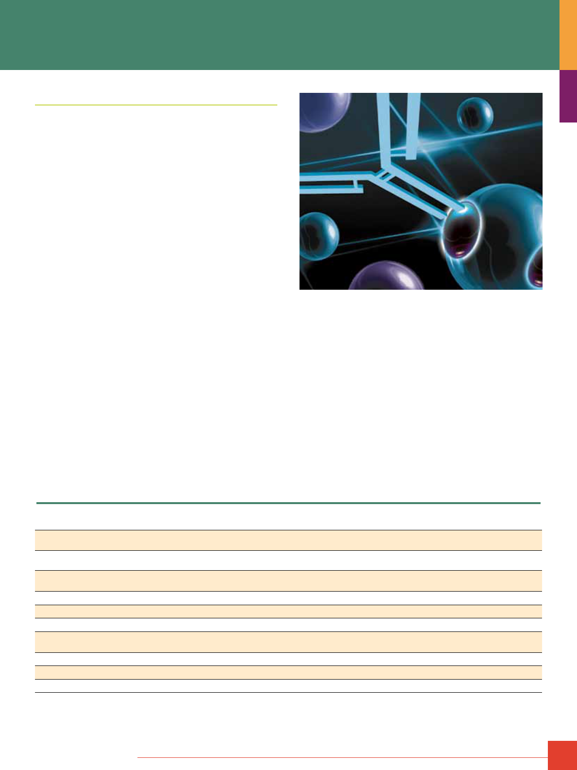
To order, call 800-874-3723 or 815-968-0747. Outside the United States, contact your local branch office or distributor.
39
IP and Co-IP Kits
The Thermo Scientific Pierce Classic and Crosslink IP Kits are
ideal when using antibodies that bind to Protein A or Protein G.
The Direct and Crosslink IP Kits are highly effective for eliminating
co-elution of IgG heavy and light chain with the antigen, which
interferes with downstream applications such as mass
spectrometry analysis or protein sequencing.
Traditional co-IP methods result in detection of the antibody with
the target proteins. Because the antibody heavy and light chains
may co-migrate with one of the relevant bands, important results
can be masked. The Pierce Co-IP Kit circumvents issues with
co-migration of antibody chains with target proteins by retain-
ing the antibody on the resin. The new kit is optimized for using
smaller amounts of sample and offers a single lysis/wash buffer
eliminating the need for a separate lysis reagent.
All four IP Kits Highlights:
• Require minimal antibody (2-10μg)
• Are highly effective and efficient in capturing antigens
• Use optimized protocols and buffers for efficient IPs and
antigen elution
• Use common lysis/binding/wash buffer
• Include spin columns and collection tubes that shorten the
protocol by minimizing handling and mixing
• Use a new elution buffer that provides milder and less denaturing
recovery of antibody:antigen complexes
• Are compatible with specialized downstream applications; e.g.,
mass spectrometry, enzyme assays and antibody production
• Are able to scale up as needed using our flexible protocol
Comparison of immunoprecipitation methods. The following table compares the key features of traditional “do-it-yourself” immunoprecipitation techniques
to the Thermo Scientific Pierce IP Kits. Consideration of these features can help to determine which method is most appropriate for the available reagents and
downstream application.
Feature
Traditional
IP method
Pierce
Classic IP Kit
Pierce
Crosslink IP Kit
Pierce
Direct IP Kit
Pierce
Co-IP Kit
Uses high binding capacity resin Variable
1
Yes
(Protein A/G Plus)
Yes
(Protein A/G Plus)
Yes
(AminoLink Plus)
Yes
(AminoLink Plus)
Crosslinker mediated immobilization No No Yes
(DSS)
No No
Requires purified antibody in amine-free and protein-free
storage solution
No No No Yes Yes
Antibody is covalently attached to agarose resin No No Yes Yes Yes
Antibody is oriented Yes Yes Yes No No
Antibody elutes with antigen Yes Yes No No No
Antigen recovery method Boiling w/SDS
(Low pH)
Low pH elution
(Boiling w/SDS)
Low pH
elution
Low pH
elution
Low pH
elution
Relative antigen recovery
2
Variable Highest Medium High High
Immobilized antibody can be reused No No Possible
3
Possible
3
Possible
3
Co-purification of interacting proteins Possible Possible Possible Optimal
1
Commercially available resins vary in binding capacity and performance for IP assays.
2
Antigen yield depends on the activity of the antibody, specific binding conditions and the immobilization method used.
3
It is possible to reuse the prepared antibody affinity resin if the antibody remains functional following low pH elution.
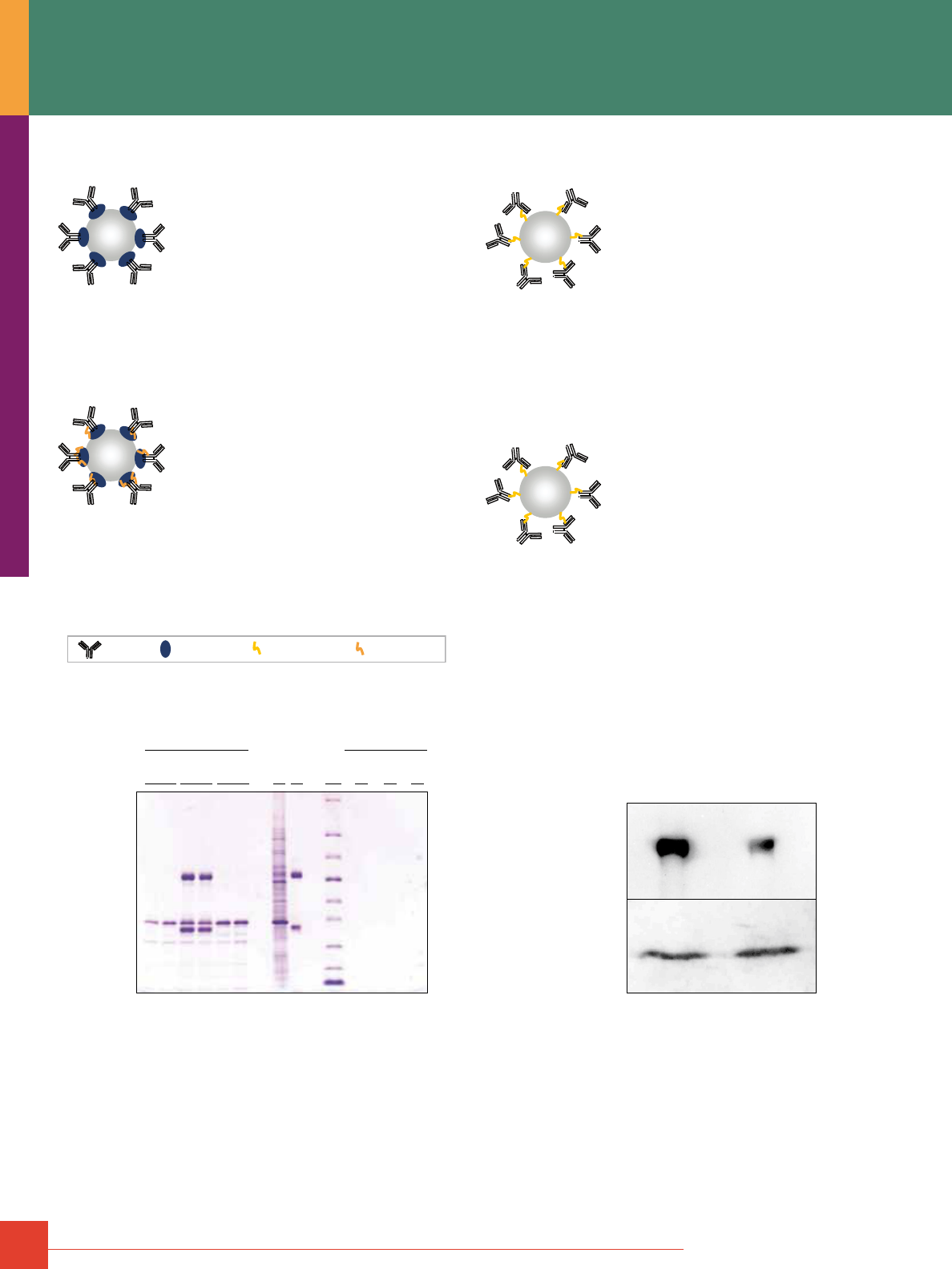
40
For more information, or to download product instructions, visit www.www.thermoscientific.com/pierce
Comparison of three different Thermo Scientific Pierce IP Kits.
Immunoprecipitations were performed according to the product instructions
using 10μg of affinity-purified goat anti-GFP antibody and the Pierce Direct,
Classic and Crosslink IP Kits. The cell lysate was prepared using IP Lysis/
Wash Buffer and pre-cleared using the Pierce Control Agarose Resin supplied
in the kits. The immune complex was formed by incubating the antibody, resin
and lysate overnight. The resin was washed with IP Lysis/Wash Buffer, 1X
Conditioning Buffer and eluted with Elution Buffer. For analysis, 4-20%
Tris-glycine gels were loaded with 20% of the eluted sample, 5% of the cell
lysate load (Lysate) and 10% of the antibody load (IgG) and stained with
Thermo Scientific Imperial Protein Stain (Product # 24615). For the resin con-
trols, the immunoprecipitation was performed without adding the antibody.
Co-immunoprecipitation of interacting proteins using the Thermo Scientific
Pierce Co-IP Kit. Epidermal Growth Factor (EGF) was co-immunoprecipitated
from 2,000μg of HeLa lysate with 40 and 20μg anti-Epidermal Growth Factor
Receptor (EGFR) mouse monoclonal IgG
1
+ IgG
2a
(Thermo Scientific) using
the Pierce Co-IP Kit. Co-immunoprecipitations were performed according to
product instructions. Two identical Western blots were probed with anti-EGFR
sheep polyclonal IgG (Millipore) or anti-EGF rabbit polyclonal IgG (Santa Cruz).
The Pierce Co-IP Kit co-immunoprecipitated EGF with the EGF-R.
The Classic IP Kit:
Product # 26146
• High binding-capacity recombinant Protein
A/G Plus Agarose Resin results in higher
antigen yields
• Recombinant Protein A/G Plus Resin offers
compatibility with a wider range of mamma-
lian IgG species for IP reactions (e.g., mouse,
rabbit, human and goat IgG subclasses)
The Crosslink IP Kit:
Product # 26147
• Able to purify target protein without con-
tamination by the antibody in the eluate
• Improved crosslinking protocol optimized
for maximum antibody functionality
• Bound immunoglobulins oriented for optimal
antigen-binding sites are more accessible
• High-capacity Protein A/G Plus Resin allows for better purifi-
cation and immobilization of antibodies
• Offers compatibility with a wider range of mammalian
IgG species
The Direct IP Kit:
Product # 26148
• Immobilize any antibodies independent
of isotype or species
• Improved antibody coupling protocol
• Purify target protein without antibody
contamination
• Activated resin for directly coupling antibodies to the
support resin
• Eliminate antibody contamination in the eluate
The Co-IP Kit:
Product # 26149
• Minimal antibody requirements for
co-IP reactions
• Shorter antibody coupling protocol
(<2 hours)
• Compatible with any antibody species
and subclass
• Requires a purified antibody in a solution free of amines and
stabilizing proteins
• Optimized protocols and buffers for efficient co-IP and
antigen elution
• Allows for selective purification of target protein
• Includes spin columns and collection tubes that shorten the
protocol by minimizing handling and mixing
• Compatible with specialized downstream applications,
e.g., mass spectrometry
• Allows for scale up
Bead
Bead
Bead
Bead
Protein A/G Plus
Antibodies
Alkylamine linkage
DSS Crosslinking
GFP (27 kDa)
IgG Heavy Chain
(55kDa)
Thermo Scientific
Pierce IP Kits
Direct
Classic
Lysate
IgG
MW
Direct
Classic
Crosslink
Crosslink
IgG Light Chain
(25kDa)
Resin Controls
EGF-R (170kDa)
µg anti-EGFR
EGF (6kDa)
40 20
IP/Co-IP

To order, call 800-874-3723 or 815-968-0747. Outside the United States, contact your local branch office or distributor.
41
Thermo Scientific Pierce Classic IP Kit
A convenient kit for a new spin on traditional immunoprecipitation.
The Pierce Classic IP Kit provides all the necessary reagents,
spin cups and collection tubes to perform successful immunopre-
cipitation (IP) experiments with ease. The kit uses high-capacity
Protein A/G agarose affinity resin for efficient binding of most
species and subclasses of IP antibodies. The included IgG elu-
tion buffer provides milder and less denaturing recovery of
antibody:antigen complexes than the traditional method of boiling
in reducing sample buffer for SDS-PAGE, facilitating a greater
variety of methods for subsequent analysis. The microcentrifuge
spin column format helps to ensure effective washing and sep-
aration of samples from the beaded agarose affinity resin.
Like traditional IP methods, the Pierce Classic IP Kit procedure
involves formation of antibody:antigen complexes in a sample
solution and then capture of that complex to an IgG-binding
protein that is covalently bound to beaded agarose resin (Protein
A/G Agarose). After washing to remove nonbound (presumably
undesired) components of the sample, the antigen and antibody
are recovered from the beaded resin with elution buffer supplied
in the kit. The entire procedure is performed in a microcentrifuge
spin column, allowing solutions to be fully separated from the
agarose resin upon brief centrifugation.
Ordering Information
Product #
Description
Pkg. Size
U.S.
Price
26146 Pierce Classic IP Kit
Sufficient reagents to perform 50 reactions.
Includes: Pierce Protein A/G Plus Agarose
IP Lysis/Wash Buffer
100X Conditioning Buffer
20X Tris-Buffered Saline
Elution Buffer
5X Lane Marker Sample
Buffer, Non-reducing
Pierce Spin Columns – Screw Cap
Microcentrifuge Collection Tubes
Microcentrifuge Sample Tubes
Pierce Control Agarose Resin
Kit
0.55mL
2 x 50mL
5mL
25mL
50mL
5mL
50 each
2mL, 100 each
1.5mL, 50 each
2mL
$259
Thermo Scientific Pierce Crosslink IP Kit
Purify target protein complexes without antibody interference!
The Crosslink IP Kit extends the functionality of traditional immuno-
precipitation (IP) methods by adding crosslinking technology and
microcentrifuge spin column sample handling to the procedure.
The primary benefits resulting from these features are the ability
to purify target protein without contamination by the antibody and
the ability to more effectively wash and separate samples from the
beaded agarose resin.
The Pierce Crosslink IP Kit method involves capturing the IP anti-
body to Protein A/G Agarose resin and covalently immobilizing it
to the support by crosslinking with disuccinmidyl suberate (DSS).
The antibody resin is then incubated with the sample that contains
the protein antigen of interest, allowing the antibody:antigen com-
plex to form. After washing to remove nonbound (presumably
undesired) components of the sample, the antigen is recovered by
dissociation from the antibody with elution buffer supplied in the kit.
The entire procedure is performed in a microcentrifuge spin cup,
allowing solutions to be fully separated from the agarose resin
upon brief centrifugation. Only antigen is eluted by the procedure,
enabling it to be identified and further analyzed without interfer-
ence from antibody fragments. Furthermore, the antibody resin
often can be reused for additional rounds of immunoprecipitation.
Ordering Information
Product #
Description
Pkg. Size
U.S.
Price
26147 Pierce Crosslink IP Kit
Sufficient reagents to perform 50 reactions.
Includes: Pierce Protein A/G Plus Agarose
20X Coupling Buffer
DSS Crosslinker, No-Weigh
™
Format
IP Lysis/Wash Buffer
100X Conditioning Buffer
20X Tris-Buffered Saline
Elution Buffer
Lane Marker Sample Buffer,
Non-reducing, (5X)
Pierce Spin Columns - Screw Cap
Microcentrifuge Collection Tubes
Microcentrifuge Sample Tubes
Pierce Control Agarose Resin
Kit
0.55mL
25mL
8 x 2mg
2 x 50mL
5mL
25mL
50mL
5mL
50 each
2mL, 100 each
1.5mL, 50 each
2mL
$305

42
For more information, or to download product instructions, visit www.www.thermoscientific.com/pierce
Thermo Scientific Pierce Direct IP Kit
Immunoprecipitate using any antibody species or subclass!
Eliminate antibody band contamination of IP products.
The Pierce Direct IP Kit represents a significant advancement in
immunoprecipitation (IP) technology by replacing the use of
immobilized Protein A or Protein G with a method for direct covalent
attachment of antibodies to the beaded agarose resin.
The primary benefits resulting from this method are the opportunity
to use any species or subclass of purified antibody (not just types
that bind to Protein A or G) and the ability to purify target protein
without contamination by the antibody. The method also makes
it possible to immunoprecipitate antigens from serum samples
without co-purifying non-target immunoglobulins. Finally, the kit
uses microcentrifuge spin cups to effectively wash and separate
samples from the beaded agarose resin.
Ordering Information
Product #
Description
Pkg. Size
U.S.
Price
26148 Pierce Direct IP Kit
Sufficient reagents to perform 50 reactions.
Includes: AminoLink Plus Coupling Resin
20X Coupling Buffer
Quenching Buffer
Wash Solution
5M Sodium Cyanoborohydride Solution
IP Lysis/Wash Buffer
100X Conditioning Buffer
20X Tris-Buffered Saline
Elution Buffer
5X Lane Marker Sample Buffer
Pierce Spin Columns – Screw Cap
Microcentrifuge Collection Tubes
Microcentrifuge Sample Tubes
Pierce Control Agarose Resin
Kit
2mL
25mL
50mL
50mL
0.5mL
2 x 50mL
5mL
25mL
50mL
5mL
50 each
2mL, 100 each
1.5mL, 50 each
2mL
$259
Thermo Scientific Pierce Co-Immunoprecipitation Kit
Perform co-immunoprecipitation experiments without
antibody interference.
The Pierce Co-Immunoprecipitation (Co-IP) Kit enables isolation
of native protein complexes from a lysate or other complex
mixture by directly immobilizing purified antibodies onto an
agarose support.
Co-IP is a common approach to study protein:protein interac-
tions that uses an antibody to immunoprecipitate the antigen (bait
protein) and co-immunoprecipitate any interacting proteins (prey
proteins). Traditional co-IP methods that use Protein A or G result
in co-elution of the antibody heavy and light chains that may
co-migrate with relevant bands, masking important results. The
Pierce Co-IP Kit resolves this issue by covalently coupling anti-
bodies onto an amine-reactive resin. The kit includes optimized
buffers for protein binding and recovery, reagents to perform con-
trol experiments and efficient spin columns and collection tubes,
which shorten the protocol and minimize handling and mixing.
Ordering Information
Product #
Description
Pkg. Size
U.S.
Price
26149 Pierce Co-Immunoprecipitation Kit*
Sufficient reagents to perform 50 reactions.
Includes: AminoLink Plus Coupling Resin
20X Coupling Buffer
5M Sodium Cyanoborohydride Solution
Quenching Buffer
Wash Solution
IP Lysis/Wash Buffer
100X Conditioning Buffer
Elution Buffer
5X Lane Marker Sample
Buffer, Non-reducing
Pierce Control Agarose Resin
20X Modified Dulbecco’s PBS Buffer
Pierce Spin Columns – Screw Cap
Microcentrifuge Collection Tubes
Microcentrifuge Sample Tubes
Kit
2mL
25mL
0.5mL
50mL
60mL
2 x 50mL
5mL
50mL
5mL
2mL
25mL
50 each
100 each
50 each
$308
* The Thermo Scientific Pierce Co-IP Kit (Product # 23600) and the Pierce Mammalian Co-IP Kit (Product
# 23605) were discontinued on December 31, 2009. The next-generation Pierce Co-IP Kit (Product #
26149) will replace these old kits. The new co-IP kit is optimized for using smaller amounts of sample and
offers a common lysis/wash buffer eliminating the need for a separate lysis reagent (Thermo Scientific
M-PER Mammalian Protein Extraction Reagent, Product # 78503) as was offered with the Mammalian
Co-IP Kit.
IP/Co-IP
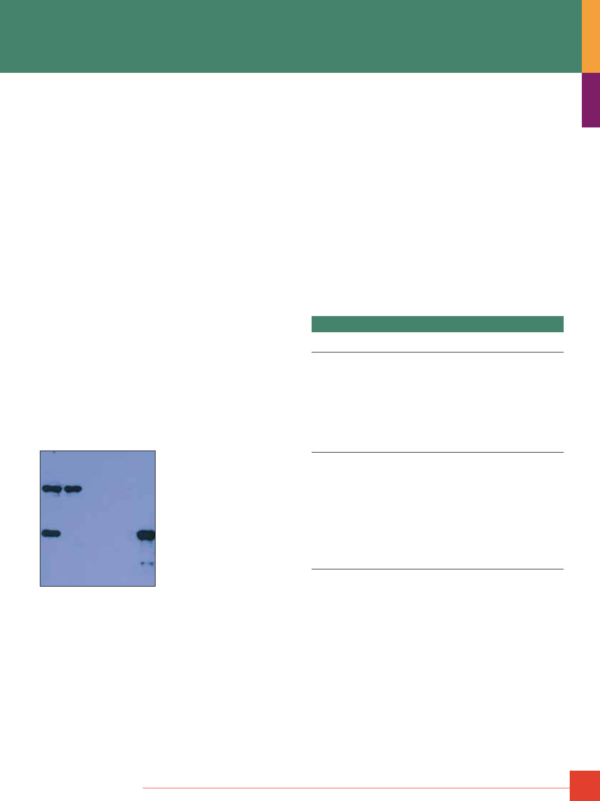
To order, call 800-874-3723 or 815-968-0747. Outside the United States, contact your local branch office or distributor.
43
Thermo Scientific Pierce Coated Plate Immunoprecipitation Kits
Pre-coated 96-well plates are easier to use and faster than
traditional microcentrifuge tube methods.
Thermo Scientific Pierce Coated Plate IP Kits enable rapid
immunoprecipitation of multiple samples without the usual tedium
of pipetting, centrifuging and separating beaded affinity resin
in individual microcentrifuge tubes. Immunoprecipitation is
accomplished using coated 96-well microplates rather than
beaded agarose resin. The plate format allows fast processing
of multiple samples. Select from Protein A/G-, Protein G- or
streptavidin-coated plates.
Highlights:
• Ready-to-use, high-quality coated plates provide high capacity
and consistency
• Plate format best suited for simultaneously processing multiple
samples and their control conditions
• Faster, easier and more thorough washing than with traditional
tube/resin IP methods
• Uses familiar and convenient ELISA tools (multichannel pipettors
and plate washing); no tedious separation of supernatant from
pelleted resin beads, and no tubes to open, close and centrifuge
• Coated plates are 96-well strip plates, convenient for experi-
ments requiring only a partial plate
• Easy-to-follow instructions, including detailed explanation of
appropriate controls
• Three kits available, suitable for most common antibody types
(mouse, rabbit, human and goat IgG subclasses) or any
biotinylated antibody or “bait” protein
3 4 521
Thermo Scientific Pierce Protein A/G Coated Plate IP Kit (Product # 45350)
immuno precipitation of CD71 (transferring receptor) from human serum using
and a goat anti-CD71 polyclonal antibody. Eluted products for the experimental
and control samples were mixed with nonreducing sample loading buffer,
separated by SDS-PAGE, transferred to nitrocellulose membrane and detected
by Western blotting with the IP antibody, Goat-anti-mouse-HRP conjugated
secondary antibody (Product # 31432) and Thermo Scientific SuperSignal West
Dura Chemiluminescent Substrate (Product # 34076).
Lane 1: Experiment (immunoprecipitation product)
Lane 2: Antibody-only control (no sample)
Lane 3: Human serum sample control (no antibody)
Lane 4: Plate control (no antibody or human sample)
Lane 5: Pure target protein (CD71) for size reference
Choosing between Protein A/G and Streptavidin Coated Plate Kits
Streptavidin is a protein that binds specifically and strongly to
biotin; therefore, the Streptavidin Coated Plate IP Kit
(Product # 45360) is appropriate for immunoprecipitation when
using a biotin-labeled (biotinylated) antibody. This kit can be
used to affinity-purify a binding partner to any antibody species
or subclass or any other protein or molecule that is biotinylated.
Because the streptavidin-biotin affinity interaction is so strong,
the elution step generally will dissociate only the antigen (binding
partner), not the biotinylated antibody or “bait” protein.
Protein A and Protein G are different proteins that bind to immuno-
globulins (primarily IgG). Typically, Protein A is preferred for use
with rabbit polyclonal antibodies, while Protein G is preferred for
use with mouse antibodies (especially monoclonals of the IgG
1
subclass). Protein A/G is a recombinant of Protein A and Protein G
that has the additive binding properties of both proteins.
Reference
Desai, S. and Hermanson, G. (1997). Previews 1(3), 2-7.
Ordering Information
Product #
Description
Pkg. Size
U.S.
Price
45350 Pierce Protein A/G Coated Plate IP Kit
Antibody binding capacity/well: 2.5µg. Sufficient
capacity for downstream analysis of IP or co-IP
proteins in-gel or by Western blot.
Includes: Protein A/G Coated 12 x 8-well strip plates
Phosphate buffered saline
Surfact-Amps
®
X-100 (10% Triton X-100)
Elution buffer
Neutralization buffer
Uncoated 96-well strip plates (white),
(for sample collection and neutralization)
Plate sealers
Kit
2 plates
2 packs
6 x 10mL
50mL
7mL
2 ea.
18 sheets
$295
45360 Pierce Streptavidin Coated Plate IP Kit
Antibody binding capacity/well: 5µg. Sufficient
capacity for downstream analysis of IP or co-IP
proteins in-gel or by Western blot.
Includes: High Binding Capacity Streptavidin
Coated Plates
Biotin blocking buffer
Phosphate buffered saline
Surfact-Amps X-100 (10% Triton X-100)
Elution buffer
Neutralization buffer
Uncoated 96-well strip plates (white),
(for sample collection and neutralization)
Plate sealers
Kit
2 plates
30mL
2 packs
6 x 10mL
50mL
7mL
2 ea.
18 sheets
$298
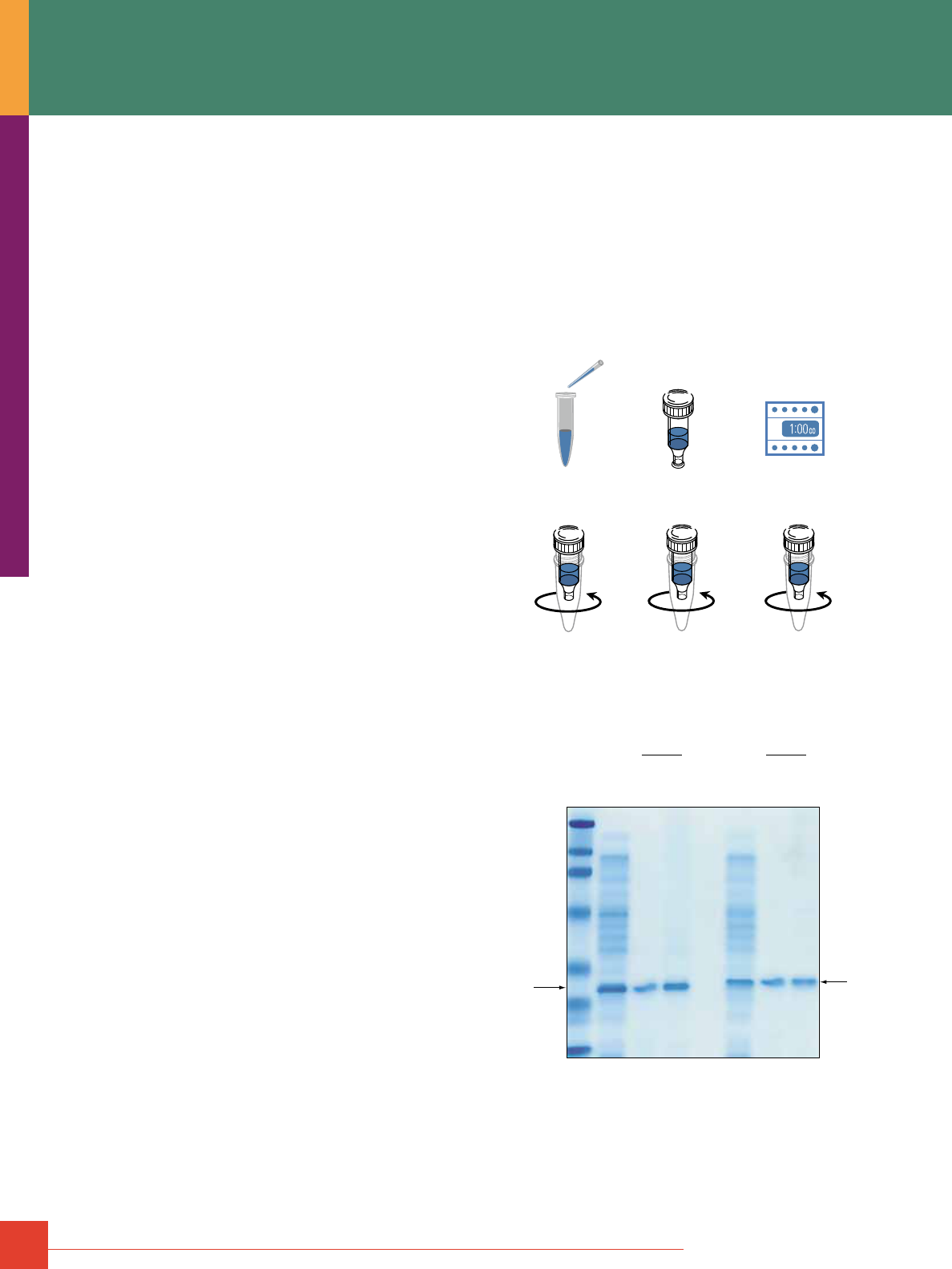
44
For more information, or to download product instructions, visit www.www.thermoscientific.com/pierce
Thermo Scientific Pierce HA- or c-Myc Tag IP/
Co-Immunoprecipitation Kits
Need to perform IP or co-IP reactions with your HA- or c-Myc-
tagged protein? Open box ... Read instructions ... Start performing
an IP or co-IP. High-specificity immobilized antibodies make it easy.
No tags are more popular for mammalian system protein expres-
sion than HA or c-Myc. Although these tags are extremely popular,
a kit that allows you to conveniently perform immunoprecipitation
(IP) or co-immunoprecipitation (co-IP) reactions using these tags
has not been available. The Pierce IP/Co-IP Kits include all neces-
sary reagents, buffers and hardware that allow efficient purifica-
tion of a tagged target protein (i.e., IP) or confirmation of potential
interactions indicated by yeast two-hybrid results (i.e., co-IP).
These four kits, which are specifically for HA- and c-Myc-tagged
proteins, allow you to easily perform an IP or co-IP experiment
with minimal optimization. High-affinity, high-specificity antibod-
ies immobilized onto an agarose matrix are at the heart of these
new kits. In addition, the kits contain a full complement of buffers,
eluents, a positive control and necessary hardware to efficiently
perform the intended application.
Highlights:
IP and co-IP for HA- or c-Myc-tagged proteins directly out of the box.
• Demonstrated utility in the IP and co-IP application benefits the
novice and expert. Our kits include all essential components to
perform the assays. There’s no need to formulate or validate
raw materials.
Immobilized high-affinity antibodies with excellent specificity
for the HA or c-Myc tag.
• The immobilized anti-HA and anti-c-Myc monoclonals precipitate
the appropriately tagged protein specifically and in high yield,
resulting in clean Western blot detection.
• Excellent results with as little as 2.5μg of anti-HA antibody and
1μg of anti-c-Myc antibody in the IP mode with the respective
positive control lysate.
• Limits possibility of nonspecific binding to other proteins in
the lysate.
• Eliminates contamination from antibody or antibody fragments
after elution of the precipitated protein or the co-IP complex.
This benefit is especially important when interpreting protein
interaction results.
Simple, flexible and easy-to-follow protocols.
• Complete kit format offers optimum convenience in both the
IP and co-IP modes.
• Eliminate Protein A or Protein G, reducing nonspecific binding
and shortening the IP procedure.
• System demonstrates excellent flexibility with respect to the
amount of antibody or amount of lysate used, enabling isolation
of low-expression HA-/c-Myc-tagged targets.
• The use of Spin Columns accelerate the IP/co-IP process.
Spin Columns.
• Spin Columns are very convenient for small sample handling.
• Allow more efficient washing.
• Eliminate resin losses
HA- or c-Myc Tag IP/Co-IP Kit Descriptions
Each kit listed at right consists of two components: an Application
Set and a Positive Control Lysate. The Application Set contains
the immobilized support appropriate for the kit and all of the
required buffers, eluents and hardware. The second component
is a bacterial lysate containing an overexpressed GST with
either HA or c-Myc as the C-terminal tag. The mammalian
version of each kit contains Thermo Scientific M-PER Protein
Extraction Reagent for use with mammalian cell-based IP or co-IP
applications. The Application Sets and Positive Control Lysates
can also be ordered separately.
1. Lyse cells expressing HA- or
c-Myc-tagged protein.
2. Combine cell lysate and
Immobilized Anti-HA or
Anti-c-Myc.
3. Incubate at 4
˚
C for
1 hour to overnight with
end-over-end mixing.
4. Pulse centrifuge for
10 seconds.
5. Wash three times with
TBS-T. Pulse centrifuge for
10 seconds after each wash.
6. Elute HA- or c-Myc-tagged
protein using Elution Buffer
or Non-Reducing Sample Buffer.
Collection
Tube
Bottom Plug
Removed
Thermo Scientific Pierce HA- or c-Myc IP/Co-IP Kit Protocol summary.
GST-c-Myc
GST-HA
IP
MW Marker
HA Lysate
Elution #1
Elution #2
c-Myc Lysate
Elution #1
Elution #2
IP
Effectiveness of elution options in the IP of GST-HA and GST-c-Myc from
bacterial lysates. IP results achieved with the Thermo Scientific Pierce HA
and c-Myc IP/Co-IP Kits using the appropriate positive control lysate provided
and suggested elution options for GST-HA and GST-cMyc, respectively.
The elution components are supplied with each kit. Elution performed with
#1. Elution Buffer or #2. 2X nonreducing sample buffer.
IP/Co-IP
To order, call 800-874-3723 or 815-968-0747. Outside the United States, contact your local branch office or distributor.
45
Ordering Information
Product #
Description
Pkg. Size
U.S.
Price
23610 HA-Tag IP/Co-IP Kit
Sufficient material to conduct 25 IP/co-IP assays using
proteins expressed with an HA tag. Kit is supplied
complete with an HA-tagged positive control lysate.
Includes: Immobilized Anti-HA (agarose resin)
BupH Tris Buffered Saline Pack
Elution Buffer, pH 2.8
Lane Marker Non-Reducing Sample Buffer (5X)
Spin Columns Accessory Pack,
27 columns with pre-inserted frit
and top and bottom caps
Collection Tubes and Caps Accessory Pack,
100 graduated 2mL tubes and plug caps
M-PER Mammalian Protein Extraction
Reagent
HA-Tagged Positive Control
Kit
150μL
1 pack
50mL
5mL
25mL
500μL
$399
23615 Mammalian HA-Tag IP/Co-IP Kit
Sufficient material to conduct 25 IP/co-IP assays using
proteins expressed with an HA tag. Kit is supplied
complete with a mammalian cell lysis buffer and
an HA-tagged positive control lysate.
Includes: Immobilized Anti-HA (agarose resin)
BupH
Tris Buffered Saline Pack
Elution Buffer, pH 2.8
Lane Marker Non-Reducing Sample Buffer (5X)
Spin Columns Accessory Pack,
27 columns with pre-inserted frit
and top and bottom caps
Collection Tubes and Caps Accessory Pack,
100 graduated 2mL tubes and plug caps
M-PER Mammalian Protein Extraction
Reagent
HA-Tagged Positive Control
Kit
150μL
1 pack
50mL
5mL
25mL
500μL
$423
23620 c-Myc-Tag IP/Co-IP Kit
Sufficient material to conduct 25 IP/co-IP assays using
proteins expressed with a c-Myc tag. Kit is supplied
complete with a c-Myc-tagged positive control lysate.
Includes: Immobilized Anti-c-Myc (agarose resin)
BupH Tris Buffered Saline Pack
Elution Buffer, pH 2.8
Lane Marker Non-Reducing Sample Buffer (5X)
Spin Columns Accessory Pack,
27 columns with pre-inserted frit
and top and bottom caps
Collection Tubes and Caps Accessory Pack,
100 graduated 2mL tubes and plug caps
c-Myc-Tagged Positive Control
Kit
250μL
1 pack
50mL
5mL
500μL
$399
23625 Mammalian c-Myc Tag IP/Co-IP Kit
Sufficient material to conduct 25 IP/co-IP assays using
proteins expressed with a c-Myc tag. Kit is supplied
complete with a mammalian cell lysis buffer and a
c-Myc-tagged positive control lysate.
Includes: Immobilized Anti-c-Myc (agarose resin)
BupH Tris Buffered Saline Pack
Elution Buffer, pH 2.8
Lane Marker Non-Reducing Sample Buffer (5X)
Spin Columns Accessory Pack,
27 columns with pre-inserted frit
and top and bottom caps
Collection Tubes and Caps Accessory Pack,
100 graduated 2mL tubes and plug caps
M-PER Mammalian Protein Extraction
Reagent
c-Myc-Tagged Positive Control
Kit
250μL
1 pack
50mL
5mL
25mL
500μL
$423
Protein Interactions Technical Handbook
Our 72-page Protein Interaction
Technical Handbook provides
protocols and technical and product
information to help maximize results
for Protein Interaction studies. The
handbook provides background,
helpful hints and troubleshooting
advice for immunoprecipitation and
co-immunoprecipitation assays, pull-
down assays, Far-Western blotting
and crosslinking. The handbook also
features an expanded section on
method to study protein:nucleic acid
interactions, including ChIP, EMSA and RNA EMSA. The hand-
book is an essential resource for any laboratory studying Protein
Interactions. (# 1601618)
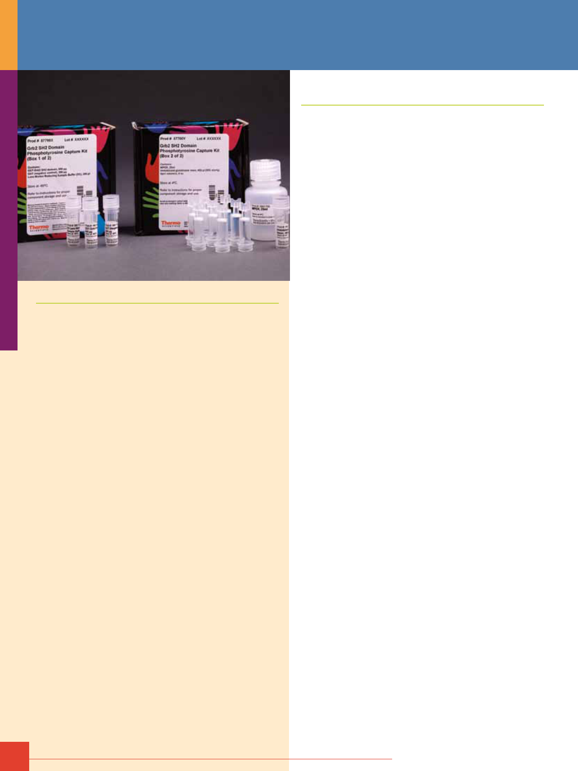
46
For more information, or to download product instructions, visit www.thermoscientific.com/pierce
Protein Enrichment
Protein Enrichment
Protein enrichment encompasses numerous techniques to
isolate subclasses of cellular proteins based on their unique
biochemical activity, post-translational modification (PTM) or
spatial localization in a cell. Protein enrichment is essential for
studying low abundant proteins and for reducing the complexity
of samples for proteomic analysis. Enrichment of specific proteins
or protein complexes can most easily be accomplished using
immunoaffinity techniques such as immunoprecipitation and
co-immunoprecipitation. Although these antibody-based tech-
niques are widely used, elution of immunoprecipitated proteins
can sometimes result in low protein recovery or antibody contami-
nation in samples.
Global protein enrichment strategies involve the selective isola-
tion of distinct protein subclasses which share a common post-
translational modification or cellular localization. Post-translational
modifications such as phosphorylation and glycosylation can be
enriched using affinity ligands such as ion-metal affinity chroma-
tography (IMAC) or immobilized lectins, respectively. In addition,
PTM-specific antibodies have been be used. Other techniques use
metabolic or enzymatic incorporation of modified amino acids or
PTMs to introduce unique protein chemistry which can be used for
enrichment. Finally, proteins can also be enriched using various
enzyme class specific compounds or cell-impermeable labeling
reagents which selectively label cell surface proteins.
Phosphoprotein Enrichment Kits
Process cell and tissue samples in less time and with greater purity.
Phosphorylation is one of the most frequently occurring post-
translational modifications in proteins. It is estimated that as many
as 30% of all cellular proteins are transiently phosphorylated on
serine, threonine and tyrosine residues.
Reversible protein phosphorylation regulates nearly all intra-
cellular biological events, including signal transduction, protein-
protein interactions, protein stability, protein localization, apoptosis
and cell-cycle control. Deregulation of protein phosphorylation is
a hallmark of numerous human diseases, including cancer and
metabolic and immune disorders.
Detecting changes in protein phosphorylation can be a difficult
task because of the transient labile state of the phosphate
group. Furthermore, low phosphoprotein abundance and poorly
developed phospho-specific antibodies contribute to difficulties in
phosphoprotein detection. Recent advances in mass spectrometry
technology in combination with phosphoprotein enrichment using
immobilized metal affinity chromatography (IMAC) have resulted in
greater resolution of the phosphoproteome.
The new Thermo Scientific Pierce Phosphoprotein Enrichment Kit
efficiently enriches phosphorylated proteins derived from mammalian
cells and tissues. The proprietary metal and buffer composition
produces superior yields with negligible nonspecific binding.
Highlights:
• Specific – low contamination from nonspecific proteins
• Fast – easy-to-use spin format enriches of phosphorylated
proteins in less than 2 hours
• Superior yield – high yield from complex biological samples, cell
culture lysate and mouse tissue extract
• Convenient format – complete kit includes pre-dispensed spin
columns, buffers, reagents and Thermo Scientific Pierce Protein
Concentrators
• Compatible – works with downstream applications, including
mass spectrometry, Western blotting and 2D-PAGE
Phospho-specific antibodies recognizing key regulatory proteins
involved in growth factor signaling were used to monitor binding
specificity of our Phosphoprotein Enrichment Kit (Figure 1).
Specificity of the kit is further demonstrated by the absence of
Cytochrome C (pI 9.6) and p15Ink4b (pI 5.5), two proteins not
predicted to be phosphorylated, in the elution fraction and their
emergence in the flow-through and wash fractions (Figure 1).
Furthermore, dephosphorylation of HeLa cell extract in vitro
resulted in diminished binding of PTEN, MAPK and GSK3β to the
Pierce Phosphoprotein Enrichment Column as evidenced by their
absence in the elution fraction. Conversely, all three proteins
were present in the elution fraction from non-treated HeLa extract
(Figure 2). Our Phosphoprotein Enrichment Kit provided superior
and efficient phosphoprotein enrichment yields when compared to
competitors’ products (Table 1). It also effectively enriched phos-
phoproteins from homogenized mouse liver tissue (Figure 3).
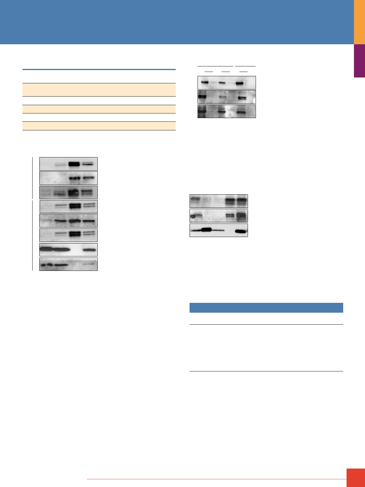
To order, call 800-874-3723 or 815-968-0747. Outside the United States, contact your local branch office or distributor.
47
Table 1. The Thermo Scientific Pierce Phosphoprotein Enrichment Kit
provides higher phosphoprotein yields in less time than competitors’ kits.
Kit Yield (%) Enrichment Time (Hours)
Thermo Scientific Pierce
Phosphoprotein Enrichment Kit
15 1.5
Supplier Q Kit 4.4 4.5
Supplier I Kit 2.6* 3.5
Supplier C Kit 8 3
Supplier E Kit Too dilute to determine 5
*Based on maximum 1mg load per manufacturer’s protocol.
Phospho-MAPK T202/Y204
Phospho-PTEN S380
Phospho-GSK3β S9
Phospho-MAPK T202/Y204
Phospho-Akt S473
Phospho-Src Y527
Cytochrome C
p15Ink4b
NIH 3T3 + PDGF HeLa + EGF
FT W E L
Figure 1. Highly pure phosphoprotein enrichment from complex biological
samples. Serum-starved HeLa and NIH 3T3 cells were stimulated with EGF and
PDGF, respectively. Cell lysate (2mg) was used for enrichment. Concentrated
flow-through, wash and elution fractions were resolved by SDS-PAGE. Gel
lanes were normalized by protein concentration (10μg/lane). Western blot
analysis was performed using antibodies that detect site-specific phosphory-
lation events. Cytochrome C (pI 9.6) and p15Ink4b (pI 5.5) served as negative
controls for nonspecific binding of non-phosphorylated proteins. FT = flow-
through fraction, W = pooled wash fractions, E = pooled elution fractions and
L = non-enriched total cell lysate.
Phospho-PTEN S380
Phospho-MAPK T202/Y204
Phospho-GSK3β S9
Total Cell Lysate Elution
25 µg 10 µg 10 µg
λ Pase
- + - + - +
Figure 2. Highly specific phosphoprotein purification from lambda
phosphatase-treated cells. Non-treated and lambda dephosphorylated
HeLa cell extract (2mg) was loaded onto separate Thermo Scientific Pierce
Phosphoprotein Enrichment Columns. Concentrated elution fractions were
resolved by SDS-PAGE. Gel lanes were normalized by protein concentration
(10μg/lane). To determine enrichment, 10μg and 25μg of non-treated or lambda
phosphatase-treated total cell extract (non-enriched) was loaded onto each
gel. Western blot analysis was performed using phospho-specific antibodies
recognizing key proteins in the Ras-MAPK and PI3K-Akt signaling cascades.
L10 FT W E L25
Phospho-PTEN S380
Phospho-Rb S795
Cytochrome C
Figure 3. Efficient enrichment of phosphoproteins from mouse liver extract.
Homogenized mouse liver extract (~2mg) was loaded onto a Thermo Scientific
Pierce Phosphoprotein Enrichment Column. Concentrated flow-through, wash
and elution fractions were resolved by SDS-PAGE. Gel lanes were normalized by
protein concentration (10μg/lane). Western blot analysis was performed using
antibodies that detect site-specific phosphorylation events. Cytochrome C
(pI 9.6) served as a negative control for nonspecific binding. L10 = non-enriched
total cell extract (10μg), FT = flow-through fraction, W = wash fraction,
E = elution fraction and L25 = non-enriched total cell extract (25μg).
Ordering Information
Product #
Description
Pkg. Size
U.S.
Price
90003
Pierce Phosphoprotein Enrichment Kit
Includes: Phosphoprotein Enrichment Column
Resin Bed (1mL)
Lysis/Binding/Wash Buffer
Elution Buffer
CHAPS
White Column Caps
Pierce Protein Concentrator
7mL/9K MWCO
Kit
10 ea.
325mL
60mL
1g
10 caps
10 Devices
$464
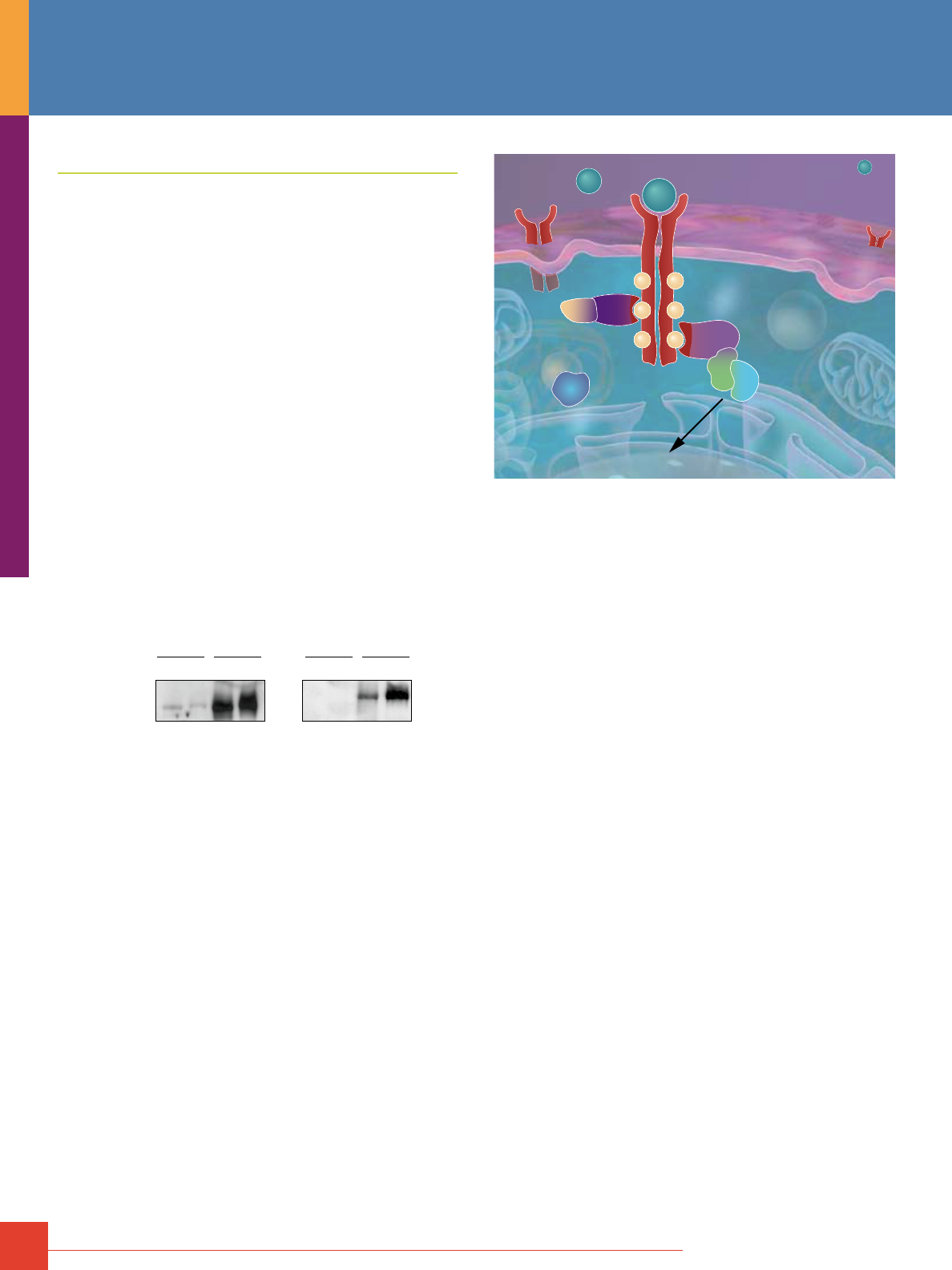
48
For more information, or to download product instructions, visit www.www.thermoscientific.com/pierce
Phosphoprotein Pull-Down with SH2 Domains
SH2 Domain Phosphotyrosine Capture Kits
SH2 domains provide an improved approach to selectively monitor
receptor tyrosine kinase signaling, as well as the binding of down-
stream effector proteins. The interactions of SH2 domain-contain-
ing proteins represent a critical interface between extracellular
stimulation of a membrane receptor and transmission of that
signal to intracellular proteins.
Our kits include optimal levels of purified GST-fused SH2 domains
of various signaling proteins integral to cell biology. Each validated
kit includes optimized buffers and columns to perform protein
pull-downs. Using SH2 domains eliminates the background
associated with low-specificity antibodies and enables analysis
of receptor targets to which antibodies are not available. Using
quality-tested purified GST-SH2 domains also ensures uniform
results without variability.
SH2 domains specifically bind phospho-tyrosine residues.
Each SH2 domain is specific for its natural target (Figure 4).
In vivo, the phosphatase Shp2 is recruited to tyrosine 1009 of the
platelet-derived growth factor receptor (PDGFR) only when it is
phosphorylated. Also, PLCg is recruited to tyrosine 1021 on PDGFR
in response to growth factor signaling. In quiescent cells both
tyrosine 1009 and 1021 are not phosphorylated and, therefore, both
domains are unable to bind.
PDGF
Antibody
GST-Shp2 GST-PLCγ
L
PD
L
PD
- +
L
PD
L
PD
- +
pPDGFR Y1009 pPDGFR Y1021
Figure 4. Site-specific interaction of GST-Shp2 and GST-PLCg SH2 on the
PDGF-receptor. NIH3T3 cells were rendered quiescent by serum withdrawal
for 48 hours followed by stimulation with PDGF (50ng/mL, Cell Signaling
Technology [CST]) for 15 minutes or no stimulation. Each cell lysate (500μg) was
incubated overnight at 4°C with either 100μg GST-PLCg1 or GST-Shp2. Protein
complexes were captured on immobilized glutathione beads and resolved
by SDS-PAGE. Western blot analysis was performed using phospho-specific
antibodies that detect known protein-protein interaction sequences (phospho-
PDGFR Y1009 and phospho-PDGFR Y1021, CST). Lanes: L = 25μg of total cell
lysate and PD = SH2 domain pull-down.
pY
pY
pY
pY
pY
pY
SH2
SH2
Receptor in cell membrane
Extracellular signal
Tyrosine phosphorylation
SH2 domains recognize pY
Other proteins recruited
Translation, Transcription, Cell Proliferation, Differentiation, Apoptosis
Cellular pathway activation
SH2 domains provided a more efficient method of capturing
site-specific phospho-tyrosine events as compared to antibody
based co-immunoprecipitation.
Co-immunoprecipitation experiments are dependent on the
quality and specificity of the antibody used. Antibodies against
phosphoproteins are notoriously difficult to work with, yielding
high background and low target protein recovery. SH2 domains
provide better capture efficiency as compared to many pan- and
site-specific phospho-antibodies because an SH2 domain is more
selective by nature. Because of this improved specificity, SH2
domains can be used to pull-down phosphotyrosine containing
proteins with improved results over traditional antibody-based
methods. In Figure 5, PDGF-stimulated NIH3T3 cells were lysed
and incubated with either a specific SH2 domain or a pan-antibody
corresponding to the SH2 domain containing protein. Binding of
each specific SH2 domain to the PDGF receptor was confirmed
by Western blot using a pan-PDGFR antibody. In all cases we
observed a higher capture efficiency using the SH2 domain
approach as compared to the traditional antibody co-immunopre-
cipitation. In many cases, we were also able to detect differential
binding of the SH2 domain in the presence of PDGF stimulation as
compared to the unstimulated state. These differences were not
apparent using the co-immunoprecipitation approach. We further
tested the interaction specificity of four SH2 domains using
phospho-antibodies which detect site-specific tyrosine
phosphorylation events which mediate each SH2 domain
interaction. We were limited to only four phospho-antibodies
against specific tyrosine sites on the PDGF receptor because
of commercial availability and/or antibody integrity. Using these
site-specific antibodies we confirmed the selectivity and specificity
of each SH2 domain tested and these interactions were much
stronger as compared to the traditional antibody immunoprecipi-
tation. Taken together these results clearly demonstrate that the
SH2 domain pull-down approach represents a robust method to
capture and monitor site specific tyrosine phosphorylation events
which are critical for cell health.
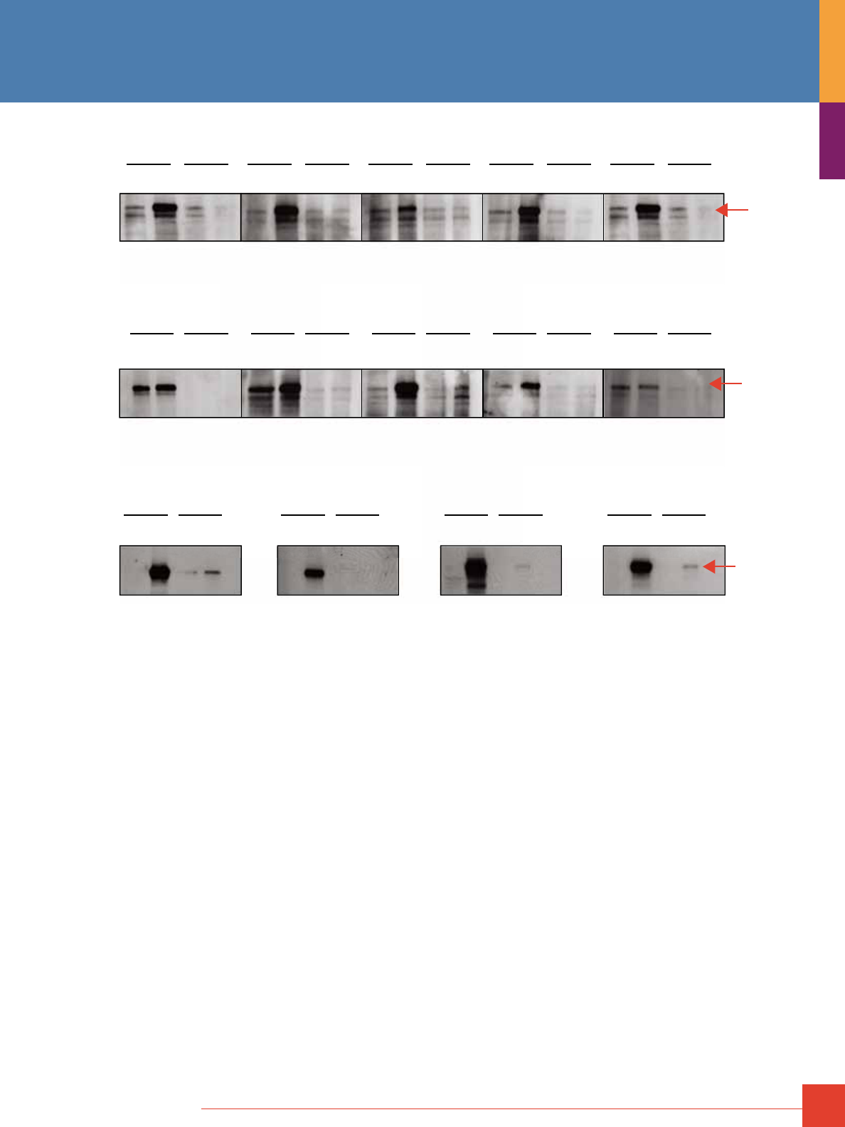
To order, call 800-874-3723 or 815-968-0747. Outside the United States, contact your local branch office or distributor.
49
Figure 5. SH2 domains provide better enrichment of phosphorylated
receptor tyrosine kinase (PDGFR) compared to antibody-based pull-down
assays. Panel A. NIH3T3 cells were rendered quiescent by serum starvation
for 24 hours with DMEM containing 0.1% FCS. Following starvation cells
were either stimulated (100ng/mL PDGF for 20 minutes) or left unstimulated.
Cells were then lysed in NP40 lysis buffer (50mm Tris pH 7.5, 150mm NaCl,
1% NP40, 5% glycerol) containing Thermo Scientific Halt Protease and
Phosphatase Inhibitor Cocktail (Product # 78440) and protein concentra-
tions determined by the Thermo Scientific Pierce 660nm Assay. SH2 and
co-immunoprecipitations were performed as follows: SH2 domain pull-downs
consisted of 100μg of SH2 domain (Abl, Crk, Grb2, Lyn, PI3K, Plc-gamma,
RasGap, Shc, Shp2, Src) added to 250μg of stimulated or unstimulated lysate.
Co-immunoprecipitations were performed by incubating 10μL of pan antibody
(Abl, Crk, Grb2, Lyn, PI3K, Plc-gamma, RasGap, Shc, Shp2, Src) with 250μg of
stimulated or non-stimulated lysate. Binding was performed for 16 hours at 4°C
on a rotating platform. Following binding 100μL of a 50% slurry of immobilized
glutathione was added to the SH2 domain samples and 100μL of a 50% slurry of
Protein A/G was added to the co-immunoprecipitation samples. Samples were
incubated for 1 hour at 4°C on a rocking platform followed by centrifugation at
1000 x g for 1 minute to collect complexes bound to the resin. Resin beds were
then washed 3 times with 500μL of Thermo Scientific M-PER Wash Buffer. To
elute bound proteins 25μL of 5X DTT loading dye was added to each sample
and boiled for 5 minutes. Protein complexes were then resolved by SDS-PAGE
on a 4-20% Tris-glycine gel. Proteins were transferred onto PVDF membrane
for 16 hours at 4°C, 30V. Membranes were blocked in 7.5% BSA in TBST and
Western blots performed using a 1:2000 dilution of mouse monoclonal
anti-PDGFR-beta antibody (CST 3175). Detection was performed using
Thermo Scientific SuperSignal Pico Chemilumescent Detection System.
Panel B. Membranes containing the Grb2, Plcg, PI3K, Src, and RasGap
pull-downs were stripped with Thermo Scientific Restore Plus Buffer and
reprobed with phospho-antibodies which detect site-specific tyrosine phos-
phorylation events on the PDGF receptor which mediate the SH2 domain
interaction. These antibodies include pPDGFRY716, pPDGFRY751,
pPDGFRY1009, pPDGFRY579 which correspond to SH2 domain docking sites
for Grb2, PI3K, Plcg, and Src respectively. Detection was performed using
SuperSignal
®
West Pico Chemilumescent Detection System.
PDGF
- + - + - + - + - + - + - + - + - + - +
PDGF
- + - + - + - + - + - + - + - + - + - +
SH2 IP SH2 IP SH2 IP SH2 IP SH2 IP
SH2 IP SH2 IP SH2 IP SH2 IP SH2 IP
Abl Crk Grb2 Lyn PI3K
Plcγ
RasGap Shc Shp2 Src
Panel A.
Panel B.
Western Blot: Pan PDGFR
Western Blot: Pan PDGFR
PDGFR
PDGFR
SH2: Grb2
IP: Pan Grb2
WB: pPDGFRY716
SH2: PI3K
IP: Pan PI3K
WB: pPDGFRY751
SH2: Plcγ
IP: Pan Plcγ
WB: pPDGFRY1009
SH2: Src
IP: Pan Src
WB: pPDGFRY579
PDGF
- + - + - + - + - + - + - + - +
SH2 IP SH2 IP SH2 IP SH2 IP
pPDGFR
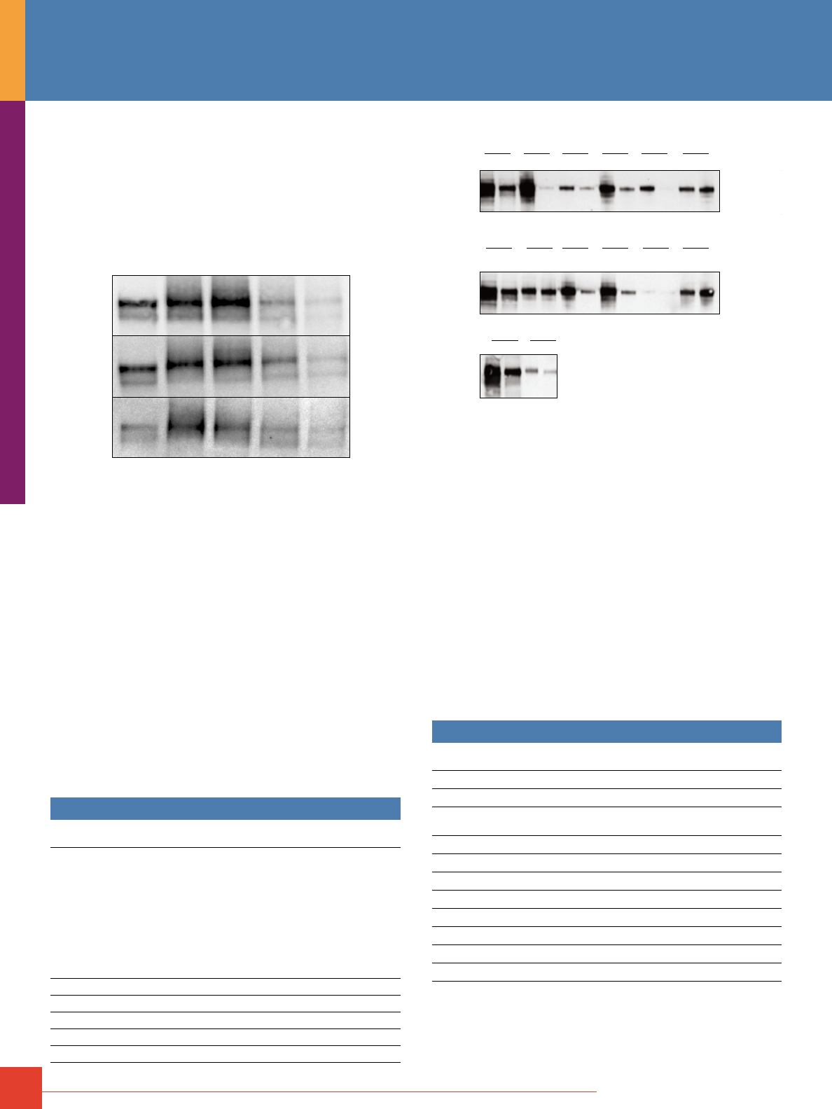
50
For more information, or to download product instructions, visit www.www.thermoscientific.com/pierce
Perform receptor phosphorylation time-course experiments.
With SH2 domain pull-downs, it is possible to monitor time-dependent
binding of SH2 domains to their target. For example, PLCg, Src
and Shc bind rapidly to the PDGF receptor in response to PDGF
stimulation (Figure 6). In nature, phosphorylation is rapid and tran-
sient, as evidenced by diminished binding at 10- and 20-minute
post-stimulation.
Minutes
Post PDGF
Stimulation
GST-PLCγ
GST-Src
GST-Shc
0 1 5 10 20
Figure 6. Time-specific binding of various SH2 domains to the PDGF receptor.
NIH3T3 cells were rendered quiescent by serum withdrawal for 48 hours. Post-
starvation cells were stimulated with PDGF (50ng/mL, CST) for the indicated
times or untreated. SH2 domain pull-downs were performed with 100μg of each
SH2 domain and 250μg of each cell lysate. Protein complexes were resolved
by SDS-PAGE and analyzed by Western blot using a pan antibody recognizing
PDGFR (CST).
Resolve specific tyrosine phosphorylation events.
SH2 domain affinity provides a level of selectivity not previously
attainable with generic anti-phosphotyrosine antibodies. Use
different SH2 domains to uncover the mechanism of how signals
are transduced or to screen multiple SH2 domains against a
particular target at one time. For example, different naturally
occurring SH2 domains efficiently bind to the epidermal growth
factor receptor (EGFR) in response to EGF stimulation (Figure 7).
There were strong interactions of many of the domains in the
presence of epidermal growth factor (EGF) and low-level binding
in quiescent, serum-starved A431 cells. RasGap binding is
independent of EGF stimulation, whereas Grb2 binding is highly
upregulated (Figure 7).
Ordering Information
Product #
Description
Pkg. Size
U.S.
Price
87700 Grb2 SH2 Domain Phosphotyrosine Capture Kit
Sufficient reagents for six pull-down reactions
(100µg/pull-down).
Includes: GST-Grb2 SH2 Domain (37 kDa)
GST (negative control, 27 kDa)
Immobilized Glutathione Resin
(50% resin slurry)
Spin Column (0.8mL)
M-PER Mammalian Protein
Extraction Reagent
Lane Marker Reducing Sample Buffer (5X)
6 pull-downs
600μg at 1mg/mL
200μg at 1mg/mL
400μL
8 each
25mL
200μL
$408
87701 Src SH2 Domain Phosphotyrosine Capture Kit
6 pull-downs
$408
87702 Abl SH2 Domain Phosphotyrosine Capture Kit
6 pull-downs
$408
87703 Crk SH2 Domain Phosphotyrosine Capture Kit
6 pull-downs
$408
87704 Fyn SH2 Domain Phosphotyrosine Capture Kit
6 pull-downs
$408
87705 Lck SH2 Domain Phosphotyrosine Capture Kit
6 pull-downs
$408
L25
*
* * *
* **
EGF
EGF
+ – + – + – + – + – + –
EGF
* = Known Interactions
Antibody: Pan EGFR
** = GST Only: Negative Control
L25
+ –
Shp2
+ –
Grb2 Src PLCγ Hck Fyn
L25
+ – + – + – + – + – + –
Gap Abl Crk GST Nck
Figure 7. High efficiency binding of different naturally occurring SH2
domains to EGFR in response to EGF stimulation. A431 cells were rendered
quiescent by serum withdrawal for 48 hours followed by stimulation with EGF
(100ng/mL) for 15 minutes or left untreated. Each cell lysate (500μg) was incu-
bated with 100μg of each GST-SH2 domain overnight at 4˚C. Protein complexes
were captured on immobilized glutathione beads and resolved by SDS-PAGE.
Western blot analysis was performed using a pan antibody that recognizes
the EGF receptor. L25 = 25μg total lysate load.
Identify binding interactions with MS.
Pull-down samples isolated with SH2 domains are compatible with
MS analysis for identifying protein interacting partners.
Improved specificity with SH2 domains and the elimination of
antibody contamination typically seen in traditional immunopre-
cipitation experiments deliver pure protein for MS analysis. Two
different SH2 domains were used to pull down interacting proteins
from NIH3T3 cell lysates, which were then analyzed by MS. Many
of the interacting proteins identified are specific to either Fgr
or PLCg.
Ordering Information
Product #
Description
Pkg. Size
U.S.
Price
87706 Nck SH2 Domain Phosphotyrosine Capture Kit
6 pull-downs
$408
87707 Shc SH2 Domain Phosphotyrosine Capture Kit
6 pull-downs
$408
87708 Ras-GAP SH2 Domain Phosphotyrosine
Capture Kit
6 pull-downs
$408
87709 Shp2 SH2 Domain Phosphotyrosine Capture Kit
6 pull-downs
$408
87710 PLC SH2 Domain Phosphotyrosine Capture Kit
6 pull-downs
$408
87711 Lyn SH2 Domain Phosphotyrosine Capture Kit
6 pull-downs
$408
87712 Hck SH2 Domain Phosphotyrosine Capture Kit
6 pull-downs
$408
87713 FGR SH2 Domain Phosphotyrosine Capture Kit
6 pull-downs
$408
87714 Cbl SH2 Domain Phosphotyrosine Capture Kit
6 pull-downs
$408
87715 Syk SH2 Domain Phosphotyrosine Capture Kit
6 pull-downs
$408
87716 PI3K SH2 Domain Phosphotyrosine Capture Kit
6 pull-downs
$408
Phosphoprotein Pull-Down with SH2 Domains
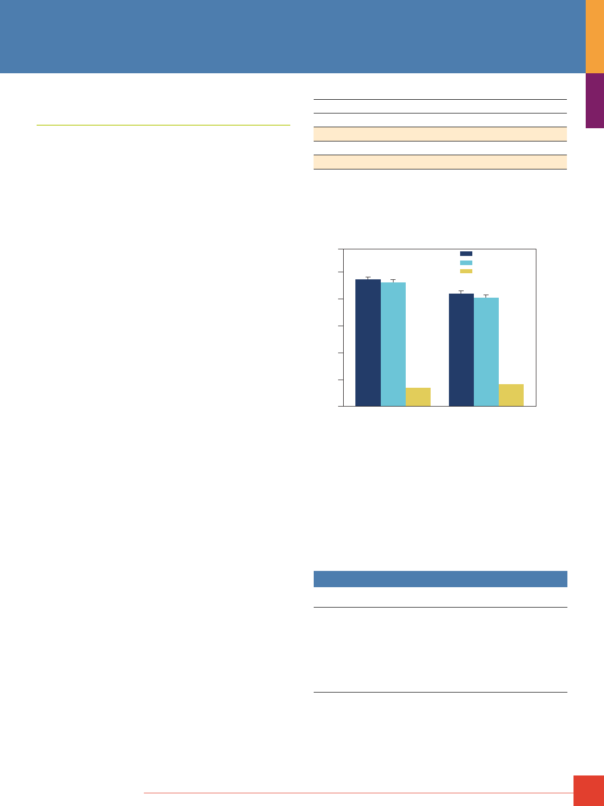
To order, call 800-874-3723 or 815-968-0747. Outside the United States, contact your local branch office or distributor.
51
Use of Titanium Dioxide for
Phosphopeptide Enrichment
Titanium dioxide enrichment of phosphopeptide samples is an
essential tool for comprehensive MS analysis of phosphopeptides.
The unique phosphopeptide binding properties of TiO
2
resin results
in different selectivity and preferential enrichment of multiply
phosphorylated peptides compared to immobilized chelated metal
ions (IMAC). TiO
2
resin typically enriches more phosphopeptides
than IMAC resins, and TiO
2
preferentially enriches monophos-
phorylated peptides.
TiO
2
resin typically binds phosphopeptides with a higher affinity
than IMAC and the preferential enrichment of singly phosphory-
lated peptides with TiO
2
is likely due to difficulty in eluting multiply
phosphorylated peptides. To enrich a more comprehensive set of
phosphopeptides, the flow-through and washes from IMAC resin
can be acidified and applied to TiO
2
resins and the resulting elu-
ates pooled for analysis. In addition, the number of phosphopep-
tides recovered can be significantly increased by pre-fractionation
of samples with strong cation exchange chromatography to
reduce sample complexity prior to enrichment with TiO
2
or IMAC
processing.
Thermo Scientific Pierce TiO
2
Phosphopeptide Enrichment and
Clean-up Kit
Selective enrichment and Clean-up of phosphopeptides in under
2 hours.
The Pierce TiO
2
Phosphopeptide Enrichment and Clean-up Kit
enables fast, selective enrichment of phosphorylated peptides.
The complete kit includes 24 titanium dioxide (TiO
2
) spin tips and
graphite spin columns with buffers to facilitate preparation of
enriched and desalted phosphopeptides for analysis by mass
spectrometry (MS).
Highlights:
• Convenient – spin-column format of TiO
2
and graphite columns
enable parallel processing and clean up of multiple samples in
less than 2 hours
• High capacity – each column enriches up to 300μg of
phosphopeptides
• Complementary – TiO
2
enriches a unique set of phosphopeptides
that complements the Pierce Fe-NTA IMAC Phosphopeptide
Enrichment Kit
• High selectivity – recover phosphopeptides with >90% selectivity
The Pierce TiO
2
Phosphopeptide Enrichment and Clean-up Kit
protocol includes stringent washing conditions that increase the
selectivity for phosphopeptides to >95%. Because some of the
salts in these stringent washes may still be present in the eluted
samples, the Pierce TiO
2
Phosphopeptide Enrichment and Clean-
up Kit includes graphite spin columns to desalt and concentrate
enriched phosphopeptides, resulting in more successful phos-
phopeptide analysis results. This kit is compatible with samples
digested in solution or after in-gel digestion with trypsin or other
MS grade proteases. Using the Pierce TiO
2
Phosphopeptide
Enrichment and Clean-up Kit and the Pierce Fe-NTA IMAC
Phosphopeptide Enrichment Kits will enable complementary sets
of phosphopeptides to be removed from complex samples.
Number of Phosphates per Pepti0de
Resin
1 2 3 4 5 6
Total
TiO
2
492 103 8 4 0 1 608
Fe-NTA IMAC
234 34 216 3 1 0 488
Overlap
155 0 1 0 0 0 156
Selective enrichment of singly and multiply phosphorylated phosphopeptides
with Thermo Scientific Pierce TiO
2
and FE-NTA IMAC resins. Average phos-
phopeptide enrichment results from duplicate experiments showing the number
of phosphopeptides containing one or more phosphate per peptide enriched
using either resin. Peptide spectrum summary results were exported from
Proteome Software Scaffold 3.0.
120
100
80
60
40
20
Total Phosphopeptides
Unique Phosphopeptides
% Acidic Residues
0
TiO2 Fe-NTA
Phosphopeptides (%)
961
240
998
238
TiO
2
selectively enriches more phosphopeptides than Fe-NTA IMAC. The num-
bers refer to the total and unique number of phosphopeptides enriched by each
method. The Y-axis is the percentage of phosphopeptides in the number of total
and unique peptides, or the percentage of acidic residues in enriched unique
phosphopeptides.
References:
Larsen M.R., et al. (2005). Highly selective enrichment of phosphorylated peptides from
peptide mixtures using titanium dioxide microcolumns. Mol Cell Proteomics. 4(7):873-86.
Carrascal, M., et al. (2008). Phosphorylation Analysis of Primary Human T Lymphocytes
Using Sequential IMAC and Titanium Oxide Enrichment. J. Proteome Res. 7(12):5167-5176.
Wilson-Grady, J.T., et al. (2008). Phosphoproteome Analysis of Fission Yeast. J. Proteome
Res. 7(3):1088-1097.
Larsen, M.R., et al. (2004). Improved detection of hydrophilic phosphopeptides using
graphite powder microcolumns and mass spectrometry: evidence for in vivo doubly phos-
phorylated dynamin I and dynamin III. Mol. Cell Proteomics. 3:456-465.
Ordering Information
Product #
Description
Pkg. Size
U.S.
Price
88301 Pierce TiO
2
Phosphopeptide Enrichment
and Clean-up Kit
Sufficient For: 24 phosphopeptide enrichment and
cleanup reactions.
Kit Contents: TiO
2
Spin Tips, 24 tips
Trifluoroacetic Acid, 1mL
90% Lactic Acid, 2mL
Pyrrolidine, 200μL
Centrifuge Column Adaptors, 24 adaptors
Pierce Graphite Spin Columns, 24 columns
24-Rxn Kit
$375
Phosphopeptide Enrichment
52
For more information, or to download product instructions, visit www.www.thermoscientific.com/pierce
Phosphopeptide Enrichment
Thermo Scientific Pierce Magnetic Titanium Dioxide
Phosphopeptide Enrichment Kit
TiO
2
magnetic particles for high throughput phosphopeptide isolation.
The Pierce Magnetic Titanium Dioxide Phosphopeptide Enrichment
Kit is for isolating phosphopeptides from complex biological sam-
ples using titanium dioxide-coated magnetic beads. The TiO
2
ligand
selectively binds peptides con-taining phosphorylated serine
(Ser), tyrosine (Tyr) or threonine (Thr), enabling phosphopeptide
enrichment from protease-digested samples. The isolated phos-
phopeptides are compatible for analysis downstream by mass
spectrometry (Table 2).
The high-performance, iron oxide, superparamagnetic particles
are validated and optimized for use with high-throughput
magnetic platforms, such as the Thermo Scientific KingFisher 96
and KingFisher
®
Flex Instruments. The beads also enable premium
performance for simple benchtop applications using an appropriate
magnetic stand.
Highlights:
• Complete MS-compatible Kits – include ready-to-use binding,
wash and elution buffers that are optimized for phosphopeptide
enrichment and downstream analysis by MALDI and ESI
mass spectrometry
• Optimized for HTS – procedure validated for processing 1 to 96
samples at a time; complete entire assay in about 15 minutes
using a KingFisher Flex Instrument
• Stable affinity ligand – titanium dioxide is specially coated as a
film on the magnetic particles
• Selective – affinity system is selective for phosphorylated Ser, Tyr
and Thr; exhibits minimal non-specific binding to acidic residues
• Sensitive – affinity provides more than 1000 times greater sensi-
tivity than traditional IMAC technologies; enables enrichment and
MS-measurement of less than 100fmol of phosphoprotein
Table 2. Phosphopeptide enrichment improves MS-identification of phospho-
proteins. Two milligrams of a tryptic digest prepared from periferal blood mono-
nuclear cells (lymphocytes) with and without phosphopeptide enrichment were
analyzed by MS. Enrichment was performed with the Thermo Scientific Pierce
Titanium Dioxide Phosphopeptide Enrichment Kit using the Thermo Scientific
KingFisher 96 Instrument. Samples were analyzed on a Thermo Scientific LTQ
Orbitrap Mass Spectrometer.
Enriched Non-Enriched
Total number of proteins identified 185 247
Total number of phosphoproteins identified 160 1
Total number of peptides identified 2347 2457
Total number of phosphopeptides identified 2009 7
Total number of unique
phosphopeptides identified 177 1
Relative enrichment for phosphopeptides (%) 86 0.3
Ordering Information
Product #
Description
Pkg. Size
U.S.
Price
88811 Pierce Magnetic Titanium Dioxide
Phosphopeptide Enrichment Kit
Includes: TiO
2
Magnetic Beads (20X)
Binding Buffer
Washing Buffer
Elution Buffer
Thermo-Fast 96 Robotic PCR Plate
(0.2mL wells)
Kit
1mL
100mL
25mL
3mL
2 plates
$427
88812 Pierce Magnetic Titanium Dioxide
Phosphopeptide Enrichment Kit, Trial Size
Includes: TiO
2
Magnetic Beads (20X)
Binding Buffer
Washing Buffer
Elution Buffer
Thermo-Fast 96 Robotic PCR Plate
(0.2mL wells)
Kit
0.25mL
100mL
25mL
3mL
2 plates
$240
To order, call 800-874-3723 or 815-968-0747. Outside the United States, contact your local branch office or distributor.
53
Thermo Scientific Pierce Fe-NTA Phosphopeptide Enrichment Kit
New Fe-NTA format optimized for capture and recovery
of phosphopeptides.
The new Pierce Fe-NTA Phosphopeptide Enrichment Kit enables
fast and efficient enrichment of phosphorylated peptides. These
spin columns are easy to use and require less than 1 hour to
process protein digests or strong cation-exchange peptide
fractions for analysis by mass spectrometry (MS).
Highlights:
• Convenient spin format for parallel processing of
multiple samples
• High-binding capacity resin for enriching up to 150μg
of phosphopeptides per column
• Excellent enrichment and recovery of phosphopeptides
Protein phosphorylation is essential to biological functions,
including cell signaling, growth, differentiation, division and
programmed cell death. Over 500 protein kinases catalyze
phosphorylation of specific targets, primarily on serine, threonine,
and tyrosine residues.
Mass spectrometry is increasingly being used to identify and
quantify phosphorylation changes; however, phosphoprotein
and phosphopeptide analysis by MS is limited by many factors,
including digestion efficiency, low stoiochiometry, low abundance,
hydrophilicity, poor ionization and poor fragmentation. As a result,
phosphopeptide enrichment is essential to successful MS analy-
sis. The new Pierce Fe-NTA Phosphopeptide Enrichment Kit is
compatible with our lysis, reduction, alkylation, and digestion
reagents and with Thermo Scientific Pierce Graphite Spin Columns
to provide a complete workflow for phosphopeptide enrichment.
To assess phosphopeptide enrichment from lysates, cultured
U2-OS cells arrested with nocodazole (100ng/mL, 25 hours)
were lysed with 6M urea in 50mM Tris, pH 8.0 containing Thermo
Scientific Halt Protease and Phosphatase Inhibitor Cocktail
(Product # 78440). Protein concentration was determined with
Thermo Scientific Pierce 660nm Protein Assay (Product # 22660).
Proteins were reduced with Thermo Scientific Bond-Breaker
TCEP Solution, Neutral pH (Product # 77720), alkylated with single-
use iodoacetamide, (Product # 90034) digested overnight with
MS-grade trypsin (Product # 90055), and desalted with Thermo
Scientific HyperSep-C18 Cartridges (Product # 60108-305). An
equivalent of 200μg of peptides were dried and dissolved in 5%
acetic acid or Sigma Phos-Select
™
Buffer. Phosphopeptides were
enriched with Pierce Fe-NTA Phosphopeptide Enrichment Kit or
Sigma Phos-Select Reagents and then desalted and concentrated
with Pierce Graphite Spin Columns (Product # 88302) according
to instructions.
Enriched phosphopeptide samples were analyzed by LC-MS/MS.
A NanoLC
™
-2D HPLC (Eksigent) with a ProteoPep II C18 Column
(75μm ID x 20cm, New Objective) was used to separate peptides
using a 4-40% gradient of solvents (A: water, 0.1% formic acid;
B: acetonitrile, 0.1% formic acid) at 250nL per minute for 60
minutes. Peptides were identified with a Thermo Scientific LTQ
Orbitrap XL ETD Mass Spectrometer using a top four experiment
consisting of high-resolution MS followed by acquisition of four
MS/MS spectra using the CID mode of fragmentation. LC-MS/
MS data were interpreted with Mascot 2.2 (Matrix Science) and
Scaffold 2.6 (Proteome Software).
To achieve robust MS results, enrichment of phosphopeptide
samples is essential because of low stoichiometry and abundance
and poor ionization relative to nonphosphorylated peptides. We
have developed an efficient means to enrich phosphopeptides
from complex samples. The new Thermo Scientific Pierce
Fe-NTA Spin Columns effectively capture, enrich, and recover
phosphopeptides. These columns enrich a higher percentage of
phosphopeptides than other resins and with an overall higher
number of total and unique phosphopeptides (Figure 8
and Table 3).
0
10
20
30
40
50
60
70
Phosphopeptides (%)
Thermo Scientific Pierce Fe-NTA Kit Sigma Phos-Select
Reagent
Total Phosphopeptides
862
430
90
178
Unique Phosphopeptides
Figure 8. Our kit enriched a greater percentage of total and unique phospho-
peptides from U2-OS cell lysate. Numbers refer to the total and unique num-
ber of phosphopeptides identified in each condition. A summary of
results is listed in Table 3.
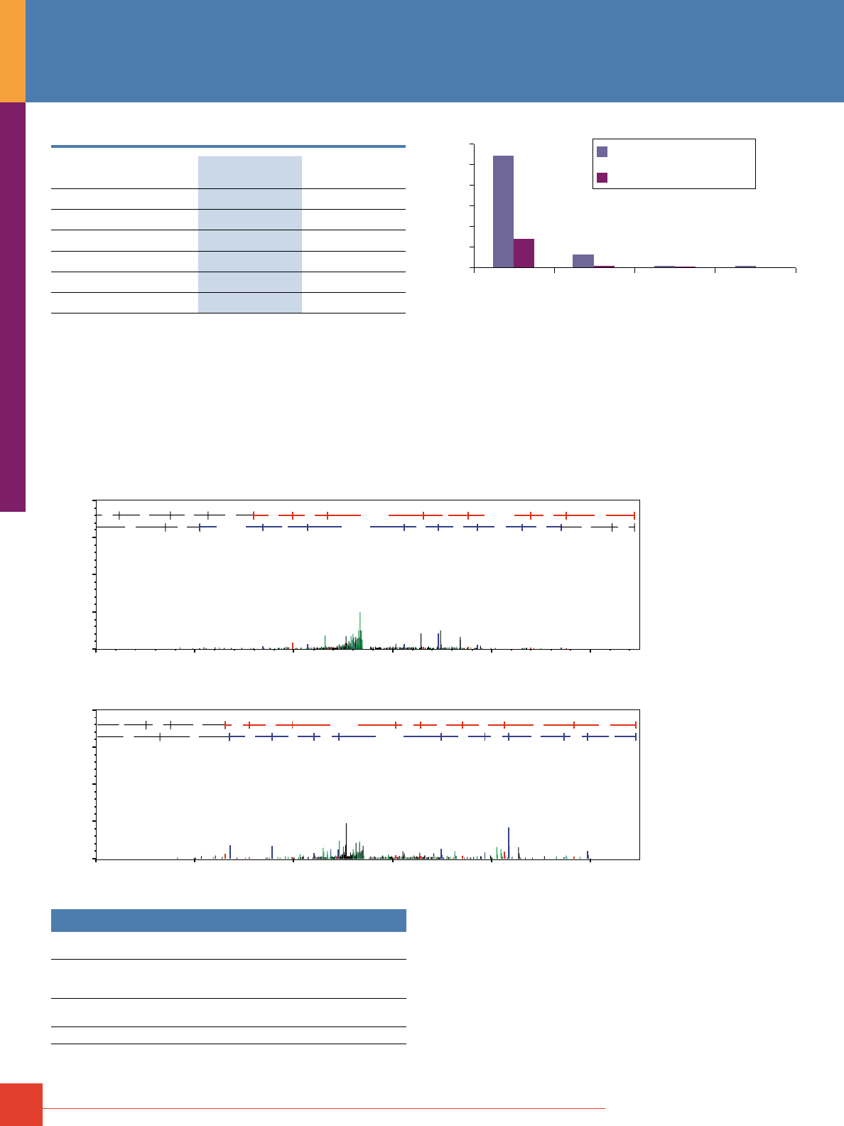
54
For more information, or to download product instructions, visit www.www.thermoscientific.com/pierce
Table 3. Average phosphopeptide enrichment results from duplicate experiments.
§
Thermo Scientific
Pierce Fe-NTA
Sigma Phos-Select
Total phosphopeptides 862 430
Total peptides 1,753 1,665
Total unique peptides 393 395
Total unique phosphopeptides 178 90
Total phosphopeptides (%) 53 31
Unique phosphopeptides (%) 50 27.5
§ Peptide summary results were exported from Scaffold and analyzed and summarized
with Microsoft Excel
®
and Access
®
.
Multiple phosphorylated amino acids within a peptide contribute
to the complexity of phosphopeptide analysis. Pierce Fe-NTA Spin
Columns enrich peptides with three or more phosphorylated sites
and significantly outperform other commercially available columns
(Figure 9). Phosphopeptide enrichment greatly reduces sample
complexity and enables effective identification and characteriza-
tion of phosphorylated peptides by MS (Figure 10).
0
20
40
60
80
100
120
Phosphopeptides (%)
Single Double
Phosphates per Peptide
Triple ≥4
Thermo Scientific Pierce Fe-NTA Kit
110
29
13
2
12 1 0
Sigma Phos-Select Reagent
Figure 9. The Thermo Scientific Pierce Fe-NTA Phosphopeptide Enrichment Kit
effectively captures phosphopeptides with multiple phosphates.
The Pierce Fe-NTA Phosphopeptide Enrichment Kit contains
detailed instructions and all necessary components to load, wash
and elute phosphopeptides within an hour. This kit is compatible
with samples digested in solution or after in-gel digestion using the
Thermo Scientific In-gel Tryptic Digestion Kit (Product # 89871).
a11+2H+1
y2 b4
y3
b5
y4
b6
y5
b7
y6
b8
y7
y8
b9 y9
b10
G E P N
V S Y+80 I C+57 S R
R S
C+57 I Y+80 S V N P
E G
m/z
0%
25%
50%
75%
100%
A.
B.
Relative Intensity
0 250 500 750 1000 1250
m/z
0%
25%
50%
75%
100%
Relative Intensity
0 250 500 750 1000 1250
GSK3A_HUMAN
Glycogen Synthase Kinase-3 alpha
(R)GEPNVSyICSR(Y)
CDC2_HUMAN
Cell division control protein 2 homolog
(K)IGEGTyGVVYK(G)
b3
y2
b4
y3
b5
y4
y5
b6
b7
y6
b8
y7
b9
y8
y9
b10
y10
I G E
G T Y+80 G V V Y K
K Y
V V G Y+80 T G E G I
Ordering Information
Product #
Description
Pkg. Size
U.S.
Price
88300 Pierce Fe-NTA Phosphopeptide
Enrichment Kit
Sufficient for 30 samples
Kit
$279
Related Products
88302 Pierce Graphite Spin Columns
30 columns
$105
Figure 10. Example MS/MS
spectra from enriched
phosphopeptides. Lowercase
letters indicate the position
of tyrosine phosphorylation
in enriched peptides.
Panel A: Glycogen synthase-3
alpha. Panel B: cdc2/cyclin
dependent kinase 1.
Phosphopeptide Enrichment
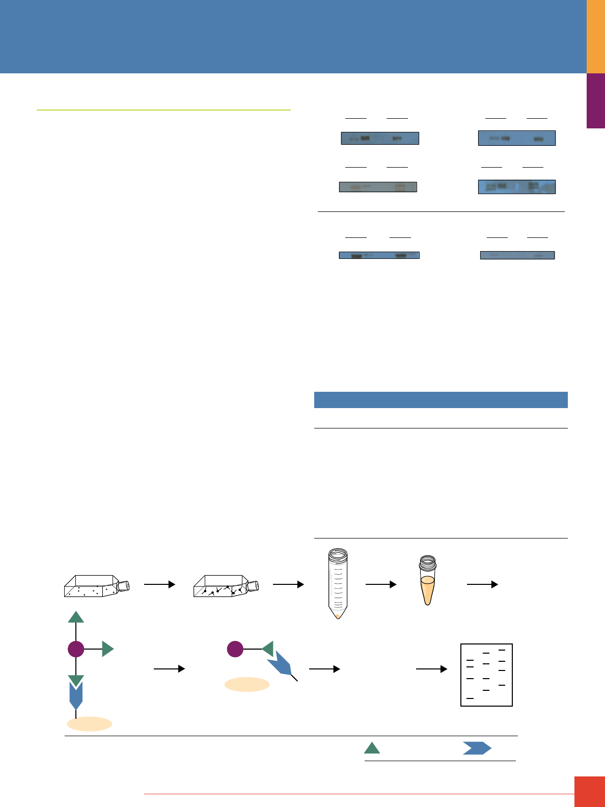
To order, call 800-874-3723 or 815-968-0747. Outside the United States, contact your local branch office or distributor.
55
Cell Surface Protein Isolation Kit
Convenient biotinylation and isolation of cell surface proteins
for Western blot analysis.
The Thermo Scientific Pierce Cell Surface Protein Isolation Kit
is a complete kit for the convenient biotinylation and isolation
of mammalian cell surface proteins, specifically targeting cell
surface proteins to the exclusion of intracellular proteins. The
kit efficiently labels proteins with accessible lysine residues and
sufficient extracellular exposure.
The isolation procedure uses a cell-impermeable, cleavable
biotinylation reagent (Sulfo-NHS-SS-Biotin) to label surface
proteins at exposed primary amines. Cells are then harvested and
lysed, and the labeled surface proteins are affinity-purified using
Thermo Scientific NeutrAvidin Agarose Resin. The isolated cell
surface proteins contain a small, nonreactive tag of the originally
labeled primary amines but are no longer biotinylated (biotin
remains bound to the resin).
Highlights:
• Isolates cell surface proteins – reduces complexity of total
cellular protein
• Efficiently recovers labeled proteins – cleavable biotin allows for
nearly 100% recovery of isolated cell surface proteins
• Convenience – all reagents are provided in one kit, along with
complete instructions for labeling, cell lysis and purification of
cell surface membrane proteins
• Western blotting applications – proteins recovered in SDS-PAGE
buffer are loaded directly onto polyacrylamide gels
• Robust system – protocol designed for diverse cell lines,
including NIH 3T3, HeLa, C6 and A431
A. Cell Surface Proteins
EGFR
IGF-1Rβ
Hsp90
Calnexin
Integrin α5
Integrin β1
R F E R F E
R F E R F E
R F E R F E R F E R F E
R F E R F E
R F E R F E
B. Intracellular Proteins
+ – + –
+ –
+ –
+ –
+ –
Protein isolation is specific to cell surface proteins. Panels are Western blot
results for known cell surface proteins (Panel A) and intracellular proteins
(Panel B) from HeLa cells tested with the Cell Surface Protein Isolation Kit.
Plus symbol (+) denotes results for cells treated with the Sulfo-NHS-SS-Biotin
reagent; minus symbol (-) denotes results for cells that were not treated with
the biotin reagent but were otherwise carried through the kit procedure.
Lanes are no-sample resin-control (R), flow-through (F) and eluted (E) frac-
tions. Presence of target cell surface proteins in the plus-E and minus-F
conditions indicate successful isolation with the kit. Presence of intracellular
proteins in F condition of both plus and minus conditions indicates that the
labeling and purification procedure is specific to cell surface proteins.
Ordering Information
Product # Description Pkg. Size
U.S.
Price
89881 Cell Surface Protein Isolation Kit
Sufficient reagents and accessories for eight
experiments, each involving four T75 flasks of
confluent cells.
Includes: EZ-Link
®
Sulfo-NHS-SS-Biotin
Quenching Solution
Lysis Buffer
NeutrAvidin Agarose
Wash Buffer
Dithiothreitol (DTT)
BupH Phosphate Buffered Saline
BupH Tris Buffered Saline
Spin Columns and Accessories
Kit
8 x 12mg vials
16mL
4.5mL
2.25mL
34mL
8 x 7.7mg
microtubes
2 packs
1 pack
$348
Biotinylate cells
30 minutes at 4˚C
Transfer cell pellet
to 1.5 ml tube
Lyse cells
30 minutes on ice
Isolate biotinylated proteins
on NeutrAvidin
®
Agarose Resin
Perform
electrophoresis
or other application
Wash gel then elute with SDS-PAGE
sample buffer + 50 mM DTT
Harvest Cells
Quench Reaction
gel
B
SH
+
protein – SH
N
N
gel
N
B
S-S-protein
N
N
Biotin
NeutrAvidin
Biotin-Binding Protein
1D gel
B
45
40
35
30
25
20
15
10
7.5
5.0
Protocol summary for the Thermo Scientific Pierce Cell Surface Protein Isolation Kit.
Protein Enrichment
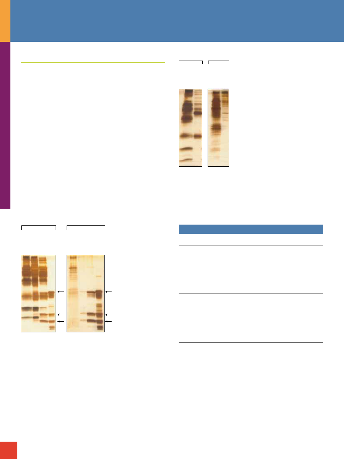
56
For more information, or to download product instructions, visit www.www.thermoscientific.com/pierce
Glycoprotein Isolation Kits
Isolate glycoproteins from complex protein mixtures.
Two lectin-based Thermo Scientific Glycoprotein Isolation Kits,
concanavalin A (ConA) and wheat germ agglutinin (WGA), allow
isolation of glycoproteins from complex protein mixtures, including
serum, tissue and cultured cell lysates, thus enabling downstream
analysis. ConA lectin recognizes a-linked mannose and terminal
glucose residues, while WGA lectin selectively binds to N-acetyl
glucosamine (GlcNAc) groups and to sialic acid.
Highlights:
• High recovery – equivalent or greater glycoprotein recovery vs.
competitor kits and lectin resins
• Fast – glycoprotein purification in less than one hour
• Versatile – isolate glycoproteins from various sample types;
e.g., human serum and cell lysate
• Robust – lectin does not leach from resin when
processing sample
• Convenient – complete kit contains lectin resins and spin
columns with all necessary reagents
• Compatible with Bradford-based protein assays – dialysis or
protein precipitation of recovered glycoproteins is not required
before protein assay
Thermo Scientific
Thermo Scientific
Supplier A
Supplier A
Supplier G
ConA
Supplier G
ConA
B.
A.
Human Serum CHO Lysate
Glycoprotein isolation from human serum and cell lysate: performance
comparison of kits using ConA resin. Human serum and CHO lysate samples
were processed with the Thermo Scientific Glycoprotein Isolation Kit, ConA
and with other commercially available ConA resins. An equivalent amount of
total protein was applied to each resin. Eluted glycoprotein fractions were
compared with ConA Resin boiled in SDS-PAGE Buffer to release lectins. All
fractions were normalized by volume and resolved on 8-16% polyacrylamide
gels. Gels were silver-stained. A. Eluted glycoprotein fractions from applied
human serum and B. eluted glycoprotein fractions from applied CHO lysate.
Arrows identify protein bands that result from ConA leaching from the resin
during the elution process.
Competitor A
Supplier A
Supplier A
B.
A.
CHO LysateHuman Serum
Thermo Scientific
Thermo Scientific
Glycoprotein isolation from human serum and cell lysate: performance
comparison of kits using WGA resin. Human serum and CHO lysate samples
were processed with the Thermo Scientific Glycoprotein Isolation Kit, WGA
and with other commercially available WGA resins. An equivalent amount of
total protein was applied to each resin. Eluted glycoprotein fractions were
normalized by volume and resolved on 8-16% polyacrylamide gels. A. Eluted
glycoprotein fraction from applied human serum and B. eluted glycoprotein
fraction from applied CHO lysate.
Ordering Information
Product #
Description
Pkg. Size
U.S.
Price
89804
Glycoprotein Isolation Kit, ConA
Sufficient reagents to isolate glycoproteins with
strong affinity for ConA from 10 samples of up to 640µL
(1-1.5mg total protein) each.
Includes: ConA Lectin Resin, 1.1mL resin supplied as
a 50% slurry
Binding/Wash Buffer, 5X Stock Solution
Elution Buffer
Column Accessory Pack, 10 Spin Columns
with Caps and 20 Collection Tubes
Kit
6.5mL
5mL
$223
89805 Glycoprotein Isolation Kit, WGA
Sufficient reagents to isolate glycoproteins with strong
affinity for WGA from 10 samples of up to 640µL
(1-1.5mg total protein) each.
Includes: WGA Lectin Resin, 1.1mL resin supplied as a
50% slurry
Binding/Wash Buffer, 5X Stock Solution
Elution Buffer
Column Accessory Pack, 10 Spin Columns
with Caps and 20 Collection Tubes
Kit
6.5mL
5mL
$282
Protein Enrichment
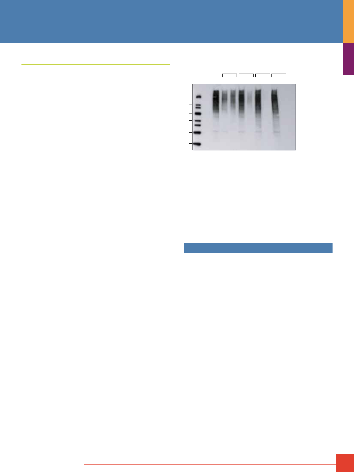
To order, call 800-874-3723 or 815-968-0747. Outside the United States, contact your local branch office or distributor.
57
Ubiquitin Enrichment Kit
Recover ubiquitin modified protein in <45 minutes.
The ubiquitin proteasome pathway is the principal non-lysosomal
pathway that controls the proteolysis of proteins. This pathway
is significantly involved in a variety of cellular processes, including
DNA repair, transcriptional regulation, signal transduction, cell
metabolism and morphogenesis. Differences in total ubiquitination
or the ubiquitination of specific proteins affect numerous pathological
conditions, including malignancies, certain genetic diseases and
neurodegenerative diseases.
1
The Thermo Scientific Ubiquitin Enrichment Kit isolates
polyubiquitin protein conjugates from cultured cells and tissue
samples. The enriched fraction is analyzed to determine the
amount of general ubiquitin conjugates present or to identify a
specific protein by Western blotting. The assay protocol is fast,
straightforward and allows isolation of polyubiquitinated proteins
and the fractionation of monoubiquitinated species in the resin
flow-through. The Ubiquitin Enrichment Kit outperforms other
suppliers’ kits and provides a clean and specific preparation of
proteins when compared to a control resin.
Highlights:
• Fast – less than 45 minutes hands-on time
• Complete – includes all reagents needed for ubiquitin-modified
protein enrichment from cultured cells and tissue samples,
including spin columns and ubiquitin antibody
• Flexible – sample incubation from 2 hours to overnight allows
assay to be completed in several hours or in less than 30 minutes
after overnight incubation
• Robust – compatible with all Thermo Scientific Cell Lysis
Solutions and standard RIPA formulations
• Multiple-sample format – easily processes 1-15 samples
concurrently
20
40
50
60
80
100
120
220
Thermo
Scientific
Kit
Supplier
C
Ab-based
Method
GSH
Resin
H F E F E F E F E
kDa
The Thermo Scientific Ubiquitin Enrichment Kit recovers more ubiquitin-
modified proteins than any other method. Epoxomicin-treated HeLa cell
lysates (EHeLa, 150μg) were enriched. After elution, all samples were
normalized to the initial load (EHeLa load). The flow-through and elution
obtained using the Thermo Scientific Ubiquitin Enrichment Kit are shown first.
The results using another supplier’s enrichment kit are shown for comparison
(Supplier C, manufacturer’s instructions for this kit were followed). Additionally,
the results obtained using an anti-ubiquitin monoclonal antibody-based
enrichment scheme (antibody-based) and a negative control resin (GSH resin)
are shown. The elution from each resin shows the amount of ubiquitin-modified
protein that was captured using that method.
Reference
1. Ciechanover, A. (1998). The ubiquitin-proteasome pathway: on protein death and cell life.
EMBO J. 17(24), 7151-1760.
Ordering Information
Product # Description Pkg. Size
U.S.
Price
89899
Ubiquitin Enrichment Kit
Contains sufficient materials for enriching up to
15 lysate samples containing ~0.15mg total protein
per sample.
Pack 1
Polyubiquitin Positive Control (1,000X), 50μL, 2mg/mL
Anti-ubiquitin Antibody, 50μL rabbit antiserum
Pack 2
Polyubiquitin Affinity Resin, 300μL, supplied as a
25% slurry
Binding Capacity: ~1μg per 20μL of slurry
BupH Tris Buffered Saline Pack, 1 ea., makes
500mL of 0.025M Tris, 0.15M NaCl; pH 7.2
Spin Columns and Accessories, 18 columns with top
and bottom caps
Kit
$399
58
For more information, or to download product instructions, visit www.thermoscientific.com/pierce
Antibody Purification Overview
Antibodies specific for an antigen of interest are one of the most
useful and important tools that biology researchers can possess.
The production and use of specific antibodies as detection
probes and purification ligands (i.e., immunotechnology) has
revolutionized bioresearch and diagnostic technologies. Animals
immunized with prepared antigens will produce specific antibodies
against the antigen. When purified from serum or hybridoma cell
lines that are prepared from tissue of the immunized animal, the
antibody can be used directly (or after labeling with enzyme or
fluorescent tags) to probe the specific antigen in Western blotting,
ELISA or a variety of other applications. Antibodies are most
commonly purified by one of two affinity purification methods:
general immunoglobulin purification (pages 58-65) or specific
antibody purification (see pages 26-35). See also Table 1.
General Purification of Immunoglobulins
Because antibodies have predictable structure, including relatively
invariant domains, it has been possible to identify certain protein
ligands that are capable of binding generally to antibodies,
regardless of the antibody’s specificity to antigen. Protein A,
Protein G and Protein L are three bacterial proteins whose
antibody-binding properties have been well characterized. These
proteins have been produced recombinantly and used routinely for
affinity purification of key antibody types from a variety of species.
A genetically engineered recombinant form of Protein A and G,
called Protein A/G, is also available. These antibody-binding
proteins are available immobilized to beaded agarose resin,
Thermo Scientific UltraLink Biosupport and coated onto microplates.
Proteins A, G, A/G and L bind to antibodies at sites other than the
antigen-binding domain. Therefore, these proteins can be used in
purification schemes such as immunoprecipitation.
Proteins A, G, A/G and L have unique properties, which make each
one suitable for different types of antibody targets (e.g., antibody
subclass or animal species). It is important to realize that use of
Protein A, G or L results in purification of general immunoglobulin
from a crude sample. Depending on the sample source, antigen-
specific antibody may account for only a small portion of the total
immunoglobulin in the sample. For example, generally only 2-5%
of total IgG in mouse serum is specific for the antigen used to
immunize the animal.
Table 1. Antibody Purification Methods.
Purification Type Description Available Thermo Scientific Support
General Negative selection Removal of all non-immunoglobulin proteins from a
serum sample
Melon Gel
IgG enrichment Immobilized globulin binding proteins to selectively
remove IgG from a serum sample
Immobilized Protein A
Immobilized Protein G
Immobilized Protein A/G
Immobilized Protein L
IgG enrichment Thiophilic adsorption Thiophilic Adsorbent
IgM enrichment Use Mannan binding protein to selectively isolate IgM Immobilized Mannan Binding Protein
IgA enrichment Use Jacalin, a D-galactose binding lectin, to selectively
isolate IgA
Immobilized Jacalin
IgY enrichment Delipidation and precipitation from egg yolks IgY Precipitation Reagent
Specific
(see page 26)
Affinity purification Create an affinity column with the antigen used to
immunize the animal.
NHS-ester Activated Agarose - for immobilizing antigens via
primary amines
AminoLink Plus Agarose - for immobilizing antigens via primary
amines
SulfoLink Resin - for immobilizing antigens via reduced sulfhydryls
CarboxyLink Resin - for immobilizing antigens via carboxylic acids
Antibody Purification
To order, call 800-874-3723 or 815-968-0747. Outside the United States, contact your local branch office or distributor.
59
w = weak binding, m = medium binding, s = strong binding, nb = no binding, – means information not available
* Data represent a summary of binding properties reported in the literature. Inevitably some discrepancies exist among reported values as a result of differences in binding
buffer conditions and form of the proteins used.
†
Binding will occur only if the appropriate kappa light chains are present. Antibodies lambda light chains will not bind, regardless of their class and subclass.
Protein A Protein G Protein A/G Protein L
†
Thiophilic
Adsorbent
Human IgG s s s s m
Mouse IgG s s s s s
Rabbit IgG s s s w m
Goat IgG w s s nb s
Rat IgG w m m s s
Sheep IgG w s s nb s
Cow IgG w s s nb s
Guinea Pig IgG s w s – s
Hamster IgG m m m s –
Pig IgG s w s s s
Horse IgG w s s – s
Donkey IgG m s s – –
Dog IgG s w s – s
Cat IgG s w s – s
Monkey IgG
(Rhesus)
s s s – s
Chicken IgY nb nb nb nb m
Human IgM w nb w s m
Human IgE m nb m s –
Human IgD nb nb nb s –
Human IgA w nb w s m
Human IgA
1
w nb w s m
Human IgA
2
w nb w s m
Human IgG
1
s s s s m
Protein A Protein G Protein A/G Protein L
†
Thiophilic
Adsorbent
Human IgG
2
s s s s m
Human IgG
3
w s s s m
Human IgG
4
s s s s m
Human Fab w w w s m
Human ScFv w nb w s m
Mouse IgG
1
w m m s s
Mouse IgG
2a
s s s s s
Mouse IgG
2b
s s s s s
Mouse IgG
3
s s s s s
Rat IgG
1
w m m s s
Rat IgG
2a
nb s s s s
Rat IgG
2b
nb w w s s
Rat IgG
2c
s s s s s
Cow IgG
1
w s s nb s
Cow IgG
2
s s s nb s
Sheep IgG
1
w s s nb s
Sheep IgG
2
s s s nb s
Goat IgG
1
w s s nb s
Goat IgG
2
s s s nb s
Horse IgG(ab) w nb w – s
Horse IgG(c) w nb w – s
Horse IgG(T) nb s s – s
Mouse IgM nb nb nb s m
Immobilized Protein L, Protein A, Protein G
and Protein A/G
We offer these popular antibody-binding proteins immobilized on
several different resins, beads and plates for use in immunoaffinity
purification techniques. All four proteins (A, G, A/G and L) are
available as purified recombinants immobilized to crosslinked 6%
beaded agarose. This is the traditional format historically used for
small-scale column purification and immunoprecipitation methods.
Our agarose resins differ from those typically offered by other
suppliers in that our immobilization method is more stable and
results in less nonspecific binding. We also offer “Plus” versions
of the Protein A, G, A/G and L agarose resins, which contain twice
the amount of protein per milliliter of resin and provide for nearly
twice the antibody binding capacity.
Protein A, G, A/G and L are also available immobilized to UltraLink
®
Biosupport, an extremely durable, polyacrylamide-based resin with
very low nonspecific binding characteristics. The UltraLink Format
is a perfect support for working with large volume samples in large-
scale purification methods requiring fast flow and high pressure.
The interaction between the various proteins and IgG is not
equivalent for all species or all antibody subclasses. The tables
on the following page will help you decide which affinity protein is
best for your application (Tables 2 and 3).
Table 2. Characteristics of immunoglobulin-binding proteins.
Recombinant
Protein L
Native
Protein A
Recombinant
Protein A
Recombinant
Protein G
Recombinant
Protein A/G
Production Source E. coli S. aureus E. coli E. coli E. coli
Molecular Weight 35,800 46,700 44,600 21,600 50,460
Number of Binding Sites for IgG 4 4 4 2 6
Albumin-Binding Site No No No No No
Optimal Binding pH 7.5 8.2 8.2 5 5-8.2
Binds to V
L
K F
C
F
C
F
C
F
C
Table 3. Binding characteristics of immunoglobulin-binding proteins and Thermo Scientific Thiophilic Adsorbent.*
60
For more information, or to download product instructions, visit www.thermoscientific.com/pierce
Protein A
Protein A Beads – Quick Reference
Protein A Native protein purified from
Staphylococcus aureus
(46.7kDa; four IgG-binding sites)
Specificity
(Table 3)
Best for polyclonal IgG from rabbit, pig, dog, cat serum;
poor for Mouse IgG1, human IgG3, rat, goat, cow
Supports
Offered
Crosslinked 6% beaded agarose
UltraLink Biosupport
Trisacryl GF-2000 Resin
Magnetic particles (1-4μm)
Package
Formats
Resin slurries (3 sizes)
Chromatography Cartridges (2 sizes)
Packed Spin Columns (3 sizes)
NAb
™
IgG Purification Kits (2 sizes)
Storage 4°C, do not freeze
Capacity Protein A: 12-19mg human IgG/mL resin
Protein A Plus: >35mg human IgG/mL resin;
16-17mg mouse IgG/mL resin
Protein A Trisacryl Resin: >15mg human IgG/mL resin
Protein A UltraLink Resin: >16mg of human IgG/mL resin
Protein A Plus UltraLink Resin: >30mg human IgG/mL resin
Protein A MagnaBind Beads: >0.2mg rabbit IgG/mL beads
Protein A Coated Plates: ~4pmol rabbit IgG/well
Recombinant Protein A: ≥12mg human IgG/mL resin using the
IgG Buffer System
Ordering Information
Product #
Description
Pkg. Size
U.S.
Price
20333 Protein A Agarose
5mL
$ 337
20334 Protein A Agarose
25mL
$1050
20356 Protein A Columns
5 x 1mL
$ 385
44667 Protein A IgG Purification Kit
Kit
$ 515
20338 Protein A Trisacryl Resin
5mL
$ 350
53139 Protein A UltraLink Resin
5mL
$ 340
22810 Protein A Plus Agarose
1mL
$ 120
22811 Protein A Plus Agarose
5mL
$ 400
22812 Protein A Plus Agarose
25mL
$1350
89924 Chromatography Cartridges, Protein A
2 x 1mL
$ 170
89925 Chromatography Cartridge, Protein A
1 x 5mL
$ 335
89952 NAb Protein A Plus Spin Columns
10 x 0.2mL
$ 222
89956 NAb Protein A Plus Spin Columns
5 x 1mL
$ 425
89960 NAb Protein A Plus Spin Column
1 x 5mL
$ 400
89948 NAb Protein A Plus Spin Purification Kit
Kit
$ 270
89978 NAb Protein A Plus Spin Purification Kit
Kit
$ 265
53142 Protein A UltraLink Plus Resin
5mL
$ 399
45202 Protein A Spin Plate
1 plate
$ 188
21348 MagnaBind Protein A Beads
5mL
$ 325
15130 Protein A, Clear 96-Well Plates
5 plates
$ 160
15132 Protein A, Clear 8-Well Strip Plates
5 plates
$ 178
15154 Protein A, White 96-Well Plates
5 plates
$ 178
15155 Protein A, Black 96-Well Plates
5 plates
$ 178
20365 Recombinant Protein A Agarose
5mL
$ 275
20366 Recombinant Protein A Agarose
25mL
$1001
Protein G
Protein G Beads – Quick Reference
Protein G Recombinant protein expressed in E. coli
(21.6kDa; two IgG binding sites)
Specificity
(Table 3)
Best for IgG from mouse, human, cow, goat and sheep;
poor for guinea pig, pig, dog and cat
Supports
Offered
Crosslinked 6% beaded agarose
UltraLink Biosupport
Magnetic particles (1-4μm)
Package
Formats
Resin slurries (3 sizes)
Chromatography Cartridges (2 sizes)
Packed Spin Columns (3 sizes)
Nab IgG Purification Kits (2 sizes)
Storage 4°C, do not freeze
Capacity Protein G: 11-15mg human IgG/mL resin
Protein G UltraLink Resin: >20mg of human IgG/mL resin
Protein G Plus: >20mg human IgG/mL resin
Protein G UltraLink Resin: >20mg of human IgG/mL resin
Protein G Plus UltraLink Resin: >25mg human IgG/mL resin
MagnaBind Protein G Beads: >0.2mg rabbit IgG/mL beads
Protein G Coated Plates: ~2pmol rabbit IgG/well
Protein G Plus: >20mg human IgG/mL resin
Protein G Plus UltraLink Resin: >25mg human IgG/mL resin
Ordering Information
Product #
Description
Pkg. Size
U.S.
Price
20398 Protein G Agarose
2mL
$ 212
20399 Protein G Agarose
10mL
$ 760
20397 Protein G Agarose
25mL
$1654
89926 Pierce Chromatography Cartridges, Protein G
2 x 1mL
$ 190
89927 Pierce Chromatography Cartridge, Protein G
1 x 5mL
$ 390
89953 NAb Protein G Spin Columns
10 x 0.2mL
$ 251
89957 NAb Protein G Spin Columns
5 x 1mL
$ 455
89961 NAb Protein G Spin Columns
1 x 5mL
$ 425
89949 NAb Protein G Spin Purification Kit
Kit
$ 310
89979 NAb Protein G Spin Purification Kit
Kit
$ 368
21193 Pierce Recombinant Protein G
5mg
$ 300
53125 Protein G UltraLink Resin
2mL
$ 270
53126 Protein G UltraLink Resin
10mL
$ 910
53127 Protein G UltraLink Columns
2 x 2mL
$ 540
45204 Protein G Spin Plate
1 plate
$ 209
22851 Protein G Plus Agarose
2mL
$ 260
22852 Protein G Plus Agarose
10mL
$ 895
53128 Protein G Plus UltraLink Resin
2mL
$ 335
21349 MagnaBind Protein G Beads
5mL
$ 325
15131 Protein G, Clear 96-Well Plates
5 plates
$ 178
15133 Protein G, Clear 8-Well Strip Plates
5 plates
$ 195
15156 Protein G, White 96-Well Plates
5 plates
$ 195
15157 Protein G, Black 96-Well Plates
5 plates
$ 195
Antibody Purification
To order, call 800-874-3723 or 815-968-0747. Outside the United States, contact your local branch office or distributor.
61
Protein A/G
Protein A/G Beads – Quick Reference
Protein A/G Recombinant protein expressed in E. coli
(50.5kDa; six IgG-binding sites)
Specificity
(Table 3)
Best for polyclonal IgG from many species; poor for individual
subclasses that have highest affinity for Protein A or Protein G.
Supports
Offered
Crosslinked 6% beaded agarose
UltraLink Biosupport
Package
Formats
Resin slurries (3 sizes)
Chromatography Cartridges (2 sizes)
Packed Spin Columns (3 sizes)
NAb IgG Purification Kits (2 sizes)
Storage 4°C, do not freeze
Capacity Protein A/G: >7mg human IgG/mL resin
Protein A/G UltraLink Resin: >20mg human IgG/mL resin
Protein A/G Plus: >50mg human IgG/mL resin
Protein A/G Plus on UltraLink Support: >28mg human IgG/mL resin
Protein A/G Coated Plates: ~5pmol rabbit IgG/well
Ordering Information
Product #
Description
Pkg. Size
U.S.
Price
15138 Protein A/G, Clear 96-Well Plates
5 plates
$ 196
20421 Protein A/G Agarose
3mL
$ 315
20422 Protein A/G Agarose
15mL
$1150
89930 Pierce Chromatography Cartridges, Protein A/G
2 x 1mL
$ 299
89931 Pierce Chromatography Cartridge, Protein A/G
1 x 5mL
$ 399
89954 NAb Protein A/G Spin Columns
10 x 0.2mL
$ 260
89958 NAb Protein A/G Spin Columns
5 x 1mL
$ 460
89962 NAb Protein A/G Spin Column
1 x 5mL
$ 435
89950 NAb Protein A/G Spin Purification Kit
Kit
$ 336
89980 NAb Protein A/G Spin Purification Kit
Kit
$ 375
53132 Protein A/G UltraLink Resin
2mL
$ 270
53133 Protein A/G UltraLink Resin
10mL
$ 910
21186 Pierce Recombinant Protein A/G
5mg
$ 303
20423 Protein A/G Plus Agarose
2mL
$ 340
53135 Protein A/G Plus on UltraLink Support
2mL
$ 367
Protein L
Protein L Beads – Quick Reference
Protein L Recombinant protein expressed in E. coli (35.8kDa;
four IgG-binding sites)
Specificity
(Table 3)
Best for human or mouse monoclonal antibodies known to
have appropriate kappa light chains; poor for general-purpose
(polyclonal) IgG purification
Supports
Offered
Crosslinked 6% beaded agarose
Package
Formats
Resin slurries (2 sizes)
Chromatography Cartridges (2 sizes)
Packed Spin Columns (3 sizes)
NAb IgG Purification Kits (2 sizes)
Storage 4°C, do not freeze
Capacity Protein L: 5-10mg human IgG/mL resin
Protein L Plus: >10-20mg human IgG/mL resin
Ordering Information
Product #
Description
Pkg. Size
U.S.
Price
20510 Protein L Agarose
2mL
$248
20512 Protein L Agarose
10mL
$950
89928 Pierce Chromatography Cartridges, Protein L
2 x 1mL
$190
89929 Pierce Chromatography Cartridge, Protein L
1 x 5mL
$375
89955 NAb Protein L Spin Columns
10 x 0.2mL
$270
89959 NAb Protein L Spin Columns
5 x 1mL
$489
89963 NAb Protein L Spin Column
1 x 5mL
$459
89951 NAb Protein L Spin Purification Kit
Kit
$365
89981 NAb Protein L Spin Purification Kit
Kit
$410
21189 Pierce Recombinant Protein L
1mg
$121
20520 Protein L Plus Agarose
2mL
$365
15190 Protein L, Clear 96-Well Plates
5 plates
$191
For complete product details,
visit www.thermoscientific.com/pierce
or request the Antibody Purification
Handbook (1601974)
Version 2
Thermo Scientific Pierce
Antibody Production and
Purification Technical Handbook
62
For more information, or to download product instructions, visit www.thermoscientific.com/pierce
IgG Binding and Elution Buffers for
Protein A, G, A/G and L
Binding and Elution Steps in Affinity Purification
Affinity purification procedures involving interaction of an antibody
with its antigen generally use binding buffers at physiologic pH
and ionic strength. However, many antibody purification methods
do not use the antibody-antigen interaction; rather, they involve
binding of antibodies by immobilized ligands that are not the
antigen. In such cases, optimal binding conditions are determined
by the unique properties of the antibody-ligand interaction, which
may be different from physiologic pH and ionic strength.
Once the binding interaction occurs (i.e., the antibody is “captured”
by the immobilized ligand), the support is washed with additional
buffer to remove nonbound components of the sample. Finally,
elution buffer is added to break the binding interaction and release
the target molecule, which is then collected in its purified form.
Elution buffer can dissociate binding partners by extremes of pH
(low or high), high salt (ionic strength), the use of detergents or
chaotropic agents that denature one or both of the molecules,
removal of a binding factor, or competition with a counter ligand.
In most cases, subsequent dialysis or desalting is required to
exchange the purified protein from elution buffer into a more
suitable buffer for storage or use.
Thermo Scientific Pierce IgG Binding and Elution Buffers have been
optimized to provide the highest possible efficiency of IgG binding
and elution using immobilized Protein A, Protein G and Protein A/G.
Use of other buffer formulations may significantly alter not only the
binding capacity but also the volumes of wash buffer required to
ensure good purification.
Binding capacities with different Thermo Scientific Buffers expressed asmg of IgG bound per 2mL of resin.
Immobilized Protein A Immobilized Protein G Immobilized Protein A/G
Serum Sample
0.1 M Tris•HCl
pH 8.0
Pierce Protein A
Binding Buffer
0.1 M Tris•HCl
pH 8.0
Pierce Protein G
Binding Buffer
0.1 M Tris•HCl
pH 8.0
Pierce Protein A
Binding Buffer
Rabbit 17.81 33.19 21.51 27.75 13.89 19.61
Sheep 2.15 10.64 25.53 33.33 9.83 15.71
Bovine 6.16 22.76 31.72 48.10 15.13 22.06
Mouse 5.25 7.15 5.65 15.05 4.32 11.49
Rat 4.99 8.30 8.43 11.80 5.20 6.66
Horse 6.25 16.50 36.19 21.46 14.88 17.12
Dog 35.77 22.27 13.38 20.55 21.96 24.60
Chicken 0.91 1.21 1.63 7.27 1.21 4.10
Pig 29.61 24.83 21.25 27.51 19.24 29.48
Human 19.88 25.53 11.68 23.59 9.92 17.67
Ordering Information
Product #
Description
Highlights
Pkg. Size
U.S.
Price
54200 Protein A/G IgG Binding Buffer
• Ensures maximum recovery of IgG from immobilized Protein A/G 240mL
$ 37
21001
21007
Protein A IgG Binding Buffer
• High-yield isolation of Mouse IgG
1
using Protein A columns
• Premixed and easy to use
1L
3.75L
$ 70
$195
21019
21011
Protein G IgG Binding Buffer
• Ensures maximum recovery of IgG from immobilized Protein G 1L
3.75L
$ 66
$194
21004
21009
IgG Elution Buffer
• High-yield isolation of IgG from Immobilized Protein A and Protein G 1L
3.75L
$ 65
$165
21020
21012
Gentle Ag/Ab Binding Buffer pH 8.0
• Specially formulated and prefiltered
• Eliminates use of harsh acidic elution conditions
1 L
3.75L
$103
$197
21030
21027
21013
Gentle Ag/Ab Elution Buffer pH 6.6
• Specially formulated for neutral elutions
• Not compatible with phosphate buffers
100mL
500mL
3.75L
$ 98
$ 90
$274
21016 IgM Binding Buffer
• Specially formulated for optimal binding of mouse IgM 800mL
$ 77
21017 IgM Elution Buffer
• Specially formulated for optimal recovery of mouse IgM 500mL
$ 63
21018 MBP Column Preparation Buffer
• Specially formulated for use with Immobilized MBP and IgM Purification Kit 50mL
$ 33
21034 Mouse IgG
1
Mild Elution Buffer
• Separate IgG
1
from other IgG subclasses 500mL
$ 80
21033 Mouse IgG
1
Mild Binding and Elution Buffer Kit
Includes: Pierce Protein A IgG Binding Buffer
Mouse IgG
1
Mild Elution Buffer
Pierce IgG Elution Buffer
• Complete kit to allow mouse IgG
1
to be separated from other mouse
IgG subclasses
Kit
1L
500mL
1L
$255
Antibody Purification
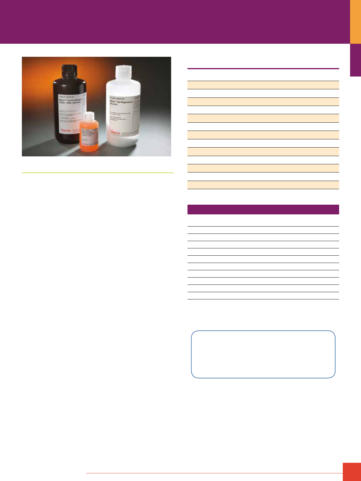
To order, call 800-874-3723 or 815-968-0747. Outside the United States, contact your local branch office or distributor.
63
Thermo Scientific Melon Gel Purification Products
Melon
™
Gel Products provide an exciting new approach to
purifying monoclonal and polyclonal antibodies from serum,
tissue culture supernatant and ascites fluid. Melon Gel works
with antibodies from a variety of species and subclasses, many of
which do not purify efficiently with Protein A or Protein G. Because
Melon Gel is not a bind-and-release support, it is extremely fast
and gentle to your antibodies, resulting in antibody preparations of
high purity and high activity!
How does it work?
Melon Gel contains a proprietary ligand that retains most protein
found in serum, ascites and culture supernatants, while allowing
IgG to pass through the support and be collected in the flow-
through fraction. The resulting recovery and purity of the IgG
isolated by this method rivals that obtained from the same samples
using bind-and-release supports such as Protein A or Protein G.
Highlights:
• Simple, one-step protocol – no tedious binding, washing, and
multiple elution steps
• Rapid purification – purifies antibodies from serum four to six
times faster than Protein A or G methods
• High recovery and purity – antibodies from many species
are recovered with greater than 90% yield and greater than
80% purity
• Robust purification – works with a wide range of antibodies
including many that do not purify well on Protein A or Protein G
• Gentle purification – no harsh elution conditions means antibod-
ies retain more activity
• Reusable support – Melon Gel Support can be used for multiple
antibody purifications
• Available in various formats – spin columns, purification kits and
chromatography cartridges for antibody purification from serum,
ascites and culture supernatant
IgG purification performance of the Thermo Scientific Melon Gel System,
Protein A and Protein G.
Source Melon Gel Protein A Protein G
Human H H H
Mouse H H H
Rabbit H H H
Rat H L M
Goat H L M
Cow M L H
Sheep M L H
Horse H L H
Guinea Pig H H L
Pig H H L
Chicken N N N
Hamster H M M
Donkey H M H
H = high recovery, M = medium recovery, L = low recover, N = no recovery
Ordering Information
Product #
Description
Pkg. Size
U.S.
Price
45206 Melon Gel IgG Spin Purification Kit
Kit
$ 243
45212 Melon Gel IgG Spin Purification Kit
Kit
$ 507
45214 Melon Gel Monoclonal IgG Purification Kit
Kit
$1106
45216 Saturated Ammonium Sulfate Solution
1L
$ 147
45219 Ascites Conditioning Reagent
5mL
$ 35
89932 Pierce Chromatography Cartridges, Melon Gel
2 x 1
$ 100
89933 Pierce Chromatography Cartridges, Melon Gel
1 x 5
$ 201
45208 Melon Gel Spin Kit Plate
2 plates
$ 311
89972 Melon Gel Purification Buffer
1 pack
$ 13
89973 Melon Gel Regenerant
1 pack
$ 10
For more data using Melon Gel and for complete
product information, please request the Antibody
Purification Technical Handbook (1601974) or visit
www.thermoscientific.com/pierce
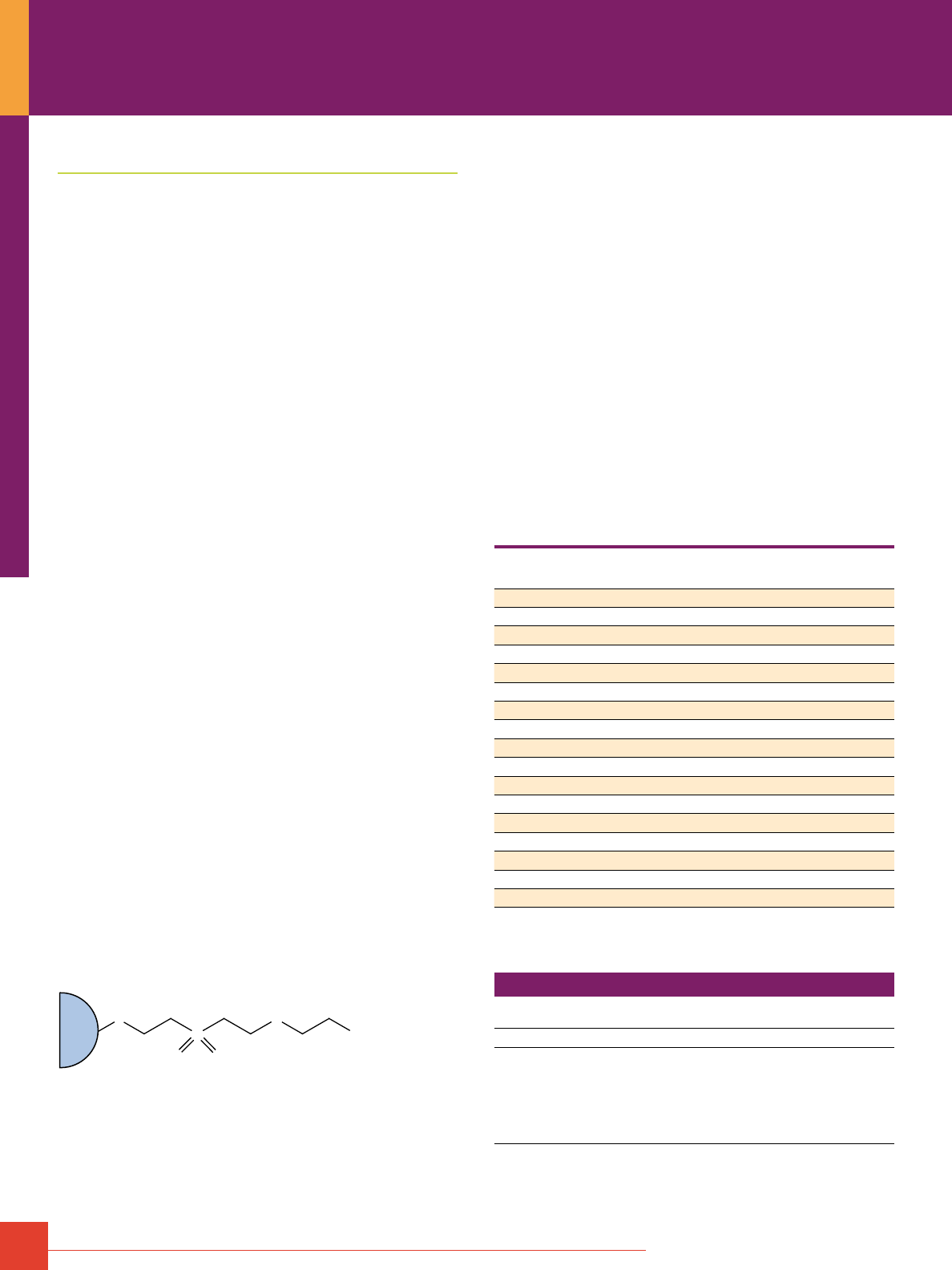
64
For more information, or to download product instructions, visit www.thermoscientific.com/pierce
Thiophilic Gel Antibody Purification
Thiophilic adsorption is a low-cost, efficient alternative to
ammonium sulfate precipitation for immunoglobulin purification
from crude samples. Ammonium sulfate precipitation must
be followed by several additional steps to completely remove
contaminants in crude samples. Thiophilic adsorption is a simple,
rapid, one-step method for antibody purification from serum,
ascites or tissue culture supernatant.
Thiophilic adsorption is a highly selective type of lyotropic
salt-promoted protein:ligand interaction phenomenon that has
been studied extensively by Porath and co-workers and other
researchers.
1
This interaction is termed thiophilic because it
distinguishes proteins that recognize a sulfone group in close
proximity to a thioether. Thiophilic adsorption incorporates properties
of both hydrophobic and hydrophilic adsorption. However, in
contrast to strictly hydrophobic systems, thiophilic adsorption
is not strongly promoted by high concentrations of sodium
chloride. Instead, thiophilic adsorption is promoted by increased
concentrations of water-interacting, non-chaotropic salts such
as potassium and ammonium sulfate.
Thermo Scientific Pierce Thiophilic Adsorbent is 6% beaded
agarose modified to contain simple sulfone/ thioether groups (see
structure at right). Our Thiophilic Adsorbent has a high binding
capacity (20mg of immunoglobulin permL of resin) and broad
specificity toward immunoglobulins derived from various animal
species. Notably, thiophilic adsorption is one of few methods
available for purification of IgY from chicken (see also subsequent
discussion of IgY purification). Among human serum proteins,
immunoglobulins and a2-macroglobulins are preferentially bound
by our Thiophilic Adsorbent.
2
Purification using Pierce Thiophilic Adsorbent results in good
protein recovery with excellent preservation of antibody activity.
Sample preparation requires the addition of 0.5M potassium
sulfate to the serum, ascites or culture fluid. Greater specificity
for immunoglobulins is obtained if the sample is buffered at pH 8.0.
The gentle elution conditions (e.g., 50mM sodium phosphate,
pH 7-8) yield concentrated, essentially salt-free, highly purified
immunoglobulins at near neutral pH.
After use, our Thiophilic Adsorbent can be regenerated by
treatment with guanidine•HCl. Our data indicate that the Adsorbent
column can be used at least 10 times without significant loss of
binding capacity.
O
S
S
OH
O
O
Agarose
Bead
Structure of Thermo Scientific Pierce Thiophilic Adsorbent.
References
1. Porath, J., et al. (1985). FEBS Lett. 185, 306-310.
2. Belew, M., et al. (1987). J. Immunol. Method 102, 173-182.
3. Hutchens, T.W. and Porath, J. (1987). Biochemistry 26, 7199-7204.
4. Lihme, A. and Heegaard, P.M.H. (1990). Anal. Biochem. 192, 64-69.
5. Unpublished internal documents.
Thermo Scientific Pierce Thiophilic Adsorbent and Purification Kit
Economical purification of mouse antibodies from ascites fluid.
Highlights:
• Binds to Fab and F(ab´)2 fragments
• Binds to ScFv1
• High-capacity (20mg/mL), good protein recovery and retention
of antibody function
• Broad specificity toward immunoglobulins derived from various
animal species (see Table 4)
• Binds chicken IgY (also IgG)
• Simple, rapid, one-step purification for monoclonal antibodies
from ascites; easy to scale up
• Used to enrich the immunoglobulin fraction from serum or tissue
culture supernatant
• Efficient alternative to ammonium sulfate precipitation for
enriching antibodies from crude samples
• Gentle elution conditions yield concentrated, salt-free
immunoglobulin at near neutral pH
• High degree of purity
Table 4. Binding characteristics of Thermo Scientific Pierce Thiophilic Adsorbent.
Species
Total A
280
Bound
from 1mL Serum
% Purity by HPLC
Human 4.8 70
Mouse 8.6 63
Mouse IgG
1
11.6 92
Mouse IgG
2a
9.3 88
Mouse IgG
2b
9.8 97
Mouse IgG
3
10.7 94
Rat 13.0 79
Bovine 17.9 90
Calf 11.1 89
Chicken 5.2 76
Dog 12.2 91
Goat 17.3 92
Guinea Pig 11.1 71
Horse 13.0 93
Pig 21.1 90
Rabbit 6.7 84
Sheep 12.3 89
Reference
1. Schulze, R.A., et al. (1994). Anal. Biochem. 220, 212-214.
Ordering Information
Product #
Description
Pkg. Size
U.S.
Price
20500 Pierce Thiophilic Adsorbent
10mL
$ 92
44916 Pierce Thiophilic Purification Kit
Includes: Thiophilic Adsorbent Columns
Binding Buffer
Elution Buffer
Column Storage Buffer (2X)
Guanidine•HCl Crystals
Column Extenders
Kit
4 x 3mL
1,000mL
1,000mL
100mL
230g
$443
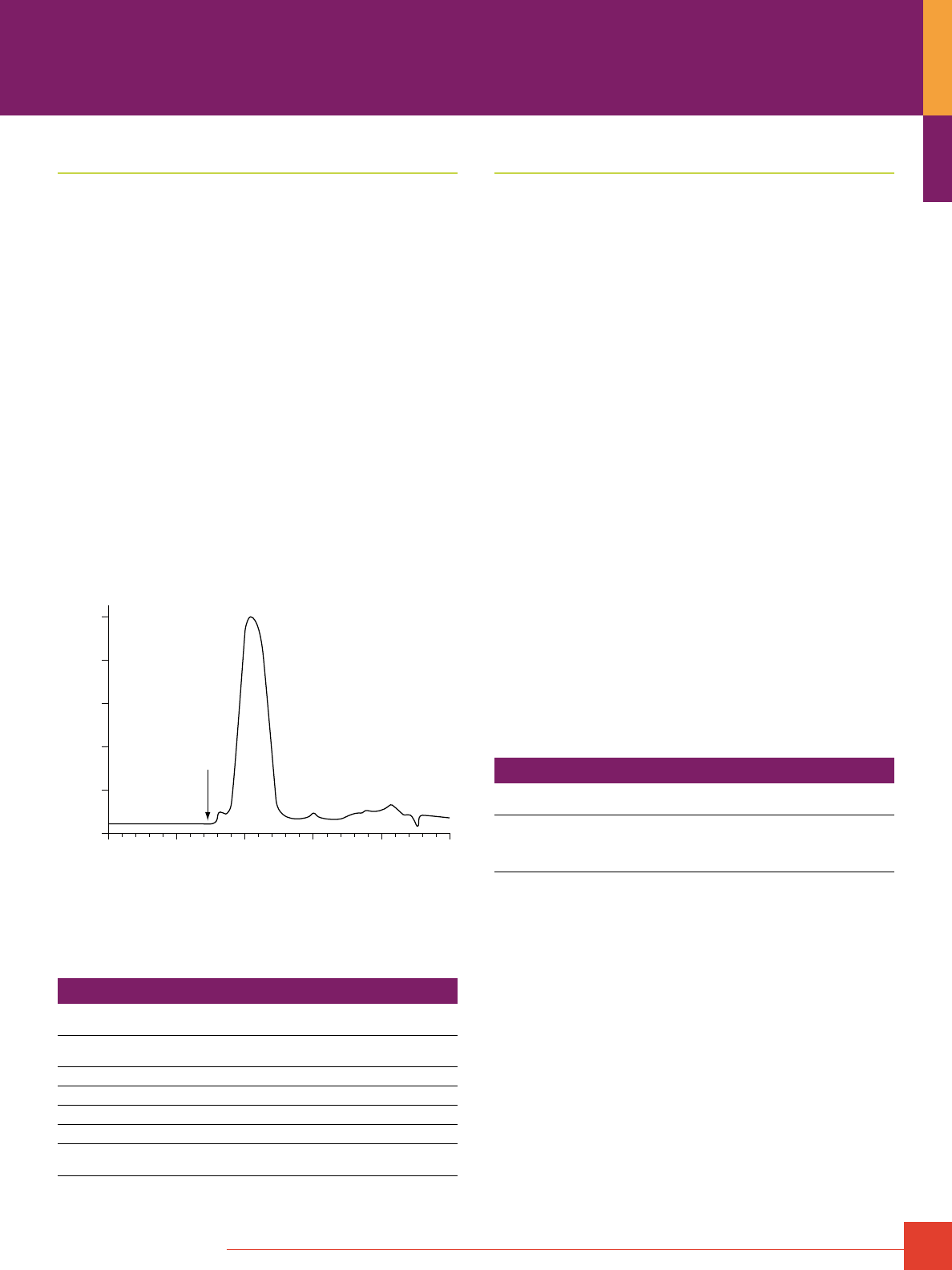
To order, call 800-874-3723 or 815-968-0747. Outside the United States, contact your local branch office or distributor.
65
IgM Purification
Immobilized Mannan Binding Protein
To develop an effective affinity matrix, our scientists examined C1q
and another similarly structured protein, mannan binding protein
(MBP). Serum MBP, like C1q, is capable of initiating carbohydrate-
mediated complement activation. MBP is a mannose and
N-acetylglucosamine-specific lectin found in mammalian sera, and
it has considerable structural homology to C1q.
8
MBP subunits are
identical, each with molecular mass of approximately 31kDa (C1q
has six each of three different polypeptide subunits of molecular
mass 24-28kDa). Studies in our labs show that MBP does not bind
F(ab´)
2
and Fab.
We have developed an easy-to-use Thermo Scientific Pierce
Immobilized Mannan Binding Protein and Buffer System to purify
IgM. It is most effective for purifying mouse IgM from ascites.
Purified IgM can be obtained from a single pass over the affinity
column. Human IgM will bind to the support, albeit with slightly
lower capacity, and yield a product at least 88% pure as assessed
by HPLC. The purification of IgM from other species and mouse
serum has not yet been optimized.
Immobilized MBP and IgM Purification Kit
Easy IgM purification with guaranteed 88% pure mouse IgM!
0 10 20 30 40 50
0
20
40
60
80
100
Time (min.)
Absorbance at 280 nm
(percent of max.)
IgM
Demonstration of the high purity of MBP-purified IgM from mouse ascites.
The bound material from mouse ascites was eluted from the 5mL MBP
column as described in the Standard Protocol. The highest 280nm absorbing
fraction from the elution was chromatographed using the conditions
described in the instructions.
Ordering Information
Product #
Description
Pkg. Size
U.S.
Price
22212 Immobilized Mannan Binding Protein
Capacity: ~1mg IgM/mL of resin
10mL
$936
44897 IgM Purification Kit
Kit
$610
21016 IgM Binding Buffer
800mL
$ 77
21017 IgM Elution Buffer
500mL
$ 63
21018 MBP Column Preparation Buffer
50mL
$ 33
53123 UltraLink Immobilized Mannan Binding Protein
Capacity: >0.75mg IgM/mL of resin
5mL
$493
IgA Purification
Human IgA Purification
Jacalin is an a-D-galactose binding lectin extracted from jack-
fruit seeds (Artocarpus integrifolia). The lectin is a glycoprotein
of approximately 40kDa composed of four identical subunits.
Jacalin immobilized on supports such as agarose has been useful
for the purification of human serum or secretory IgA
1
. IgA can be
separated from human IgG and IgM in human serum or colostrum.
1
IgD is reported to bind to jacalin.
2
Immobilized jacalin is also useful
for removing contaminating IgA from IgG samples.
Binding of IgA to immobilized jacalin occurs at physiologic pH
and ionic strength, as in phosphate buffered saline (PBS). Elution
of bound IgA occurs with competitor ligand (e.g., 0.1 M melibiose
or 0.1 M a-D-galactose) in PBS. We offer immobilized jacalin on
crosslinked 6% agarose.
References
1. Roque-Barreira, M.C. and Campos-Neto, A. (1985). J. Immunol. Method 134(30), 1740-1743.
2. Aucouturier, P., et al. (1987). Mol. Immunol. 24(5), 503-511.
Immobilized Jacalin
Ideal for human IgA purification.
Highlights:
• Ideal for preparing human IgA that is free of contaminating IgG
• Found to bind human IgA1, but not human IgA
2
– useful for sepa-
rating the two subclasses
References
Kumar, G.S., et al. (1982). J. Biosci. 4, 257-261.
Roque-Barreira, M.C. and Campos-Neto, A. (1985). J. Immunol. 134, 1740-1743.
Mestecky, J., et al. (1971). J. Immunol. 107, 605-607.
Van Kamp, G.J. (1979). J. Immunol. Method 27, 301-305.
Kondoh, H., et al. (1986). J. Immunol. Method 88, 171-173.
Ordering Information
Product #
Description
Pkg. Size
U.S.
Price
20395 Immobilized Jacalin
Capacity: 1-3mg human IgA/mL of resin
Support: Crosslinked 6% beaded agarose
Loading: 4.5mg of jacalin/mL of resin
5mL
$201
Antibody Purification
66
For more information, or to download product instructions, visit www.thermoscientific.com/pierce
Biotin
Biotin, also known as vitamin H, is a small molecule (MW 244.3)
that is present in tiny amounts in all living cells. The valeric
acid side chain of the biotin molecule can be derivatized to
incorporate various reactive groups that are used to attach biotin
to other molecules. Once biotin is attached to a molecule, the
molecule can be affinity-purified using an immobilized version of
any biotin-binding protein. Alternatively, a biotinylated molecule
can be immobilized through interaction with a biotin-binding protein,
then used to affinity-purify other molecules that specifically
interact with it. We offer biotin-labeled antibodies and a number
of other biotinylated molecules, as well as a broad selection of
biotinylation reagents to label any protein.
O
Biotin
MW 244.3
S
H
H
Valeric Acid Side Chain
H
H
O
HO
Biotin-Binding Proteins
Avidin – The extraordinary affinity of avidin for biotin allows
biotin-containing molecules in a complex mixture to be discretely
bound with avidin. Avidin is a glycoprotein found in the egg white
and tissues of birds, reptiles and amphibians. It contains four identi-
cal subunits having a combined mass of 67,000–68,000 daltons.
Each subunit consists of 128 amino acids and binds one molecule
of biotin. The extent of glycosylation on avidin is high; carbohy-
drate accounts for about 10% of the total mass of the tetramer.
Avidin has a basic isoelectric point (pI = 10–10.5) and is stable
over a wide range of pH and temperature. Extensive chemical
modification has little effect on the activity of avidin, making it
especially useful for protein purification. However, because of
its carbohydrate content and basic pI, avidin has relatively high
nonspecific binding properties.
Streptavidin – Another biotin-binding protein is streptavidin, which
is isolated from Streptomyces avidinii and has a mass of 75,000
daltons. In contrast to avidin, streptavidin has no carbohydrate and
has a mildly acidic pI (5.5). Thermo Scientific Pierce Streptavidin
is a recombinant form having a mass of 53,000 daltons and a
near-neutral pI. Streptavidin is much less soluble in water than
avidin. There are considerable differences in the composition of
avidin and streptavidin, but they are remarkably similar in other
respects. Streptavidin is also a tetrameric protein, with each
subunit binding one molecule of biotin with affinity similar to that of
avidin. Guanidinium chloride will dissociate avidin and streptavidin
into subunits, but streptavidin is more resistant to dissociation.
Streptavidin contains an RYD sequence similar to the RGD
sequence that binds cell surface receptors. The RYD sequence
can cause background in some applications.
NeutrAvidin Protein – We also offer a deglycosylated version of
avidin, known as NeutrAvidin Protein, with a mass of approximate-
ly 60,000 daltons. As a result of carbohydrate removal, lectin bind-
ing is reduced to undetectable levels, yet biotin-binding affinity is
retained because the carbohydrate is not necessary for this activ-
ity. NeutrAvidin Protein offers the advantages of a near-neutral pI
(6.3) to minimize nonspecific adsorption, along with lysine residues
that remain available for derivatization or conjugation. NeutrAvidin
Protein yields the lowest nonspecific binding among the known
biotin-binding proteins due to its near-neutral pI and lack of both
carbohydrate and RYD sequence.
Avidin:Biotin Binding
To order, call 800-874-3723 or 815-968-0747. Outside the United States, contact your local branch office or distributor.
67
A Comparison of Biotin-Binding Proteins
The strong association between avidin and biotin can be used in
the field of affinity separations. By attaching avidin to a solid
support, a biotinylated product can be anchored to the same solid
support. The attachment is stable over a wide range of pH, salt
concentrations and temperatures. To dissociate biotin from avidin,
8M guanidine•HCl, pH 1.5 or boiling in SDS-PAGE sample buffer
must be used.
Avidin
Streptavidin
Thermo Scientific
NeutrAvidin Protein
Molecular Weight
67K 53K 60K
Biotin-binding Sites
4 4 4
Isoelectric Point (pl)
10 6.8–7.5 6.3
Specificity
Low High Highest
Affinity for Biotin (K
d
)
10
-15
M 10
-15
M 10
-15
M
Nonspecific Binding
High Low Lowest
Immobilized Avidin Products
Strong biotin interaction creates a nearly irreversible bond.
Immobilized avidin can be used in a variety of applications for
the affinity purification of biotinylated macromolecules. In one
variation, an antibody that has an affinity for a particular antigen
is labeled with biotin. Cells containing the antigen are lysed, then
incubated with the biotinylated antibody to form a typical antigen/
antibody complex. To isolate the antigen, the crude mixture is
passed through an immobilized avidin or streptavidin column,
which will bind the complex. After appropriate washes, the antigen
can be eluted from the column with a low pH elution buffer. The
biotinylated antibody is retained by the column.
Applications:
• Binding biotinylated anti-transferrin for purifying transferrin
from serum
1
• Binding biotinylated peptides and elution with an SDS/urea solution
2
• Hybridization of biotinylated RNA to its complementary DNA
and binding to immobilized avidin, with subsequent elution of the
single-stranded DNA
3
• Purification of double-stranded DNA
4
References
1. Wilchek, M. and Bayer, E.A. (1989). Protein Recognition of Immobilized Ligands.
Hutchins, T.W., ed. Alan R. Liss, Inc., pp. 83–90.
2. Swack, J.A., et al. (1978). Anal. Biochem. 87, 114–126.
3. Manning, J., et al. (1977). Biochemistry 16, 1364–1370.
4. Pellegrini, M., et al. (1977). Nucleic Acids Res. 4, 2961–2973.
Ordering Information
Product #
Description
Pkg. Size
U.S.
Price
20219 Avidin Agarose Resin
Support: Crosslinked 6% beaded agarose
Capacity: ≥20μg biotin/mL resin
5mL
$150
20225 Avidin Agarose Resin
Support and Capacity: Same as above
5 x 5mL
$533
20362 Avidin Agarose Columns
Support and Capacity: Same as above
5 x 1mL
$190
Immobilized Streptavidin Products
Same high biotin-binding affinity as avidin with low
nonspecific binding.
Applications:
• Purification of membrane antigens in conjunction with
biotinylated monoclonal antibodies
1,2
• Cell-surface labeling with biotinylation reagents, followed by
precipitation with immobilized streptavidin
3
• Purification of cell-surface glycoproteins using biotinylated
Concanavalin A
4
• Recovery of single-stranded DNA for dideoxy sequencing
5
References
1. Gretch, D.R., et al. (1987). Anal. Biochem. 163, 270–277.
2. Updyke, T.V. and Nicolson, G.L. (1984). J. Immunol. Method 73, 83–95.
3. Lisanti, M.P., et al. (1989). J. Cell Biol. 109, 2117–2127.
4. Buckie, J.W. and Cook, G.M. (1986). Anal. Biochem. 156(2), 463–472.
5. Baqui, M., et al. (2003). J. Biol. Chem. 278, 1206–1211.
Ordering Information
Product #
Description
Pkg. Size
U.S.
Price
20347 Streptavidin Agarose Resin
Support: Crosslinked 6% beaded agarose
Capacity: 1–3mg biotinylated BSA/mL resin
15–28μg biotin/mL resin
2mL
$178
20349 Streptavidin Agarose Resin
Support and Capacity: Same as above
5mL
$310
20353 Streptavidin Agarose Resin
Support and Capacity: Same as above
10mL
$440
20351 Streptavidin Agarose Columns
Support and Capacity: Same as above
5 x 1mL
$318
53113 Streptavidin UltraLink Resin
Support: UltraLink Biosupport
Capacity: ≥2mg biotinylated BSA/mL resin
≥24μg biotin/mL resin
2mL
$212
53114 Streptavidin UltraLink Resin
Support and Capacity: Same as above
5mL
$370
53116 Streptavidin Plus UltraLink Resin
Support: UltraLink Biosupport
Capacity: ≥4mg biotinylated BSA/mL resin
≥48μg biotin/mL resin
2mL
$285
53117 Streptavidin Plus UltraLink Resin
Support and Capacity: Same as above
5mL
$410
20357 High Capacity Streptavidin Agarose Resin
Support: Crosslinked 6% beaded agarose
Capacity: >10mg biotinylated BSA/mL of resin
2mL
$225
20359 High Capacity Streptavidin Agarose Resin
Support and Capacity: Same as above
5mL
$350
20361 High Capacity Streptavidin Agarose Resin
Support and Capacity: Same as above
10mL
$600
21344 MagnaBind Streptavidin Beads
Support: 1–4μm, iron oxide particles
Capacity: 2μg biotin/mL beads
5mL
$271
88816 Pierce Streptavidin Magnetic Beads
1mL
$213
88817 Pierce Streptavidin Magnetic Beads
5mL
$721

68
For more information, or to download product instructions, visit www.thermoscientific.com/pierce
Immobilized NeutrAvidin Products
Less nonspecific binding produces cleaner results and better yields.
When nonspecific binding is a problem in your application,
Thermo Scientific Immobilized NeutrAvidin Products are superior
alternatives to avidin or streptavidin. NeutrAvidin Biotin-Binding
Protein is a modified avidin derivative that combines several
key features to provide biotin-binding with exceptionally low
nonspecific binding properties.
Highlights:
• Carbohydrate-free – just like streptavidin, NeutrAvidin Biotin-
Binding Protein has no carbohydrate, eliminating nonspecific
binding problems due to sugars
• No interaction with cell surface molecules – absence of the
Arg-Tyr-Asp sequence (present in streptavidin), which mimics
the universal cell surface recognition sequence present in a
variety of molecules, eliminates cross-reactivity of cell surface
molecules
• Neutral pl – with a pl of 6.3, NeutrAvidin Protein has a pl that is
closer to neutrality than avidin or streptavidin, eliminating
electrostatic interaction that contributes to nonspecific binding
Applications:
• Immunoprecipitation
• Purifying proteins that bind to biotinylated ligands
• Capturing biotinylated cell-surface proteins
1-3
• Purifying biotinylated peptides
4
Ordering Information
Product #
Description
Pkg. Size
U.S.
Price
29200 NeutrAvidin Agarose Resin
Support: Crosslinked 6% beaded agarose
Capacity: >20μg or 80nmol biotin/mL resin
(approx. 1–2mg biotinylated BSA/mL resin)
5mL
$205
29201 NeutrAvidin Agarose Resin
Support and Capacity: Same as above
10mL
$350
53150 NeutrAvidin UltraLink Resin
Support: UltraLink Biosupport
Capacity: 12–20μg biotin/mL gel
5mL
$255
53151 NeutrAvidin Plus UltraLink Resin
Support: UltraLink Biosupport
Capacity: ≥30μg biotin/mL gel
5mL
$340
29202 High Capacity NeutrAvidin Agarose Resin
Support: Crosslinked 6% beaded agarose
Capacity: >75μg biotin/mL resin
>8mg biotinylated BSA/mL resin
5mL
$250
29204 High Capacity NeutrAvidin Agarose Resin
Support and Capacity: Same as above
10mL
$410
Immobilized Monomeric Avidin and Kit
Ideal affinity support for gentle, reversible binding of
biotinylated proteins.
To break the avidin-biotin interaction, 8M guanidine•HCl at pH 1.5
or boiling in SDS-PAGE sample buffer is required. These elution
methods may result in denaturation of the biotinylated protein and
cause irreversible damage to the support. In addition, avidin or
streptavidin will be irreversibly denatured and lose the ability to
bind subsequent biotinylated samples.
When avidin is coupled to a solid support as the subunit monomer,
the specificity for biotin is retained, but the affinity for biotin bind-
ing substantially decreases (K
a
~10
8
M
-1
). The Monomeric Avidin
Agarose Resin and Kit can be used to bind biotinylated molecules,
and the bound material can be competitively eluted using 2mm
biotin in phosphate-buffered saline (PBS). This technique provides
the gentlest elution conditions without contamination of the avidin
subunits or substantial loss of column-binding capacity.
Highlights:
• Purifies biotinylated products under mild elution conditions
• Can be regenerated and reused at least 10 times
• Exhibits little nonspecific binding (3% or less)
Ordering Information
Product #
Description
Pkg. Size
U.S.
Price
20228 Monomeric Avidin Agarose Resin
Support: Crosslinked 4% beaded agarose
Capacity: ≥1.2mg biotinylated BSA/mL resin
5mL
$355
20267 Monomeric Avidin Agarose Resin
Support and Capacity: Same as above
10mL
$570
20227 Monomeric Avidin Agarose Kit
Support and Capacity: Same as above
Includes: 1 x 2mL Column, Binding and Elution buffers
Kit
$341
53146 Immobilized Monomeric Avidin UltraLink Resin
Support: UltraLink Biosupport
Capacity: ≥1.2mg biotinylated BSA/mL resin
5mL
$399
29129 Biotin
1g
$ 54
Thermo Scientific Products for Avidin:Biotin Binding
To order, call 800-874-3723 or 815-968-0747. Outside the United States, contact your local branch office or distributor.
69
Immobilized Iminobiotin and Biotin
Iminobiotin offers mild dissociation conditions at pH 4.
O
NH
HN
S
NH
Immobilized Iminobiotin
H
N
H
N
H
N
Agarose
Bead
Iminobiotin is the guanido analog of biotin. The dissociation
constant of the avidin-iminobiotin complex is pH-dependent.
At pH 9.5-11.0, the avidin-iminobiotin complex will bind tightly.
At pH 4, the avidin-iminobiotin complex will dissociate. Because
denaturing agents such as 8 M guanidine•HCI or 4 M urea are
not used in the purification, an avidin conjugate has a better
chance of maintaining its activity during purification.
Use immobilized D-Biotin as an “irreversible linkage” to bind
streptavidin conjugates. The biotin-streptavidin interaction can
withstand extremes in pH, salt and detergents.
Ordering Information
Product #
Description
Pkg. Size
U.S.
Price
20221 Iminobiotin Agarose Resin
Support: Crosslinked 6% beaded agarose
Spacer: Diaminodipropylamine
Capacity: ≥1mg of avidin/mL resin
5mL
$119
20218 Biotin Agarose Resin
Support: Pierce CDI Support
Spacer: Diaminodipropylamine
Capacity: ≥2mg of avidin/mL resin
5mL
$ 99
70
For more information, or to download product instructions, visit www.thermoscientific.com/pierce
Thermo Scientific Pierce Chromatography Cartridges are
convenient, reliable, ready-to-use, pre-packed 1mL and 5mL
columns of sample-prep and affinity purification resins for
manual or automated liquid chromatography (LC).
The cartridge fittings are compatible with the popular automated
liquid-chromatography systems or for manual syringe processing.
The cartridges attach directly to ÄKTA or FPLC Systems without
additional connectors. Cartridges can be used individually or
connected in a series to obtain even higher column capacity. Each
product supplied in the Pierce Chromatography Cartridge format
accessory pack that readily adapts cartridges for use using
Luer-Lok Syringe Fittings or tubing. The cartridges provide fast,
easy and reproducible chromatographic separations and can be
regenerated for multiple uses.
Thermo Scientific FPLC Cartridges shematic.
Chromatography Cartridge Highlights
• Two sizes – 1mL and 5mL, convenient for typical research scales
• Compatible – fittings allow connection with popular LC systems
or a standard syringe
• Versatile – use singly or connected in series to service different
capacity requirements
• Validated – available cartridges have been tested to ensure
performance in the format
• Reusable – accessory pack includes caps for convenient
storage between uses
• Economical – comparable performance at lower cost than other
commercially available cartridges
Thermo Scientific Pierce Chromatography Cartridge properties.
1mL Cartridge 5mL Cartridge
Dimensions 0.7 x 2.7cm 1.3 x 3.8cm
Recommended Flow Rate 1-4mL/min 1-7mL/min
Maximum Pressure 0.3 mPa (43 psi or 3 bar) 0.3 mPa (43 psi or 3 bar)
Cartridge Material polypropylene polypropylene
Frit Material polyethylene polyethylene
Applications for Pierce Chromatography Cartridges:
• His-tagged protein purification
• GST-tagged protein purification
• Antibody purification
• Biotin binding
• Phosphoprotein enrichment
• Protein desalting
FPLC Cartridge Overview
20mm
Port Diameter: 4.2mm
10-32 threaded coned port
14mm
4mm
17.5 mm17.5mm
89mm
13.5mm
Port Diameter: 4.2mm
10-32 threaded coned port
8.5mm
4mm
12.5mm
75mm
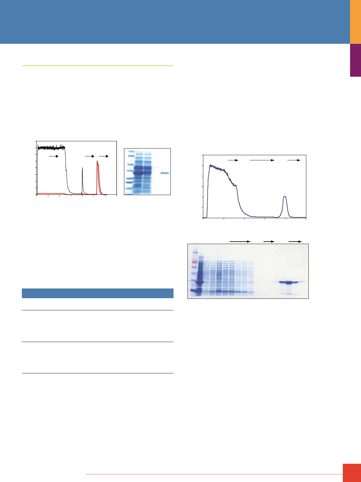
To order, call 800-874-3723 or 815-968-0747. Outside the United States, contact your local branch office or distributor.
71
His-tagged Protein FPLC Purification
Thermo Scientific HisPur Nickel-NTA Cartridges
Delivers the highest yield of His-tagged protein with a
re-useable resin.
Highlights:
• Bind up to 60mg of 6xHis-tagged protein per milliliter of resin
• Purify proteins using native or denaturing conditions
• Use with Pierce Cell Lysis Reagents and a variety of buffer additives
• Reuse cartridges several times
0 20 40 60 80 100 120 140mL
0
500
1000
1500
2000
2500
3000
3500
4000
A
280
M L FT W E
FT W E
Purification of 6xHis-GFP from E. coli lysate using a Thermo Scientific HisPur
Nickel-NTA Cartridge. Bacterial lysate (130mg total protein) containing over-
expressed 6xHis-GFP (green fluorescent protein) was diluted 1:1 with equilibra-
tion buffer and applied to a HisPur Ni-NTA Chromatography Cartridge at a flow
rate of 1mL/min. The cartridge was washed with PBS, 68mM imidazole until the
baseline absorbance was reached. The 6xHis-GFP was eluted with PBS,
300mM imidazole. Left panel: 6xHis-GFP elution was monitored at 280nm (black
line) and 485nm (red line; GFP-specific). Right panel: Selected fractions were
analyzed by SDS-PAGE. Gel lanes were normalized to equivalent volume.
M = MW marker, L = lysate load, FT = flow-through and E = elution.
See page 14 for more information and other packaging formats of
HisPur Ni-NTA Resin.
Ordering Information
Product #
Description
Pkg. Size
U.S.
Price
90098 HisPur Ni-NTA
Chromatography Cartridges, 1mL
Formulation: 1mL resin cartridges with
LC and Luer-Lok fittings.
Sufficient for: Binding up to 60mg of His-tagged
protein per cartridge.
5 cartridges
$155
90099 HisPur Ni-NTA
Chromatography Cartridges, 5mL
Formulation: 5mL resin cartridges with
LC and Luer-Lok fittings.
Sufficient for: Binding up to 300mg of His-tagged
protein per cartridge.
2 cartridges
$200
Thermo Scientific HisPur Cobalt Cartridges
Highly selective binding for the highest purity His-tagged
protein purification.
Highlights:
• Obtain more than 10mg of pure His-tagged protein per milliliter
of resin without optimizing imidazole washing conditions
• Cobalt-chelate coordination core binds fewer host protein con-
taminants, resulting in lower background than nickel resins
• No metal contamination in eluted histidine-tagged protein sample
• Purify proteins under native or denaturing conditions; compatible
with Pierce Cell Lysis Reagents and a variety of buffer additives
• Reuse cartridges several times
0
1000
2000
3000
4000
5000
6000
0 5 10 15 20 25mL
Flow-through Wash Elution
A
280
Flow-through (FT)M S Wash Elution
Purification of 6xHis-GFP from E. coli lysate using a Thermo Scientific HisPur
Cobalt Cartridge. His-tagged green fluorescent protein (GFP) was extracted
from E. coli using Thermo Scientific B-PER Bacterial Protein Extraction
Reagent in Phosphate Buffer (Product # 78266) containing Thermo Scientific
Halt Protease Inhibitor Cocktail, EDTA-Free (Product # 78415). The lysate
was diluted 1:1 with equilibration/wash buffer (50mM sodium phosphate,
300mM sodium chloride, 10mM imidazole, pH 7.4) and applied to a HisPur
Cobalt Chromatography Cartridge at a flow rate of 0.3mL/min. The cartridge
was washed with equilibration/wash buffer until the baseline absorbance at
280nm was reached. His-tagged GFP was eluted (50mM sodium phosphate,
300mM sodium chloride, 150mM imidazole; pH 7.4) and selected fractions
were analyzed by SDS-PAGE and Thermo Scientific GelCode Blue Stain
Reagent (Product # 24592). M = MW Marker; S = non-fractionated lysate;
FT = flow-through.
See page 16 for more information and other packaging formats of
HisPur Ni-Cobalt Resin.
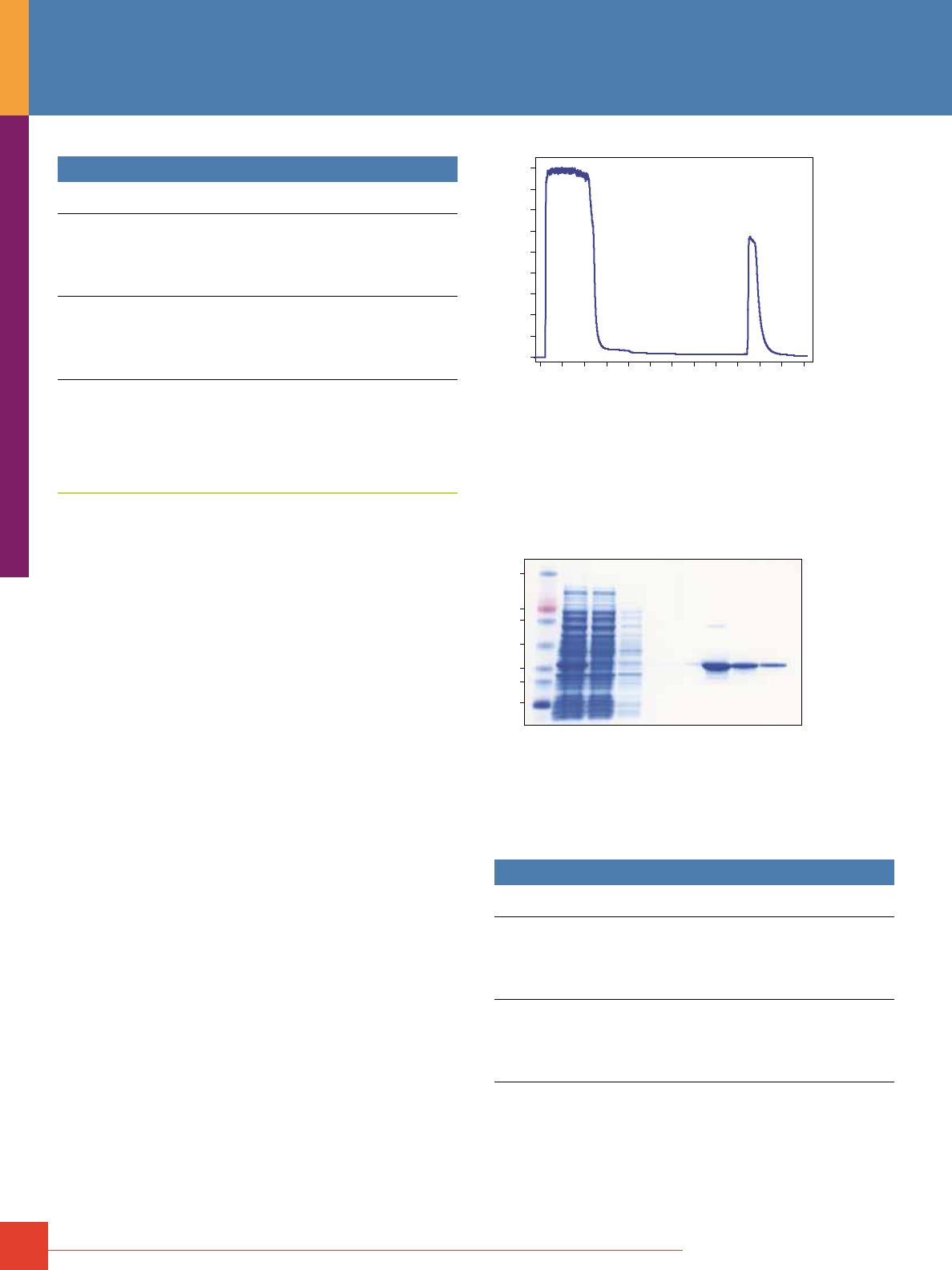
72
For more information, or to download product instructions, visit www.www.thermoscientific.com/pierce
Ordering Information
Product #
Description
Pkg. Size
U.S.
Price
90093 HisPur Cobalt
Chromatography Cartridges, 1mL
Formulation: 1mL resin cartridges with
LC and Luer-Lok fittings.
Sufficient for: Binding > 10mg of His-tagged
protein per cartridge.
5 cartridges
$160
90094 HisPur Cobalt
Chromatography Cartridges, 5mL
Formulation: 5mL resin cartridges with
LC and Luer-Lok fittings.
Sufficient for: Binding > 50mg of His-tagged
protein per cartridge.
2 cartridges
$267
GST-Tagged Protein FPLC Purification
Thermo Scientific Pierce Glutathione Agarose Cartridges
Cost-effective and high-performance GSH resins for purification
of recombinant GST fusion proteins.
Highlights:
• Binds at least 25mg of recombinant GST protein per milliliter
of resin
• Consistently purifies at least 10mg of GST-tagged protein per
milliliter of resin with greater than 90% purity
• Economically priced and can be reused at several times without
reduction in binding capacity and purification performance
• Works well to purify GST-fusion proteins from bacterial lysates
or use with pre-purified GST-tagged proteins to pull down protein
interactions
• Validated and effective for use with Pierce Cell Lysis Reagents to
extract and purify from bacterial or mammalian cell cultures
See page 18 for more information and other packaging formats of
Pierce Immobilized Glutathione Agarose Resin.
0
1000
2000
A
280
3000
4000
mAU
Sample
Application
Wash Elution
of
11.8 mg pure
GST-fusion protein
0.0 10.0 20.0 30.0 40.0 50.0 60.0mL
Purification of Pak 1-GST fusion protein on Thermo Scientific
Pierce Glutathione Chromatography Cartridge 1mL.
Sample: 50mg (10mL) clarified E. coli lysate containing expressed GST-Pak 1,
M 34,000
Binding Buffer: 5mM Tris, 150mM sodium chloride, pH 8.0
Elution Buffer: 50mM Tris, 150mM sodium chloride, 10mM Glutathione, pH 8.0
Flow Rate: Load at 0.5mL/min, Wash, Elute 1mL/min
Instrument: AKTApurifier
16.5
47
32
25
110
80
210
M Load FT Wash Elution
Purification of Pak 1-GST fusion protein using the Pierce GST Spin Purification
Kit (Product # 16106). Pak 1-GST lysate (2.4mg total protein) was applied in
Glutathione Binding Buffer to a 0.2mL Pierce Glutathione Spin Column and
eluted with 10mM Glutathione Elution Buffer, pH 8.0. Fractions were resolved
by SDS-PAGE using a 4-20% Tris-Glycine gel. Gel was stained with GelCode
Blue Stain Reagent (Product #24590). Pierce 3-Color Protein Molecular Weight
Marker Mix (Product # 26691) was used.
Ordering Information
Product #
Description
Pkg. Size
U.S.
Price
16109 Pierce Glutathione
Chromatography Cartridges, 1mL
Formulation: 1mL resin cartridges with
LC and Luer-Lok fittings.
Sufficient for: Binding approx. 10mg of GST-tagged
protein per cartridge.
5 cartridges
$230
16110 Pierce Glutathione
Chromatography Cartridges, 5mL
Formulation: 5mL resin cartridges with
LC and Luer-Lok fittings.
Sufficient for: Binding approx. 50mg of GST-tagged
protein per cartridge.
2 cartridges
$400
FPLC Cartridges
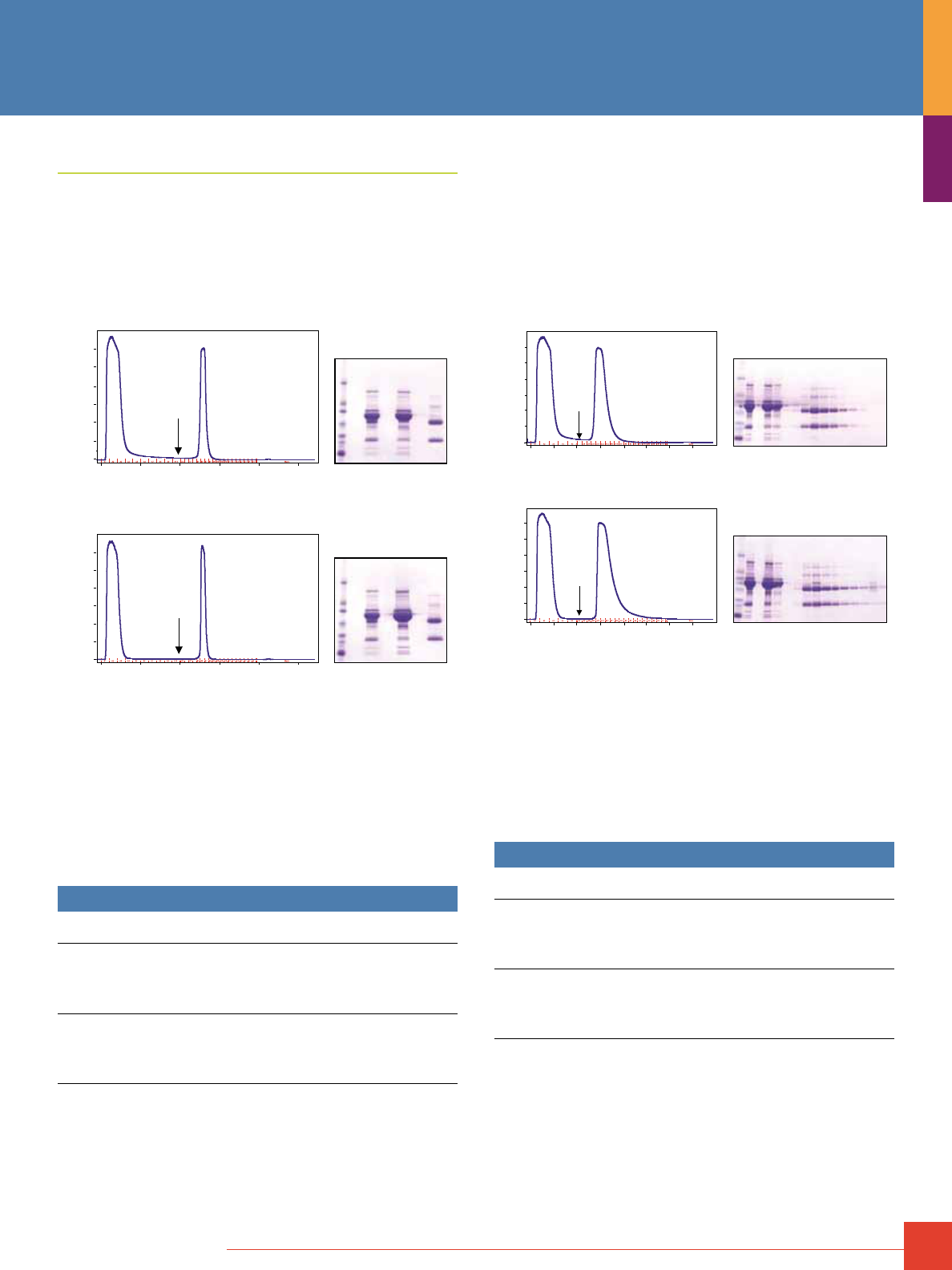
To order, call 800-874-3723 or 815-968-0747. Outside the United States, contact your local branch office or distributor.
73
Antibody FPLC Purification
Thermo Scientific Pierce Protein A Agarose Cartridges
Ideal for purification of IgG from serum and other fluids.
Highlights:
• Binds to a wide range of antibodies – especially good for
purification of rabbit IgG
• Less expensive than Protein G agarose
M
L FT E
FT
FT E
E
A
280
A
280
M
L FT E
50.0mL40.030.020.010.00.0
50.0mL40.030.020.010.0
0
500
1000
1500
2000
2500
3000
mAU
0.0
0
500
1000
1500
2000
2500
3000
mAU
Comparable antibody yield and purity achieved with Thermo Scientific
Pierce Protein A Chromatography Cartridge. Normal human serum (60mg)
was applied in PBS to a 1mL Thermo Scientific Pierce Protein A Cartridge (top)
and a HiTrap
®
Column (bottom) and eluted with 0.1M glycine, pH 2.8, using a
flow rate of 1mL/minute. The arrow denotes the start of the low-pH elution.
The yield of human IgG was 6.85mg and 6.88mg, respectively. Fractions were
separated by SDS-PAGE and the gels were stained with Thermo Scientific
Imperial Protein Stain (Product # 24615). M = MW marker, L = sample load,
FT = flow-through and E = elution.
See page 60 for more information and other packaging formats of
Pierce Protein A Agarose Resin.
Ordering Information
Product #
Description
Pkg. Size
U.S.
Price
89924 Pierce Protein A
Chromatography Cartridges, 1mL
Formulation: 1mL resin cartridges with
LC and Luer-Lok fittings.
Sufficient for: Binding 35mg human IgG per cartridge.
2 cartridges
$170
89925 Pierce Protein A
Chromatography Cartridges, 5mL
Formulation: 5mL resin cartridge with
LC and Luer-Lok fittings.
Sufficient for: Binding 175mg human IgG per cartridge.
1 cartridge
$335
Thermo Scientific Pierce Protein G Agarose Cartridges
Especially suited for purification of monoclonal antibodies from
mouse and the broadest spectrum of species and IgG subclasses
from human, goat and sheep samples.
Highlights:
• Albumin and cell surface binding site have been removed from
Protein G to provide higher antibody purity
• Binds to a wider range of antibodies than Protein A beads
M
L FT Elution
FT
FT E
E
A
280
A
280
M
L FT Elution
35.0mL30.020.0 25.010.0 15.05.00.0
35.0mL30.020.0 25.010.0 15.05.00.0
0
500
1000
1500
2000
2500
3000
mAU
0
500
1000
1500
2000
2500
3000
mAU
Comparable antibody yield and purity acheived with Thermo Scientific
Pierce Protein G Chromatography Cartridge. Normal human serum (60mg)
was applied in PBS to a 1mL Thermo Scientific Pierce Protein G Cartridge
(top) and a 1mL HiTrap Column (bottom) and eluted with 0.1M glycine, pH 2.8,
using a flow rate of 1mL/minute. The arrow denotes the start of the low pH
elution. Fractions were separated by SDS-PAGE and the gels were stained
with Imperial
®
Protein Stain (Product # 24615). M = MW marker, L = sample
load, FT = flow-through and Elution = elution fractions.
See page 60 for more information and other packaging formats of
Pierce Protein G Agarose Resin
Ordering Information
Product #
Description
Pkg. Size
U.S.
Price
89926 Pierce Protein G
Chromatography Cartridges, 1mL
Formulation: 1mL resin cartridges with
LC and Luer-Lok Fittings.
Sufficient for: Binding 11 to 15mg human IgG per cartridge.
2 cartridges
$190
89927 Pierce Protein G
Chromatography Cartridges, 5mL
Formulation: 5mL resin cartridge with
LC and Luer-Lok Fittings.
Sufficient for: Binding 55 to 75mg human IgG per cartridge.
1 cartridge
$390
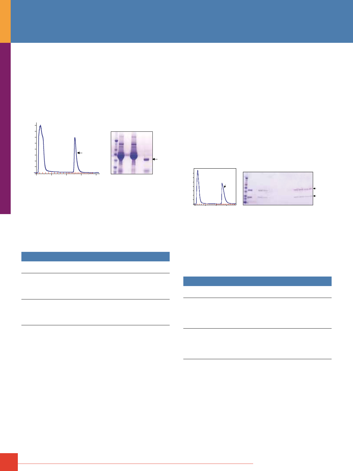
74
For more information, or to download product instructions, visit www.www.thermoscientific.com/pierce
Thermo Scientific Pierce Protein A/G Agarose Cartridges
Genetically-engineered protein that combines the IgG binding
domains of both Protein A and Protein G.
Highlights:
• Single support provides all benefits of Protein A and Protein G
• Binds a wider range of antibodies than Protein A and
Protein G beads
0
500
1000
1500
2000
2500
3000
3500
4000
mAU
0.0 10.0 20.0 30.0 40.0mL
A1 A2 A3 A4 A5 A6 A7 A8 A9 A10 A12 B11 B9 B8 B7 B6 B5 B4 B3 B2 B1 C1 C2 C3 C4
A)
Pure
Rabbit
IgG
B)
M S FT E
Rabbit
IgG
kDa
210
110
80
47
32
25
A
280
Thermo Scientific Pierce Protein A/G Chromatography Cartridges are effec-
tive for affinity purification of immunoglobins from serum. A) 2mL of rabbit
serum was applied to the cartridge and the resulting chromatogram recorded.
B) Fractions were analyzed by SDS-PAGE on a 4-20% Tris Glycine gel stained
with Imperial Protein Stain (Product # 24615). M= Marker proteins, S= Sample
applied, FT= Flow-through during sample load and wash, E= Eluted rabbit IgG.
The arrow indicates the location of the isolated IgG.
See page 61 for more information and other packaging formats of
Pierce Protein A/G Agarose Resin.
Ordering Information
Product #
Description
Pkg. Size
U.S.
Price
89930 Pierce Protein A/G Chromatography
Cartridges, 1mL
Formulation: 1mL resin cartridges with
LC and Luer-Lok Fittings.
Sufficient for: Binding 7mg human IgG per cartridge.
2 cartridges
$200
89931 Pierce Protein A/G Chromatography
Cartridges, 5mL
Formulation: 5mL resin cartridge with
LC and Luer-Lok Fittings.
Sufficient for: Binding 35mg human IgG per cartridge.
1 cartridge
$399
Thermo Scientific Pierce Protein L Agarose Cartridges
Great for purifying ScFv or Fab fragments and monoclonal
antibodies containing kappa light chains.
Highlights:
• Binds kappa light chains from a wide range of species without
interfering with antigen-binding sites
†
• Binds to all classes of Ig (e.g., IgG, IgM, IgA, IgE and IgD)
†
• Binds single-chain variable fragments (ScFv)
†
• Does not bind bovine, goat or sheep Igs
• Does not bind to bovine antibodies, making it ideal for purification
of mouse IgG
†
from cell culture supplemented with bovine serum
†
Note: Lambda light chains and some kappa light chains will not bind. Binding will only
occur if the appropriate kappa light chains are present.
0
50
100
150
200
250
300
350
mAU
0.0 10.0 20.0 30.0mL
A1 A2 A3 A4 A5 A6 A7 A8 A9 A10 A12 B11 B9 B8 B7 B6 B5 B4 B3 B2 B1 C1 C2 C3 C4
M L Flow-through Wash Eluate
Sample
Application
kDa
210
80
47
32
25
16.5
IgA
A)
A
280
B)
IgA
heavy
chain
kappa light
chain
110
Thermo Scientific Pierce Protein L Chromatography Cartridges isolate and purify
immunoglobulin classes IgG, IgM, IgA, IgE and IgD via their kappa light chains.
A) Human IgA Serum (2mg) was applied in 100mM sodium phosphate, 150mM
sodium chloride, pH 7.2 to a 1mL Pierce Protein L Chromatography Cartridge
and purified at a flow rate of 1mL/minute. Target was eluted in 0.1M glycine,
pH 2.8. B) Fractions were analyzed by SDS-PAGE on a 4-20% Tris-Glycine gel
stained with GelCode Blue Stain Reagent (Product # 24590).
See page 61 for more information and other packaging formats of
Pierce Protein L Agarose Resin.
Ordering Information
Product #
Description
Pkg. Size
U.S.
Price
89928 Pierce Protein L
Chromatography Cartridges, 1mL
Formulation: 1mL resin cartridges with
LC and Luer-Lok fittings.
Sufficient for: Binding 5 to 10mg human IgG
per cartridge.
2 cartridges
$190
89929 Pierce Protein L
Chromatography Cartridges, 5mL
Formulation: 5mL resin cartridge with
LC and Luer-Lok Fittings.
Sufficient for: Binding 25 to 50mg human IgG
per cartridge.
1 cartridge
$375
FPLC Cartridges
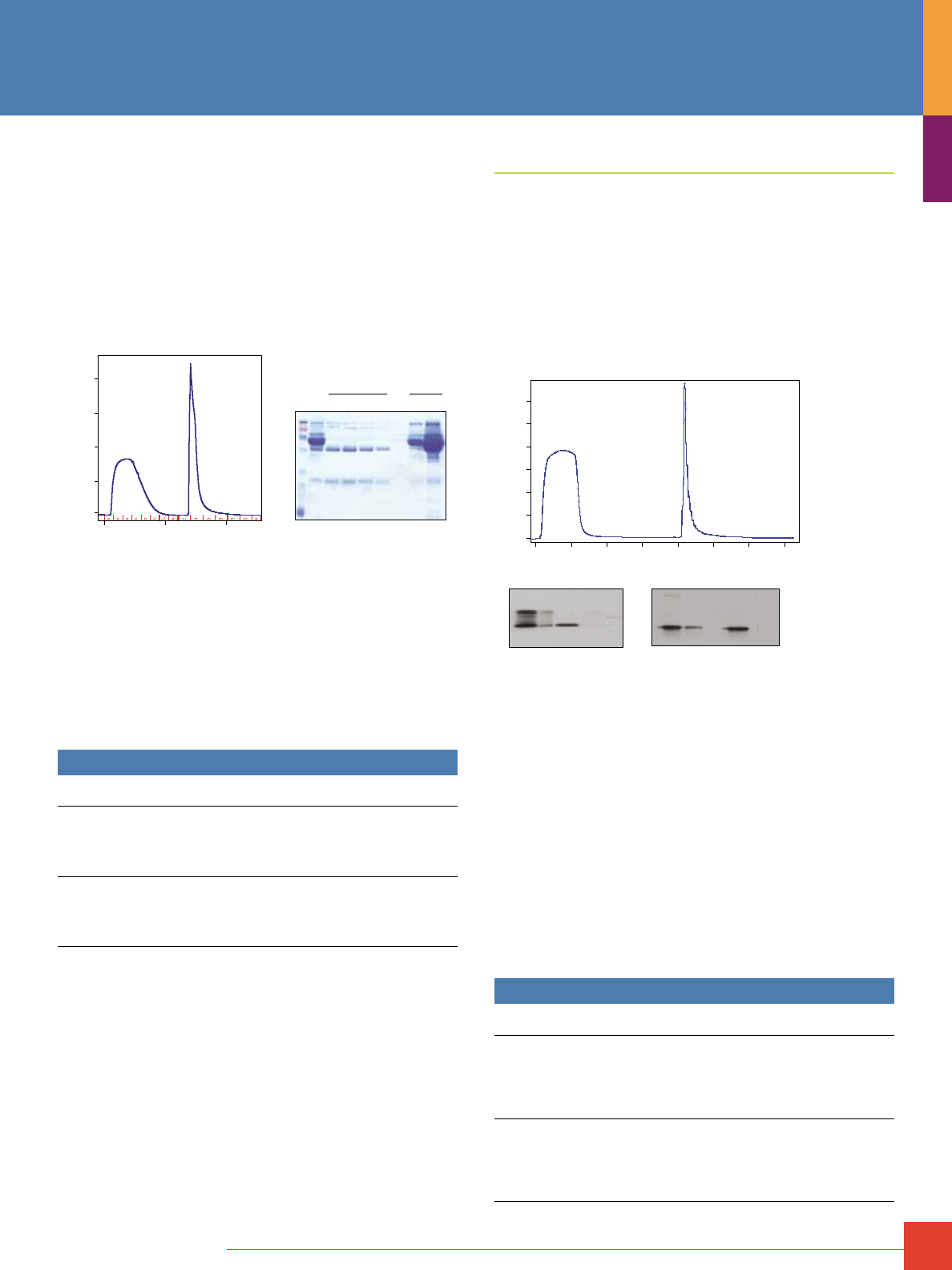
To order, call 800-874-3723 or 815-968-0747. Outside the United States, contact your local branch office or distributor.
75
Thermo Scientific Melon Gel IgG Purification Resins
Easily purify IgG from serum in 15 minutes.
Highlights:
• No tedious binding, washing and multiple elution steps
• Purifies antibodies from serum four to six times faster than
Protein A or G methods
• Recover antibodies from many species with >80% purity
• No harsh elution conditions means antibodies retain more activity
• Reusable support
M L
10.00.0
0
1000
2000
3000
4000
mAU
20.0mL
1 2
FT
R
A
280
FT R
3 4 1 2
Effective reverse-affinity purfication of antibody with Thermo Scientific Melon
Gel Cartridges. Normal human serum (1mL) was applied to a 1mL Melon Gel
Cartridge at a flow rate of 1mL/minute. Protein contaminants bind to the resin
while the antibodies flow through (FT). Volume of recovered antibody = 4 to
5mL. Regeneration (R) solution strips the bound protein contaminants so that
the cartridge can be reused multiple times. The purity of the isolated antibody
was evaluated by SDS-PAGE (right panel) and stained with Thermo Scientific
GelCode Blue Stain Reagent (Product # 24590). M = MW Marker, L = sample
loaded, FT = flow-through and R = regeneration fractions.
See page 63 for more information and other packaging formats of
Melon Gel Resin.
Ordering Information
Product #
Description
Pkg. Size
U.S.
Price
89932 Melon Gel Chromatography Cartridges, 1mL
Formulation: 1mL resin cartridges with
LC and Luer-Lok Fittings.
Sufficient for: IgG purification from 1 to 2mL
serum per cartridge.
2 cartridges
$100
89933 Melon Gel Chromatography Cartridges, 5mL
Formulation: 5mL resin cartridge with
LC and Luer-Lok Fittings.
Sufficient for: IgG purification from 5 to 10mL
serum per cartridge.
1 cartridge
$201
Phosphoprotein FPLC Purification
Thermo Scientific Pierce Phosphoprotein Enrichment Kit
Purify and isolate phosphoproteins for analysis by Western blotting
or mass spectrometry.
Highlights:
• Low nonspecific protein contamination from complex biological
samples, such as cell culture lysate and mouse tissue extract
• Easy spin format enables enrichment of phosphorylated proteins
in less than 2 hours
• Achieves higher yields than other commercially available kits
Phosho-p42/44 MAPK
Cytochrome-C (12KD)
Non-phosphorylated protein
25ug 10ug FT El25ug 10ug FT El
TCL TCL
A)
B)
0
200
400
600
800
1000
1200
mAU
10.00.0 20.0 30.0 40.0 50.0 60.0 70.0mL
Non-phosphorylated
Proteins
Flow-Through
Concentrated
Phosphorylated
Proteins In Eluate
A
280
Figure A. Enrichment of phosphorylated proteins from K562 cell lysate,
4mg (10mL), processed using a Thermo Scientific Pierce Phosphoprotein
Enrichment Chromatography Cartridge, 1mL. Binding/Wash Buffer: 50mM
MES 2-(N-morpholino) ethanesulfonic acid, monohydrate), 250mM sodium
chloride, 25mM adipic acid, 0.25% CHAPS; pH 5.0; Elution Buffer: 100mM
sodium phosphate, 500mM sodium chloride, 0.25% CHAPS; pH 7.5; Flow rate
during sample application 0.3mL/min; 1mL/min during wash and elution.
Figure B. Thermo Scientific Pierce Phosphoprotein Enrichment
Chromatography Cartridge, 1mL, provides high specificity for the enrichment
of phosphoproteins from complex biological samples. Concentrated Flow-
through and Elution fractions were resolved on SDS-PAGE. Gel lanes were
normalized by protein concentration, 10mg/lane. Western blot analysis was
performed using phospho-specific antibodies. Cytochrome C is a negative
control for nonspecific binding of non-phosphorylated proteins. High specific-
ity of binding affinity for the phosphorylated target was demonstrated by the
presence of Phospho-p42/44 MAPK in the Eluate and absence, thereof, in the
Flow-through.
See page 53 for more information and other packaging formats of
Pierce Protein Phosphoprotein Enrichment Resin.
Ordering Information
Product #
Description
Pkg. Size
U.S.
Price
87743 Pierce Phosphoprotein Enrichment
Chromatography Cartridges, 1mL
Formulation: 1mL resin cartridges with
LC and Luer-Lok fittings.
Sufficient for: Purifying samples containing 4mg total
protein (400µg phosphoprotein) per cartridge.
2 cartridges
$124
87744 Pierce Phosphoprotein Enrichment
Chromatography Cartridges, 5mL
Formulation: 5mL resin cartridge with
LC and Luer-Lok fittings.
Sufficient for: Purifying samples containing 20mg total
protein (2mg phosphoprotein) per cartridge.
1 cartridge
$310
76
For more information, or to download product instructions, visit www.www.thermoscientific.com/pierce
Biotinylated Protein FPLC Purification
Thermo Scientific Streptavidin Agarose and
Neutravidin Agarose Cartridges
Resins with high biotin affinity and low nonspecific binding.
Highlights:
• Exhibit high binding affinity towards the vitamin Biotin (vitamin H)
• Captures biotinylated proteins
• Derivatives of the protein avidin from chicken egg whites that
differ by their source and glycosylation level
Pierce High Capacity Streptavidin Chromatography Cartridge
0
500
1000
1500
2000
2500
3000
3500
mAU
0.0 20.0 40.0 60.0 80.0mL
Yield
655 µg
Target
GE HiTrap Streptavidin HP
0
500
1000
1500
2000
2500
3000
mAU
0.0 20.0 40.0 60.0 80.0mL
Yield
469 µg
Target
Pierce High Capacity NeutrAvidin Chromatography Cartridge
0
500
1000
1500
2000
2500
3000
mAU
0.0 20.0 40.0 60.0 80.0mL
Yield
388 µg
Target
Thermo Scientific High Capacity Streptavidin Chromatography Cartridge
achieves comparable yield and performance in binding of small biotinylated
molecule, Biotin 4 Nitrophenyl ester (BpNPE), as compared to HiTrap.
Sample: 4.8mg BpNPE (in 24mL)
Column Size: 1mL
Binding Buffer: 0.5M Sodium Acetate, pH 5.0
Elution Buffer: 0.5M NaOH Streptavidin cartridges; 0.1M NaOH NeutrAvidin
cartridge
Flow Rate: Sample Application 0.3mL/minute; Wash and Elution 1mL/minute
Monitored 2 wavelengths: Flow through/ Wash in Na Acetate A 270nm (red);
Elution in NaOH A 410nm (pink)
Instrument: AKTApurifier
Yield determined by OD A 410nm (Extinction Coefficient for BpNPE 18.3 A410nm)
Supplier
Cartridge
Size
Biotinylated
BSA Bound
Pierce High Capacity Streptavidin
Chromatography Cartridge
1mL 12.9mg
5mL 75.9mg
GE HiTrap Streptavidin HP
1mL 10.7mg
5mL
(Not offered
in 5mL size)
Pierce High Capacity NeutrAvidin
Chromatography Cartridge
1mL 12.8mg
5mL 70.mg
Binding capacity of Thermo Scientific High Capacity Streptavidin
Chromatography Cartridges is comparable to that of HiTrap. Columns were
overloaded with Biotinylated BSA and purified per manufacturer's instructions.
Binding capacity was determined using the Thermo Scientific Pierce BCA
Protein Assay Kit (Product # 23225).
Note: Capacity for the avidin resins was determined indirectly by subtracting the
unbound biotinylated BSA present in the flow-through fractions from the total amount
applied to the column.
Comparison of biotin-binding proteins.
Biotin-Binding Protein
Avidin Streptavidin NeutrAvidin
Molecular Weight
67K 53K 60K
Biotin-binding Sites
4 4 4
Isoelectric Point (pI)
10 6.8-7.5 6.3
Specificity
Low High Highest
Affinity for Biotin (K
d
)
10
-15
M 10
-15
M 10
-15
M
Nonspecific Binding
High Low Lowest
See pages 67-68 for more information and other packaging formats
of Steptavidin and Neutravidin Agarose Resin
Ordering Information
Product #
Description
Pkg. Size
U.S.
Price
87739 High Capacity Streptavidin Chromatography
Cartridges, 1mL
Formulation: 1mL crosslinked 6% beaded agarose
cartridges with LC and Luer-Lok fittings.
Sufficient for: Binding > 100µg biotin per cartridge
(> 10mg biotinylated BSA per cartridge).
2 cartridges
$176
87740 High Capacity Streptavidin Chromatography
Cartridge, 5mL
Formulation: 5mL crosslinked 6% beaded agarose
cartridge with LC and Luer-Lok fittings.
Sufficient for: Binding > 500µg biotin per cartridge
(> 50mg biotinylated BSA per cartridge).
1 cartridge
$240
87741 High Capacity NeutrAvidin Chromatography
Cartridges, 1mL
Formulation: 1mL crosslinked 6% beaded agarose
cartridges with LC and Luer-Lok fittings.
Sufficient for: Binding > 75µg biotin per cartridge
(> 8mg biotinylated BSA per cartridge).
2 cartridges
$149
87742 High Capacity NeutrAvidin Chromatography
Cartridge, 5mL
Formulation: 5mL crosslinked 6% beaded agarose
cartridge with LC and Luer-Lok fittings.
Sufficient for: Binding > 375µg biotin per cartridge
(> 40mg biotinylated BSA per cartridge).
1 cartridge
$206
FPLC Cartridges
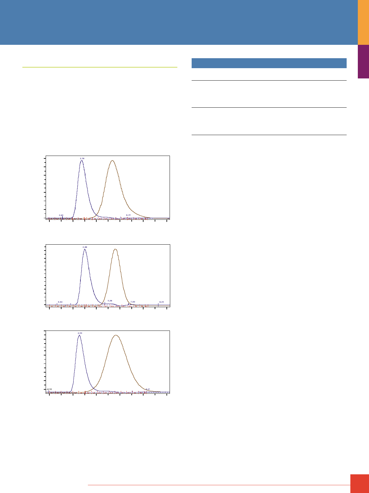
To order, call 800-874-3723 or 815-968-0747. Outside the United States, contact your local branch office or distributor.
77
Protein Desalting
Thermo Scientific Zeba Desalting Chromatography Cartridges
The best protein desalting resin in cartridge format.
Highlights:
• Exceptional desalting and protein-recovery characteristics
compared to other commercially available resins
• Successful with very dilute (25μg/mL) protein samples
• Greater than 95% retention (removal) of salts and other
small molecules and good recovery of proteins and other
macromolecules
A
280
(blue)
Conductivity (brown)
A
280
(blue)
Conductivity (brown)
A
280
(blue)
Conductivity (brown)
Thermo Scientific Pierce Chromatography Cartridge
Supplier G Chromatography Cartridge
Supplier B Chromatography Cartridge
mAU
600
500
400
300
200
100
0
0.0 2.0 4.0 6.0 8.0 10.0mL
0.0 2.0 4.0 6.0 8.0 10.0mL
0.0 2.0 4.0 6.0 8.0 10.0mL
0
100
200
300
400
500
600
700
mAU
mAU
600
500
400
300
200
100
0
Efficient salt removal and protein recovery with desalting chromatography
cartridge. Bovine serum albumin (1mg) in 1M NaCl was applied to 5mL Thermo
Scientific Zeba Desalting Cartridge (top) at a flow rate of 5mL/minute. Cartridge
profile shows isocratic elution of BSA (blue) and NaCl detected by conductivity
(brown). Greater than 95% of the BSA was recovered and more than 95% of
the was salt removed. Results were comparable to those obtained with desalt-
ing cartridges from other suppliers (results for the Thermo Scientific Cartridge
were essentially identical to that obtained with more expensive GE Healthcare
and Bio-Rad Products (suppliers G and B, respectively).
Ordering Information
Product #
Description
Pkg. Size
U.S.
Price
89934 Zeba Desalting Chromatography Cartridges,
7K MWCO, 1mL
Formulation: 1mL resin cartridges with
LC and Luer-Lok fittings.
Sufficient for: Desalting 50 to 250µL samples per use.
5 cartridges
$147
89935 Zeba Desalting Chromatography Cartridges,
7K MWCO, 5mL
Formulation: 5mL resin cartridges with
LC and Luer-Lok fittings.
Sufficient for: Desalting 100 to 1500µL samples per use.
5 cartridges
$168
78
For more information, or to download product instructions, visit www.www.thermoscientific.com/pierce
In addition to the few affinity supports whose ligands have broad
application to many different protein methods, there are many
others whose applications are more narrowly defined or are
incorporated into kits for very specific purposes. These include
kits to isolate cell surface proteins using biotinylation and
NeutrAvidin Agarose, kits to enrich for phosphoproteins, glyco-
proteins and ubiquitinated proteins. The stand-alone resins on
this page and the next include those that can be used to remove
trypsin from protein digest, isolate blood proteins, purify C-reactive
protein and capture ribonucleosides. In addition, page 80 presents
our complete line of magnetic beads, together with their magnet
accessories.
Immobilized Soybean Trypsin Inhibitor
For effective removal of trypsin, chymotrypsin and elastase from
protein digests.
Applications:
• Purifying trypsin, chymotrypsin and elastase
1,2
• Removing proteases from activated pancreatic juices
3
References
1. Feinstein, G., et al. (1974). Euro. J. Biochem. 43(3), 569–581.
2. Peterson, L.M., et al. (1976). Biochemistry 15(12), 2501–2508.
3. Reeck, G.R., et al. (1971). Biochemistry 10(25), 4690–4698.
Ordering Information
Product #
Description
Pkg. Size
U.S.
Price
20235 Immobilized Soybean Trypsin Inhibitor
Support: 4% beaded agarose
Capacity: ≥6mg trypsin/mL of resin
2mL
$98
Immobilized Pepstatin
An excellent cathepsin binding matrix.
H
N
H
N
H
N
Pepstatin =
R = (4-amino-3-hydroxy-6-methyl) heptanoic acid
Thermo Scientific Immobilized Pepstatin
i-ValValValR
AlaR
Pepstatin
Agarose
Bead
Immobilized Heparin
Reference
Helseth, Jr., D.L. and Veis, A. (1984). Proc. Natl. Acad. Sci. USA 81, 3302–3306.
Ordering Information
Product #
Description
Pkg. Size
U.S.
Price
20215 Immobilized Pepstatin
Support: Crosslinked 6% beaded agarose
Spacer: Diaminodipropylamine
Capacity: 1–2mg of pepsin/mL of resin
5mL
$159
Additional Thermo Scientific Affinity Supports and Kits
To order, call 800-874-3723 or 815-968-0747. Outside the United States, contact your local branch office or distributor.
79
Immobilized Heparin
Use to isolate many blood proteins that have enzymatic activities.
Applications:
• Enrich lysates for nucleic acid-binding proteins
• Isolate many blood proteins
Agarose
Bead
O
OSO
3
-
OH
COO
-
O
NHOSO
3
-
OH
CH
2
OSO
3
-
O
O
n
O
Thermo Scientific Immobilized Heparin
Reference
Smith, P.K., et al. (1980). Anal. Biochem. 109, 466–473.
Ordering Information
Product #
Description
Pkg. Size
U.S.
Price
20207 Immobilized Heparin
Support: 4% beaded agarose
Loading: ≥0.2mg of heparin/mL of resin
(determined by the colorimetric method)
Kit
$73
Immobilized p-Aminophenyl Phosphoryl Choline
For C-reactive protein binding.
O
O
N
+
P
O
O
O
-
HN
Thermo Scientific Immobilized p-Aminophenyl Phosphoryl Choline
Agarose
Bead
Reference
Robey, F.A. and Liu, T.Y. (1981). J. Biol. Chem. 256, 969–975.
Ordering Information
Product #
Description
Pkg. Size
U.S.
Price
20307 Immobilized p-Aminophenyl
Phosphoryl Choline
Support: Crosslinked 6% beaded agarose
Capacity: ≥3mg of human C-reactive protein/mL of resin
5mL
$202
Immobilized D-Galactose
For lectin and galactosidase binding.
Applications:
• Human alpha-galactosidase A purification
1
• E. coli heat labile enterotoxin purification
2
• C-type lectin purification
3
• Cholera toxin (CT) purification
4,5
O
S
O
O
O
OH
OH
O
HO
OH
Thermo Scientific Immobilized Thio-alpha-D-Galactose
Agarose
Bead
References
1. Yasuda, K., et al. (2004). Protein Expression and Purification. 37, 499–506.
2. Okamoto, K., et al. (1998). J. Bacteriol. 180, 1368–1374.
3. Matsumoto, J., et al. (2001). Development. 128, 3339–3347.
4. Bowman, C.C. and Clements, J.D., et al. (2001). Infec. Immun. 69, 1528–1535.
5. Tinker, J.K., et al. (2003). Infec. Immun. 71, 4093–4101.
Ordering Information
Product #
Description
Pkg. Size
U.S.
Price
20372 Immobilized D-Galactose
Support: 6% beaded agarose
Capacity: ≥20mg jacalin/mL resin
5mL
$145
Immobilized Boronic Acid
For ribonucleoside isolation.
B
OH
OH
H
N
Thermo Scientific Immobilized Boronic Acid
O
OH
Resin
Bead
References
Vlassara, H., et al. (1981). Proc. Natl. Acad. Sci. USA 78, 5190–5192.
Gehrke, C.W., et al. (1978). J. Chromatogr. 150, 455–476.
Ordering Information
Product #
Description
Pkg. Size
U.S.
Price
20244 Immobilized Boronic Acid
Support: Polyacrylamide resin beads
Capacity: ≥99% binding and recovery of 110μmol
AMP/mL of resin
Spacer: m-aminophenyl group
Loading: 100μmol boronate/mL of resin
10mL
$190
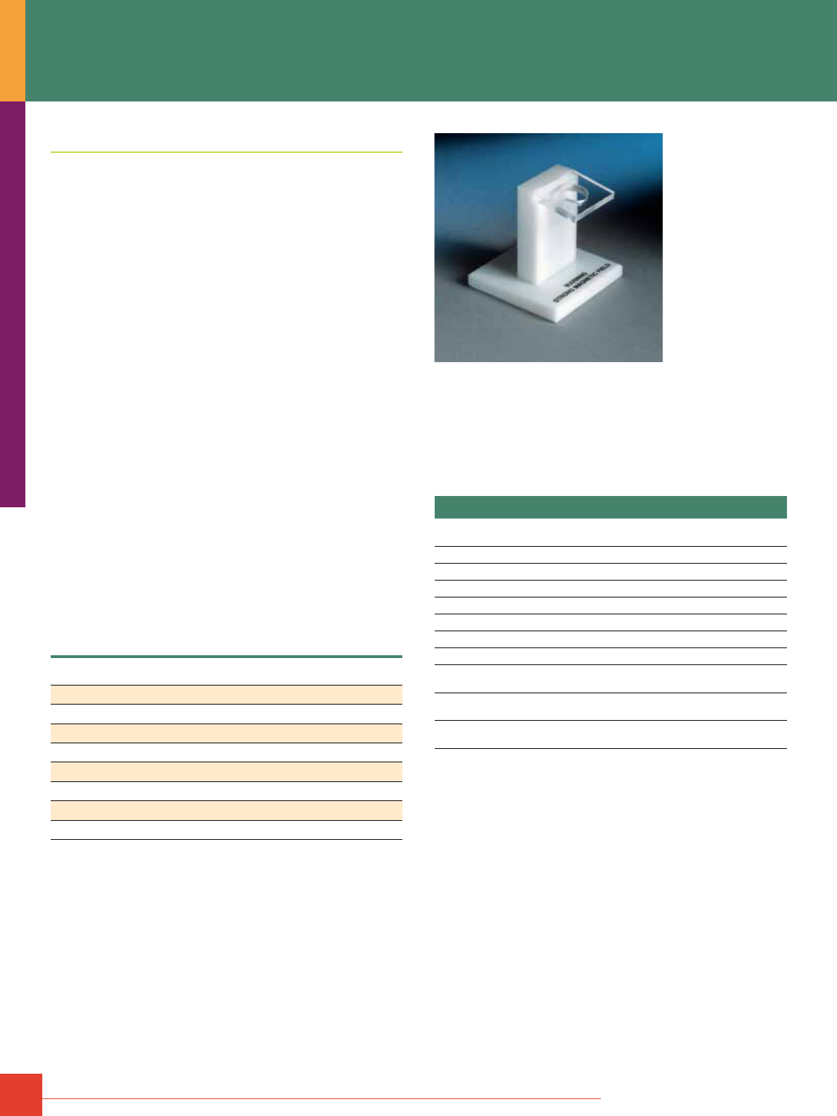
80
For more information, or to download product instructions, visit www.www.thermoscientific.com/pierce
Thermo Scientific MagnaBind Beads and Supports
A convenient method for isolating biomolecules using affinity
binding, while retaining biological activity.
Magnetic beads are a convenient affinity support for a variety
of assays, which allow easy purification of the target without
columns or centrifugation. After a binding step in an affinity
purification procedure, the magnetic particles are easily and
rapidly collected by placing the microcentrifuge tube or reaction
vessel next an appropriate rare-earth magnet (Figure 1).
Thermo Scientific MagnaBind beads respond rapidly to
MagnaBind Magnets but can be easily dispersed and regathered
multiple times (i.e., they will not irreversibly aggregate) because
they do not have any magnetic memory. MagnaBind Beads are
available pre-coated with Protein A, Protein G, streptavidin, anti-
mouse or anti-rabbit antibodies. Activated beads, with either
amine or carboxyl groups, are also available for attaching other
proteins or affinity ligands to a magnetic particle.
Highlights:
• Available pre-coated with popular affinity ligands or derivatized
for covalent attachment of proteins and other specific ligands
• Beads do not irreversibly aggregate because they have no
magnetic memory; collect and disperse the beads multiple times
if needed
• Most separations require a short five- to 10-minute bench-top
procedure
Applications:
• Cell sorting using positive or negative selection
• Protein purification or immunoassays using direct or indirect methods
Characteristics of underivatized Thermo Scientific MagnaBind Beads.
Composition
Silanized iron oxide
Magnetization
25–35EMU/g
Type of Magnetization
Superparamagnetic (no magnetic memory)
Surface Area
>100m
2
/g
Settling Rate
4% in 30 minutes
Effective Density
2.5g/mL
Number of Beads
1 x 10
8
beads/mg
pH Stability
Aqueous solution, above pH 4.0
Concentration
5mg/mL
Note: To establish a microbe-free preparation, MagnaBind Beads can be washed with
antibiotic medium or g-irradiated.
Figure 1. Thermo Scientific MagnaBind Magnet for 1.5mL Microcentrifuge
Tube. Thermo Scientific MagnaBind Beads in solutions within a microcentrifuge
tube are rapidly “pelleted” when the tube is placed in the magnetized holder.
Magnets for six microcentrifuge tubes and 96-well microplates are also available.
References
Chaudhuri, T.K., et al. (2001). Cell 107, 235–246.
Newey, S.E., et al. (2001). J. Biol. Chem. 276, 6645–6655.
Xu, X., et al. (2001). J. Biol. Chem. 276, 43221–43230.
Ordering Information
Product #
Description
Pkg. Size
U.S.
Price
21344 MagnaBind Streptavidin Beads
5mL
$271
21348 MagnaBind Protein A Beads
5mL
$325
21349 MagnaBind Protein G Beads
5mL
$325
21354 MagnaBind Goat Anti-Mouse IgG Beads
50mL
$256
21356 MagnaBind Goat Anti-Rabbit IgG Beads
50mL
$256
21353 MagnaBind Carboxyl Derivatized Beads
5mL
$127
21352 MagnaBind Amine Derivatized Beads
5mL
$130
21358 MagnaBind Magnet for
96-Well Plate Separator
1 magnet
$392
21357 MagnaBind Magnet for 1.5mL
Microcentrifuge Tube
1 magnet
$152
21359 MagnaBind Magnet for 6 x 1.5mL
Microcentrifuge Tubes
1 magnet
$305
Thermo Scientific Affinity Supports

To order, call 800-874-3723 or 815-968-0747. Outside the United States, contact your local branch office or distributor.
81
ÄKTA, ÄKTApurifier
™
, HiTrap
®
and pGEX
®
are trademarks of GE Healthcare.
Phos-Select
™
is a tradmark of Sigma-Aldrich Co.
NanoLC
™
is a trademark of Eskigent.
Trisacryl
®
is a trademark of Pall Corporation.
Luer-Lok
™
is a trademark of Becton, Dickinson and Company.
Brij
®
is a registered trademark of ICI Americas.
Cibacron
®
is a registered trademark of Ciba Specialty Chemicals, Inc.
Triton
®
is a registered trademark of Rohm & Haas Company.
Tween
®
is a trademark of ICI Americas.

Contact Information
Belgium and Europe,
the Middle East
and Africa Distributors
Tel: +32 53 85 71 84
France
Tel: 0 800 50 82 15
The Netherlands
Tel: 076 50 31 880
Germany
Tel: 0228 9125650
United Kingdom
Tel: 0800 252 185
Switzerland
Tel: 0800 56 31 40
Email: perbio.euromarketing@thermofisher.com
www.thermoscientific.com/perbio
United States
Tel: 815-968-0747 or 800-874-3723
Customer Assistance E-mail:
Pierce.CS@thermofisher.com
www.thermoscientific.com
© 2010 Thermo Fisher Scientific Inc. All rights reserved.
These products are supplied for laboratory or manufacturing
applications only. Unless indicated otherwise on the inside
back cover, all trademarks are property of Thermo Fisher
Scientific Inc. and its subsidiaries.
1602015 11/10 Printed in the U.S.
