INSTRUCTIONS
Pierce Biotechnology
PO Box 117
(815) 968-0747
www.thermo.com/pierce
3747 N. Meridian Road
Rockford, lL 61105 USA
(815) 968-7316 fax
Number Description
46110
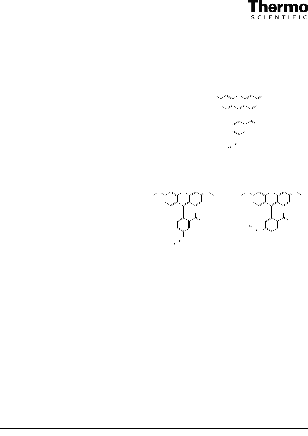
Fluorescein Isothiocyanate (FITC), 1 g
OHO O
O
OH
N
C
S
Molecular Weight: 389.38
Exact Mass: 389.04
Extinction Coeff: 72,000 M
-1
cm
-1
(at 594 nm in aqueous buffer, pH 8)
Ex/Em Wavelength : 494/520 nm
CAS #3326-32-7
46112
Tetramethylrhodamine-5-(and 6)-isothiocyanate (TRITC), 10 mg
Molecular Weight: 478.97
Exact Mass: 478.10
Extinction Coeff: 100,000 M
-1
cm
-1
(at 544 nm in methanol)
Ex/Em Wavelength : 541/572 nm
CAS #6749-36-6
ON N+
O
O
N
C
S
ON N+
O
O
N
C
S
and
Cl
-
Cl
-
Storage: Upon receipt store at -20°C. Product is shipped at ambient temperature.
Store fluorescent dye in foil pouch with desiccant to protect from light and moisture.
Introduction
FITC and TRITC are among the most simple and commonly used reagents for protein fluorescent labeling. These
isothiocyanates will crosslink to amino, sulfhydryl, imidazoyl, tyrosyl or carbonyl groups on a protein. However, only the
derivatives of primary and secondary amines generally yield stable products. Reactions are most efficient at pH 8-9, and must
be performed in an amine-free buffer such as carbonate/bicarbonate. Avoid Tris buffer, which is a primary amine that will
compete with the intended labeling reaction. Antibody and other proteins can be effectively labeled with several fluorophore
tags per protein molecule when reacted with a 20- to 25-fold molar excess of isothiocyanate-activated fluorophore. Excess
nonreacted and hydrolyzed reagent can be removed by dialysis or gel filtration.
Procedure for Labeling Streptavidin with FITC
This method was adapted from Horisberger.
1
Materials Required
• Conjugation buffer: 100 mM carbonate/bicarbonate buffer, pH 9.0
• Streptavidin solution: dissolve 1 mg salt-free streptavidin (Product No. 21122) in 1 ml conjugation buffer
• Zeba Desalt Spin Column (Product No. 89891) or other gel filtration column with a 5,000-7,000 MW cut-off
0365.2
46110 46112
FITC and TRITC

Pierce Biotechnology
PO Box 117
(815) 968-0747
www.thermo.com/pierce
3747 N. Meridian Road
Rockford, lL 61105 USA
(815) 968-7316 fax
2
Procedure
1. Dissolve 200 µg of FITC in 200 µl of conjugation buffer and immediately mix it with 1 ml of streptavidin solution.
Note: When substituting TRITC for FITC, a similar protocol can be followed; however, the TRITC must first be
dissolved in dimethylsulfoxide (DMSO) at 100 µg/100 µl instead of 200 µg/200 µl in conjugation buffer.
2. Incubate for 1 hour at 37°C in the dark.
3. Remove excess and hydrolyzed FITC by gel filtration.
Procedure for Labeling an Antibody with FITC
Materials Required
• Conjugation buffer: 100 mM carbonate/bicarbonate buffer, pH 9.0
• Antibody solution: dissolve ~1 mg of antibody in 1 ml of conjugation buffer
• Zeba Desalt Spin Column (Product No. 89891) or other gel filtration column with a 5,000-7,000 MW cut-off
Procedure
1. Dissolve FITC in conjugation buffer at a final concentration of 1 mg/ml immediately before use.
2. Add 10 µl of FITC solution to the 1 ml of the antibody solution; mix thoroughly.
3. Incubate for 1 hour at room temperature in the dark.
4. Remove excess and hydrolyzed FITC by gel filtration.
Procedure for Labeling an Antibody with TRITC
This method is adapted from Larsson.
2
Materials Required
• TRITC: dissolve in DMSO at 1 mg/ml
• Conjugation buffer: 100 mM carbonate/bicarbonate buffer, pH 9.0
• Antibody solution: dialyze antibody into conjugation buffer at 6 mg/ml
• Zeba Desalt Spin Column (Product No. 89891) or other gel filtration column with a 5,000-7,000 MW cut-off
Procedure
1. While stirring, slowly add 35 µl of TRITC to 1 ml of the 6 mg/ml antibody solution; mix thoroughly.
2. Incubate for 2 hours at room temperature in the dark.
3. Remove excess and hydrolyzed TRITC by gel filtration.
Additional Information
• Visit the web site for a listing of related Thermo Scientific products (other fluorophores and convenient labeling kits) and
technical resources such as the Tech Tip: Calculate dye:protein (F/P) ratios.
• Fading (photobleaching in tissue sections) can sometimes be reduced by mounting in an alkaline buffered media (pH 9).
2
There are several reagents that may be used with FITC and/or TRITC derivatives to prevent fading including n-propyl
gallate at 0.1-0.25 M dissolved in glycerol for FITC or TRITC.
3
For FITC derivatives, o- or p-phenylenediamine added
to the mounting buffer from 1 µg/ml to 1 mg/ml in glycerin also may be used.
2,3
References
1. Horisberger, M. (1984). In Immunolabeling for Electron Microscopy. Polak, J., Varndel, I. Ed. Elsevier: Amsterdam, p. 98.
2. Larsson, L. (1988). Immunocytochemistry: Theory and Practice. CRC. Boca Raton, 77-83, 224-225.
3. Goding, J. (1986). Monoclonal Antibodies: Principles and Practice, 2nd ed. Academic, London.

Pierce Biotechnology
PO Box 117
(815) 968-0747
www.thermo.com/pierce
3747 N. Meridian Road
Rockford, lL 61105 USA
(815) 968-7316 fax
3
This product (“Product”) is warranted to operate or perform substantially in conformance with published Product specifications in effect at the time of sale,
as set forth in the Product documentation, specifications and/or accompanying package inserts (“Documentation”) and to be free from defects in material and
workmanship. Unless otherwise expressly authorized in writing, Products are supplied for research use only. No claim of suitability for use in applications
regulated by FDA is made. The warranty provided herein is valid only when used by properly trained individuals. Unless otherwise stated in the
Documentation, this warranty is limited to one year from date of shipment when the Product is subjected to normal, proper and intended usage. This
warranty does not extend to anyone other than the original purchaser of the Product (“Buyer”).
No other warranties, express or implied, are granted, including without limitation, implied warranties of merchantability, fitness for any particular
purpose, or non infringement. Buyer’s exclusive remedy for non-conforming Products during the warranty period is limited to replacement of or
refund for the non-conforming Product(s).
There is no obligation to replace Products as the result of (i) accident, disaster or event of force majeure, (ii) misuse, fault or negligence of or by Buyer, (iii)
use of the Products in a manner for which they were not designed, or (iv) improper storage and handling of the Products.
Current versions of product instructions are available at
www.thermo.com/pierce. For a faxed copy, call 800-874-3723 or contact your local distributor.
© 2010 Thermo Fisher Scientific Inc. All rights reserved. Unless otherwise indicated, all trademarks are property of Thermo Fisher Scientific Inc. and its
subsidiaries. Printed in the USA.
