
Female Organs of the
Reproductive System
Unit. Human Structure and Function: The Reproductive System
Problem Area. Understand the Organs of the Reproductive System
Lesson. Female Organs of the Reproductive System
¢
Student Learning Objectives.
Instruction in this lesson should result in students
achieving the following objectives:
1
Describe the anatomy of the female reproductive organs.
2
Describe the functions of the female reproductive organs.
3
Describe the hormones of the female reproductive system.
¢ List of Resources.
The following resources may be useful in teaching this lesson:
Badasch, S., and Chesebro, D. (2004). Introduction to Health Occupations,
6th ed. Upper Saddle River, NJ: Prentice Hall.
Black, J., and Matassarin-Jacobs, E. (1993). Luckmann and Sorenson’s
Medical-Surgical Nursing: A Psychophysiologic Approach, 4th ed. Philadel
-
phia: W.B. Saunders.
Gerdin, J. (2003). Health Careers Today, 3rd ed. St. Louis: Mosby.
Miller, B., and Keane, C. (1992). Encyclopedia and Dictionary of Medicine,
Nursing, and Allied Health, 5th ed. Philadelphia: W.B. Saunders.
Simmers, L. (2004). Diversified Health Occupations, 6th ed. Clifton Park, NY:
Delmar Learning.
Lesson: Female Organs of the Reproductive System
Page 1 u www.MyCAERT.com
Copyright © by CAERT, Inc. | Reproduction by subscription only. | L630072

Smeltzer, S., and Bare, B. (2004). Textbook of Medical-Surgical Nursing,
10th ed. Philadelphia: Lippincott Williams & Wilkins.
Sorrentino, S. (2000). Mosby’s Textbook for Nursing Assistants, 5th ed. St.
Louis: Mosby.
Utah State Office of Education (2006). Reproductive System. Retrieved from
MAP CD.
¢
List of Equipment, Tools, Supplies, and Facilities
ü
Overhead or PowerPoint projector
ü
Visual(s) from accompanying master(s)
ü
Copies of sample test, lab sheet(s), and/or other items designed for duplication
ü
Materials listed on duplicated items
ü
Computers with Internet access
ü
Classroom resource and reference materials
¢ Terms.
The following terms are presented in this lesson (shown in bold italics):
>
alveolar glands
>
androgens
>
areola
>
Bartholin glands
> body
>
broad ligament
>
cervix
>
cilia
>
clitoris
>
corpus luteum
>
endometrium
>
estrogen
>
fallopian tubes
>
fimbriae
>
follicle-stimulating hormone
>
fornix
>
fundus
>
gonadotropin-releasing hormone
>
greater vestibular glands
>
hymen
>
labia majora
>
labia minora
>
lobules
>
luteinizing hormone
Lesson: Female Organs of the Reproductive System
Page 2 u www.MyCAERT.com
Copyright © by CAERT, Inc. | Reproduction by subscription only. | L630072

>
mammary glands
>
mons pubis
>
myometrium
>
ova
>
ovarian follicles
>
ovarian ligament
>
ovaries
>
oviducts
>
perimetrium
>
progesterone
>
relaxin
>
rugae
>
uterine tubes
>
uterus
>
vagina
>
vaginal orifice
>
vestibule
>
vulva
¢ Interest Approach.
Use an interest approach that will prepare the students for the
lesson. Teachers often develop approaches for their unique class and student situation. A
possible approach is included here.
Tell the students to look around the classroom and notice the males and
females in the room. Tell them to notice the different hairstyles between the
guys and girls, the different clothing styles, and the different body builds. Now
ask them if they know what is different between the insides of the males and
females. Tell them that this lesson will focus on the female organs of the
reproductive system.
Lesson: Female Organs of the Reproductive System
Page 3 u www.MyCAERT.com
Copyright © by CAERT, Inc. | Reproduction by subscription only. | L630072

SUMMARY OF CONTENT AND
TEACHING STRATEGIES
Objective 1: Describe the anatomy of the female reproductive organs.
Anticipated Problem: What is the anatomy of the female reproductive system?
I. The female reproductive system is composed of individual organs that work together.
A. The ovaries are the paired female gonads.
1. They are about the size and shape of almonds and are located within the pel
-
vic cavity.
2. The ovaries measure 4 cm long, 2 cm wide, and 1 cm thick.
3. The ovaries are held in place by several ligaments which attach them to the
abdominal wall and the uterus.
a. The broad ligament and the ovarian ligament are two of the ligaments
that hold the ovaries in place.
4. The ovaries contain small sacs called ovarian follicles in which the ovum, or
egg, matures.
5. The corpus luteum is a solid glandular mass that is left after the follicle
releases the ovum.
6. The ovaries contain an immense number of ova.
B. The oviducts are tubes approximately 12.5 cm (5 in.) long.
1. The oviducts are also called the fallopian tubes or uterine tubes.
2. These tubes are made of muscle tissue.
3. The oviducts are not directly attached to the ovaries but are attached to the
uterus.
4. The ends of the tube by the ovaries have finger-like projections called
fimbriae.
5. The oviduct is lined with cilia, which are tiny hair-like projections.
C. The uterus is a pear-shaped muscular organ located in the pelvic cavity.
1. It is divided into three sections called the fundus, body, and cervix.
a. The fundus is the superior dome-shaped area of the uterus above the
openings to the uterine tubes.
b. The body is the major, tapering, central portion of the uterus that contains
the hollow interior uterine cavity.
c. The cervix is the narrow, thick muscular area that opens into the vagina.
Lesson: Female Organs of the Reproductive System
Page 4 u www.MyCAERT.com
Copyright © by CAERT, Inc. | Reproduction by subscription only. | L630072

2. The uterus also contains three layers called the perimetrium, the myometrium,
and the endometrium.
a. The perimetrium is the outermost layer of the uterus which provides a
small amount of protection to the uterus.
b. The myometrium is the middle, smooth muscle layer of the uterus, which
makes up the majority of the uterus.
c. The endometrium is the innermost layer of the uterus.
3. The uterus is also supported by the broad ligament.
D. The vagina is a muscular tube that opens to the outside of the body.
1. It is approximately 7.5 cm (3 in.) long.
2. The top of the vagina that attaches to the cervix has a recessed area called
the fornix.
3. The vagina is lined with mucous membrane that has folds in it.
a. These folds are called rugae, and they allow the vagina to enlarge during
childbirth.
4. The vaginal orifice is the distal end of the vagina that opens into the external
environment.
5. The hymen is a thin fold of vascular mucous membrane that forms a border
around the vaginal orifice, partially closing it.
6. The greater vestibular glands can be found near the opening of the vagina.
a. These are also called the Bartholin glands.
b. These are mucus-producing glands that provide lubrication during
intercourse.
E. The vulva is the term used to describe the external genitalia of the female and
includes several parts.
1. Mons pubis is an elevation of adipose tissue covered by skin and coarse
pubic hair.
a. The labia majora is an area of lateral, longitudinal folds extending from
front to back.
(1) These have much adipose tissue and are covered by pubic hair.
(2) They are homologous (sharing the same origin, but different function)
to the male scrotum.
b. The labia minora are the medial, longitudinal folds of the vulva.
(1) These have no adipose tissue or pubic hair.
2. The clitoris is a small, cylindrical mass of nervous and erectile tissue.
3. The vestibule is the cleft between the labia minora.
F. The mammary glands, or breasts, are considered accessory organs of the female
reproductive system.
1. The mammary glands are actually modified sweat glands.
2. Each gland consists of 15 to 20 lobes or compartments separated by adipose
tissue.
Lesson: Female Organs of the Reproductive System
Page 5 u www.MyCAERT.com
Copyright © by CAERT, Inc. | Reproduction by subscription only. | L630072

3. The amount of adipose tissue between the lobes determines the size of the
breast.
4. Breast size is not related to the ability to produce milk.
5. Each lobe is broken down into smaller compartments called lobules which
contain milk-secreting glandular cells called alveolar glands (milk-producing
glands of the breast).
6. The areola is the dark, circular, pigmented area that encircles the nipple.
7. The nipple is the raised area on the breast that an infant suckles to receive
milk.
Many techniques can be used to help students master this objective. As an
example, students could use Chapter 6 in Mosby’s Textbook for Nursing
Assistants. Also, use VM–A.
Objective 2: Describe the functions of the female reproductive organs.
Anticipated Problem: What are the functions of the female reproductive organs?
II. The female reproductive organs work together to allow production of offspring to
continue the species.
A. The ovaries are responsible for producing ova (eggs).
1. A newborn female infant’s ovaries contain a large amount of potential ova.
2. Once that female reaches puberty, several eggs per month will ripen, but usu-
ally only one egg is released.
3. Once the ovum has matured in the ovarian follicle, the follicle will rupture and
release the egg.
4. The ovaries are also responsible for releasing hormones.
B. The oviducts, or fallopian tubes, help to transport ova from the ovaries to the
uterus.
1. The ovum is carried from the ovaries to the fallopian tubes by the sweeping
action of the fimbriae.
2. Once in the fallopian tubes, the ovum is transported down the tube by the
movement of the cilia and by the action of peristalsis (waves of involuntary
muscle contractions).
3. The oviduct is also the site of egg fertilization by a sperm.
C. The function of the uterus is to serve as the site of gestation or pregnancy for the
developing embryo/fetus.
1. The inner lining of the uterus, the endometrium, changes throughout a
female’s menstrual cycle.
a. The endometrium first prepares to house the fertilized egg and give it
nourishment.
Lesson: Female Organs of the Reproductive System
Page 6 u www.MyCAERT.com
Copyright © by CAERT, Inc. | Reproduction by subscription only. | L630072

b. If fertilization does not occur, this lining breaks down and is released as
menstrual flow.
D. The vagina functions as a passageway for the spermatozoa.
1. It also serves as the canal for the release of menstrual flow.
2. The vagina is the lower portion of the birth canal.
3. It also functions as a receptacle for the penis during sexual intercourse.
E. The vulva has three functions.
1. The vulva serves as protection for the sexual organs and the urethra.
2. The clitoris is vital for women’s sexual arousal.
3. The vulva stretches to accommodate childbirth.
F. The mammary glands provide milk to the infant after its birth.
1. The suckling action of the infant stimulates milk production and release by the
breast.
Many techniques can be used to help students master this objective. As an
example, students could use Chapter 6 in Mosby’s Textbook for Nursing
Assistants. Also, use VM–B.
Objective 3: Describe the hormones of the female reproductive system.
Anticipated Problem: What are the hormones of the female reproductive system?
III. There are several hormones that are essential to the function of the female
reproductive system.
A. Estrogen is one of the better known hormones of the female reproductive system.
1. Estrogen is produced by the ovaries.
2. There at least six different types of estrogen.
3. Estrogen is responsible for the development and maintenance of the female
reproductive system.
4. It initiates the growth of the endometrium of the uterus.
5. It helps control fluid and electrolyte balance.
6. It maintains blood calcium levels and bone density.
7. It is also responsible for body fat distribution associated with females (buttocks
and thighs).
B. Progesterone works in conjunction with estrogen to prepare the endometrial
lining for implantation of a fertilized ovum.
1. It also stimulates milk secretion.
2. Progesterone maintains the uterine linings during pregnancy.
Lesson: Female Organs of the Reproductive System
Page 7 u www.MyCAERT.com
Copyright © by CAERT, Inc. | Reproduction by subscription only. | L630072

C. Androgens, mostly testosterone, are produced by the ovaries, but only in small
amounts.
1. These are vital to maintain bone density.
2. They are also important in maintaining the female sex drive.
D. Gonadotropin-releasing hormone (GnRH) is a hormone produced by the
hypothalamus in the brain.
1. This hormone controls the production of estrogen in the body.
E. The follicle-stimulating hormone (FSH) and the luteinizing hormone (LH) are
both produced by the pituitary gland.
1. FSH helps to stimulate follicles, thus causing egg production and estrogen pro
-
duction.
2. LH is responsible for the stimulation of the production of progesterone.
F. Relaxin is a hormone produced by the corpus luteum during pregnancy.
1. This hormone is most prominent during the final trimester of pregnancy.
2. Relaxin relaxes the pelvic bones.
3. It also helps dilate the uterine cervix to facilitate delivery of the fetus.
Many techniques can be used to help students master this objective. As an
example, use VM–C.
¢ Review/Summary.
Use the student learning objectives to summarize the lesson.
Have students explain the content associated with each objective. Student responses can
be used in determining which objectives need to be reviewed or taught from a different
angle. Questions at the ends of chapters in the textbook may also be used in the
review/summary.
¢ Application.
Use the included visual masters and lab sheet to apply the information
presented in the lesson.
¢
Evaluation.
Evaluation should focus on student achievement of the objectives for the
lesson. Various techniques can be used, such as student performance on the application
activities. A sample written test is provided.
¢
Answers to Sample Test:
Part One: Listing
1. fundus, body, cervix
2. Any two of the following: passageway for the spermatozoa, serves as the canal for
the release of menstrual flow, lower portion of the birth canal, receptacle for the
penis during sexual intercourse
3. follicle-stimulating hormone, luteinizing hormone
Lesson: Female Organs of the Reproductive System
Page 8 u www.MyCAERT.com
Copyright © by CAERT, Inc. | Reproduction by subscription only. | L630072

4. relaxes the pelvic bones, helps dilate the uterine cervix to facilitate delivery of the
fetus
Part Two: Matching
1. b
2. e
3. f
4. g
5. a
6. c
7. d
Lesson: Female Organs of the Reproductive System
Page 9 u www.MyCAERT.com
Copyright © by CAERT, Inc. | Reproduction by subscription only. | L630072

Sample Test
Name ________________________________________
Female Organs of the
Reproductive System
u
Part One: Listing
Instructions: Complete the following.
1. List the three sections of the uterus.
2. List two functions of the vagina.
3. List the two hormones affecting the reproductive system that are produced by the pituitary
gland.
4. List the two functions of the hormone relaxin.
Lesson: Female Organs of the Reproductive System
Page 10 u www.MyCAERT.com
Copyright © by CAERT, Inc. | Reproduction by subscription only. | L630072

u
Part Two: Matching
Instructions: Match the definition with the correct term.
a. estrogen e. relaxin
b. progesterone f. gonadotropin-releasing hormone
c. follicle-stimulating hormone g. androgens
d. luteinizing hormone
_____1. Maintains the uterine linings during pregnancy
_____2. Helps dilate the uterine cervix to facilitate delivery of the fetus
_____3. Hormone produced by the hypothalamus in the brain
_____4. Important in maintaining the female sex drive
_____5. Responsible for body fat distribution associated with females (buttocks and thighs)
_____6. Helps to stimulate follicles
_____7. Responsible for the stimulation of the production of progesterone
Lesson: Female Organs of the Reproductive System
Page 11 u www.MyCAERT.com
Copyright © by CAERT, Inc. | Reproduction by subscription only. | L630072
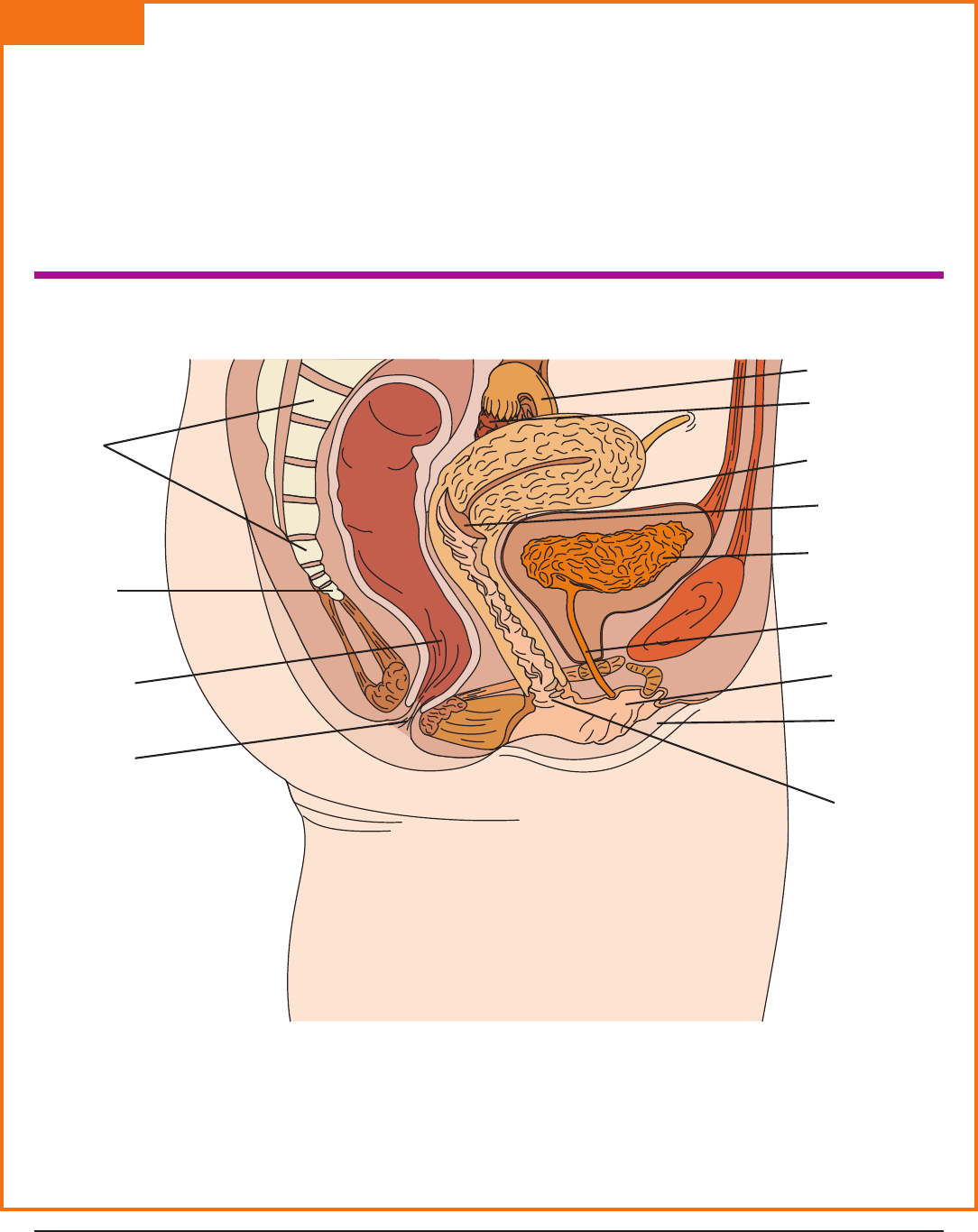
VM–A
FEMALE REPRODUCTIVE
ORGANS AND SURROUNDING
PELVIC STRUCTURES
Lesson: Female Organs of the Reproductive System
Page 12 u www.MyCAERT.com
Copyright © by CAERT, Inc. | Reproduction by subscription only. | L630072
Sacrum
Coccyx
Rectum
Anus
Cervix
Vagina
Labia minora
Labia majora
Urethra
Urinary bladder
Body of uterus
Ovary
Fallopian tube
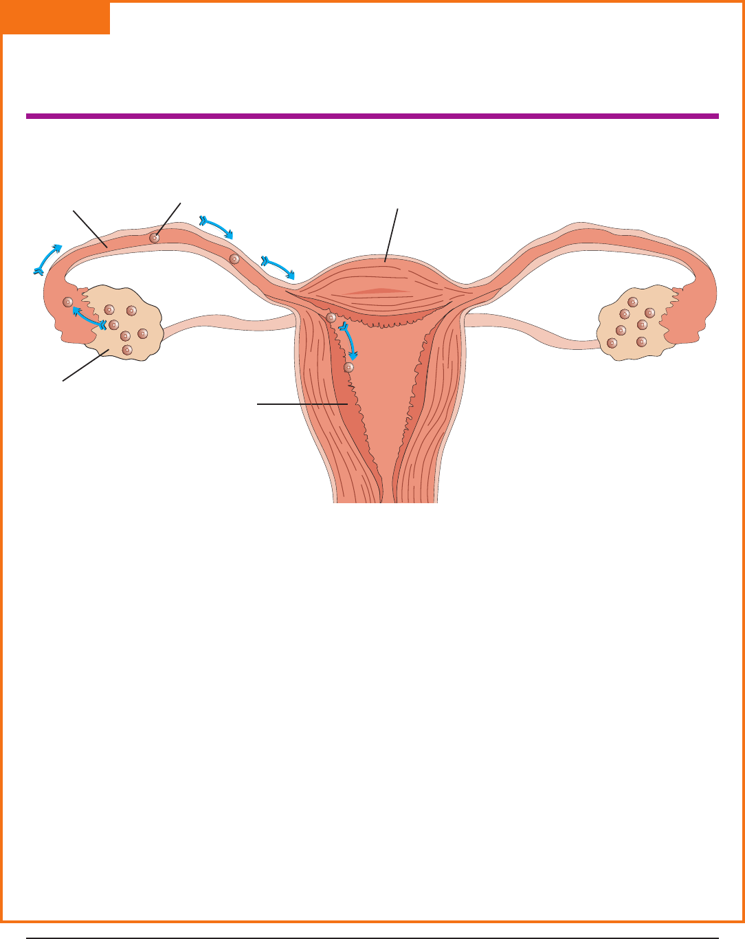
VM–B
FUNCTION OF THE OVARIES
Lesson: Female Organs of the Reproductive System
Page 13 u www.MyCAERT.com
Copyright © by CAERT, Inc. | Reproduction by subscription only. | L630072
Uterus
Endometrium
Fallopian tube
Egg in fallopian tube
Ovary
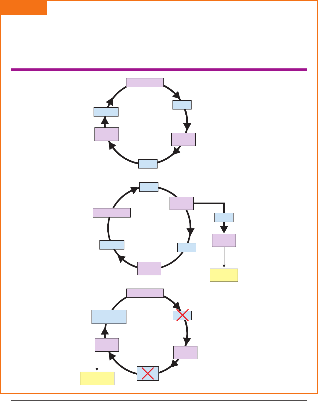
VM–C
FEMALE REPRODUCTIVE
HORMONES
Lesson: Female Organs of the Reproductive System
Page 14 u www.MyCAERT.com
Copyright © by CAERT, Inc. | Reproduction by subscription only. | L630072
Hypothalamus
Hypothalamus
Hypothalamus
Estrogen
Progesterone
and estrogen
Estrogen
GnRH
GnRH
GnRH
LH
FSH
FSH
and LH
FSH
Anterior
Pituitary
Anterior
Pituitary
Anterior
Pituitary
Ovarian
follicle
Corpus
luteum
Ovarian
follicle
Ovarian
follicle
Ovulation
by day 14
Degenerates
after day 28
Day 1–10
Day 14–28
Day 10–14
i
n
h
i
b
i
t
s
i
n
h
i
b
i
t
s

LS–A: Teacher Information
Labeling the Structures of the
Female Reproductive System
Purpose
The purpose of this activity is to allow students to identify the structures of the female
reproductive system.
Objectives
1. Visually learn the location of the various structures of the female reproductive system.
2. Label the diagram of the structures involved with the female reproductive system.
Materials
t
copies of VM–A (with labels removed)
t
writing utensil
Procedure
1. Remove the labels from VM–A.
2. Print the diagram and distribute one to each student.
3. Ask the students to fill in the names of the appropriate structures of the reproductive
system.
Answer Key
The following structures must be labeled:
1. Ovary
2. Fallopian tube/oviduct
3. Uterus
4. Cervix
5. Vagina
6. Labia majora
7. Labia minora
Lesson: Female Organs of the Reproductive System
Page 15 u www.MyCAERT.com
Copyright © by CAERT, Inc. | Reproduction by subscription only. | L630072
