Kenneth A. Krackow, M.D.
Clinical Director,
Department of Orthopaedic Surgery
Kaleida Health System
Buffalo General Hospital
Professor and Full Time Faculty
State University of New York at Buffalo
Department of Orthopaedic Surgery
THE
MEASUREMENT
AND ANALYSIS
OF AXIAL
DEFORMITY
AT THE KNEE

Copyright © 2008 Stryker.
Copyright © 2008 Stryker.
Kenneth A. Krackow, M.D.
Dr. Krackow is a graduate of the Duke
University Medical School in Durham,
North Carolina. After a General Surgery
Internship, he completed his Orthopaedic
Residency at Johns Hopkins University
in Baltimore, Maryland.
Currently, Dr. Krackow is Professor
of Orthopaedic Surgery at the State
University of New York at Buffalo. He is
also Clinical Director of Orthopaedic
Surgery, Kaleida Health, Buffalo, New
York. He practices at both Kaleida Health
and Buffalo General Hospital.
PREFACE i
U
NIT 1
Lower Extremity Alignment Terminology 1
U
NIT 2
Measurement of Overall Varus/Valgus Deformity at the Knee 7
UNIT 3
Medial Lateral Tibiofemoral Translation – Subluxation 15
UNIT 4
Extra-articular Deformity 17
UNIT 5
Characterizing Deformity About the Knee 23
UNIT 6
Instructional Examples 30
EXAMPLE 1
Varus Deformity of the Femur and Tibia 31
EXAMPLE 2
Varus Deformity of the Tibia 41
EXAMPLE 3
Varus Deformity at the Femur with Minor 49
“Compensation” at the Tibia
EXAMPLE 4
Valgus Deformity at Both the Femur and Tibia 55
EXAMPLE 5
Valgus Deformity at the Femur and Tibia 61
EXAMPLE 6
Valgus Deformity at the Femur and Tibia 67
EXAMPLE 7
Extra-articular Varus Angulation of the Tibia 73
EXAMPLE 8
Valgus Deformity at the Femur and 81
Extra-articular Varus Tibial Angulation
EXAMPLE 9
Extra-articular Varus Angulation of the Femur 89
EXAMPLE 10
Valgus Deformity of the Femur with 97
Extra-articular Valgus Tibial Angulation
UNIT 7
Online Interactive Practice 105
REFERENCES 106
TABLE OF CONTENTS

i
6
Copyright © 2008 Stryker.
Copyright © 2008 Stryker.
PREFACE
This booklet is intended to be used principally by
orthopaedic residents and fellows as an instructional
tool. By combining instruction, illustrated examples,
and problems, it provides a comprehensive overview
of knee alignment—a difficult topic to teach and
explain successfully. The content of this booklet
appears to be quite clear; however, it is in practical
application where the challenges arise. Repetition and
practice are the keys not only to learning how to
assess an X-ray and to perform a proper alignment
analysis but, more importantly, they are critical to
retaining these skills. This booklet provides the reader
with the ability to practice application of knee
alignment principles within the textbook itself as well
as within an interactive format provided by an online
computer simulation module. We hope this booklet
will provide the reader with the information and
experience-based opportunity to help achieve a level
of mastery on this subject that will continue
throughout his or her entire career.
ABOUT THE HOMER
STRYKER CENTER
The Homer Stryker Center is dedicated to improving
patient outcomes through education and research.
The Center offers courses in orthopaedic bioskills
and surgical simulation as well as didactic education
and discussion groups. Our intention is to work
with an internationally recognized faculty to develop
exceptional educational material using modern
education technologies.
2
1
Copyright © 2008 Stryker.
Copyright © 2008 Stryker.
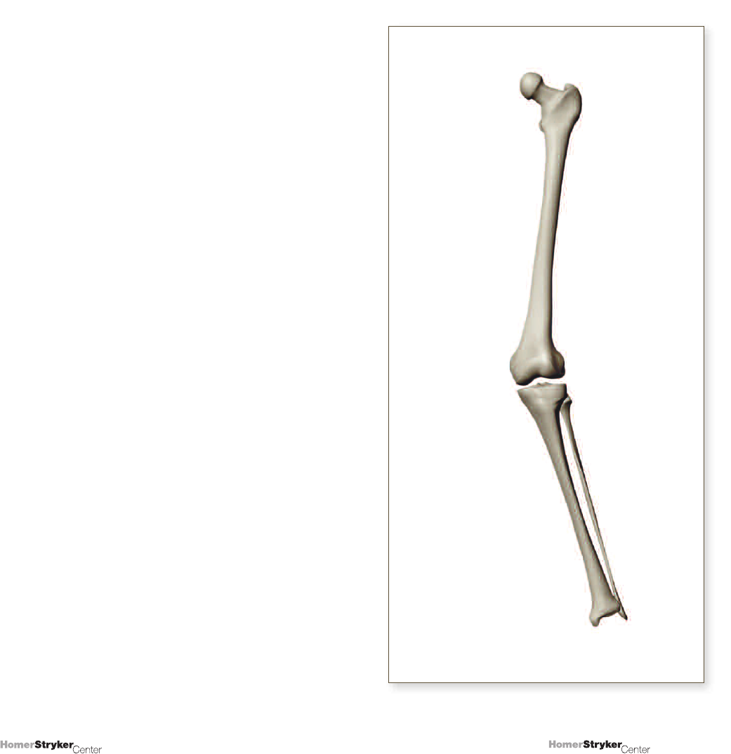
UNIT 1
Lower Extremity Alignment Terminology
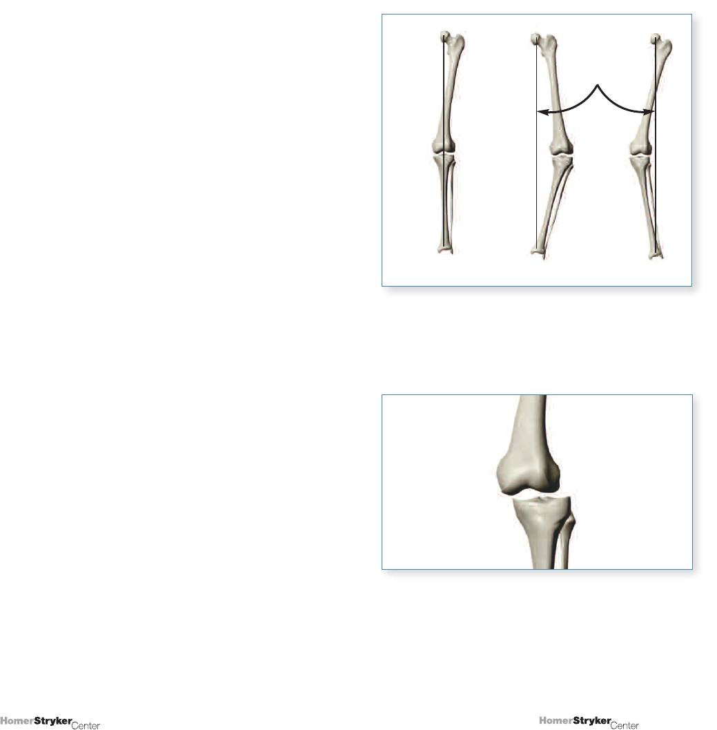
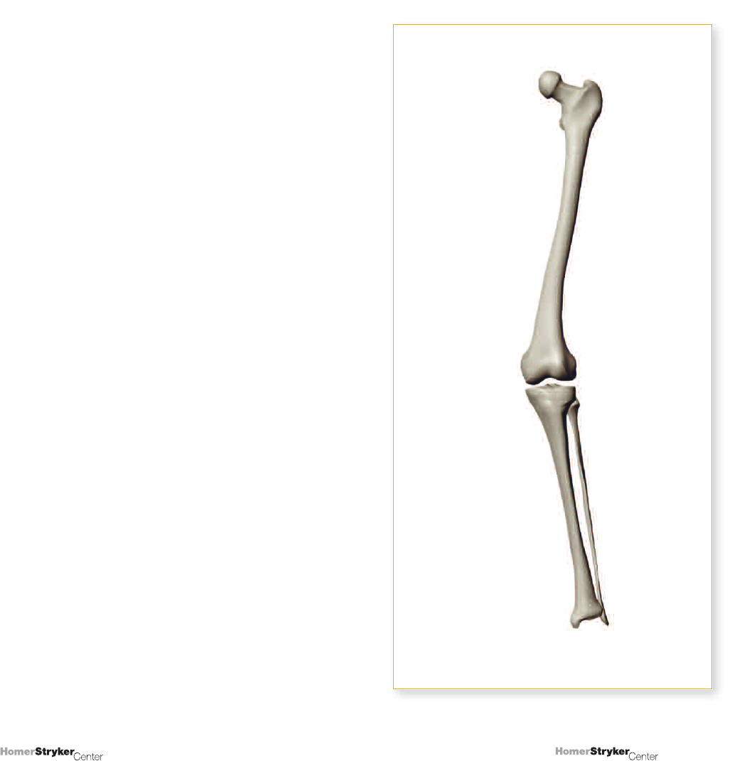
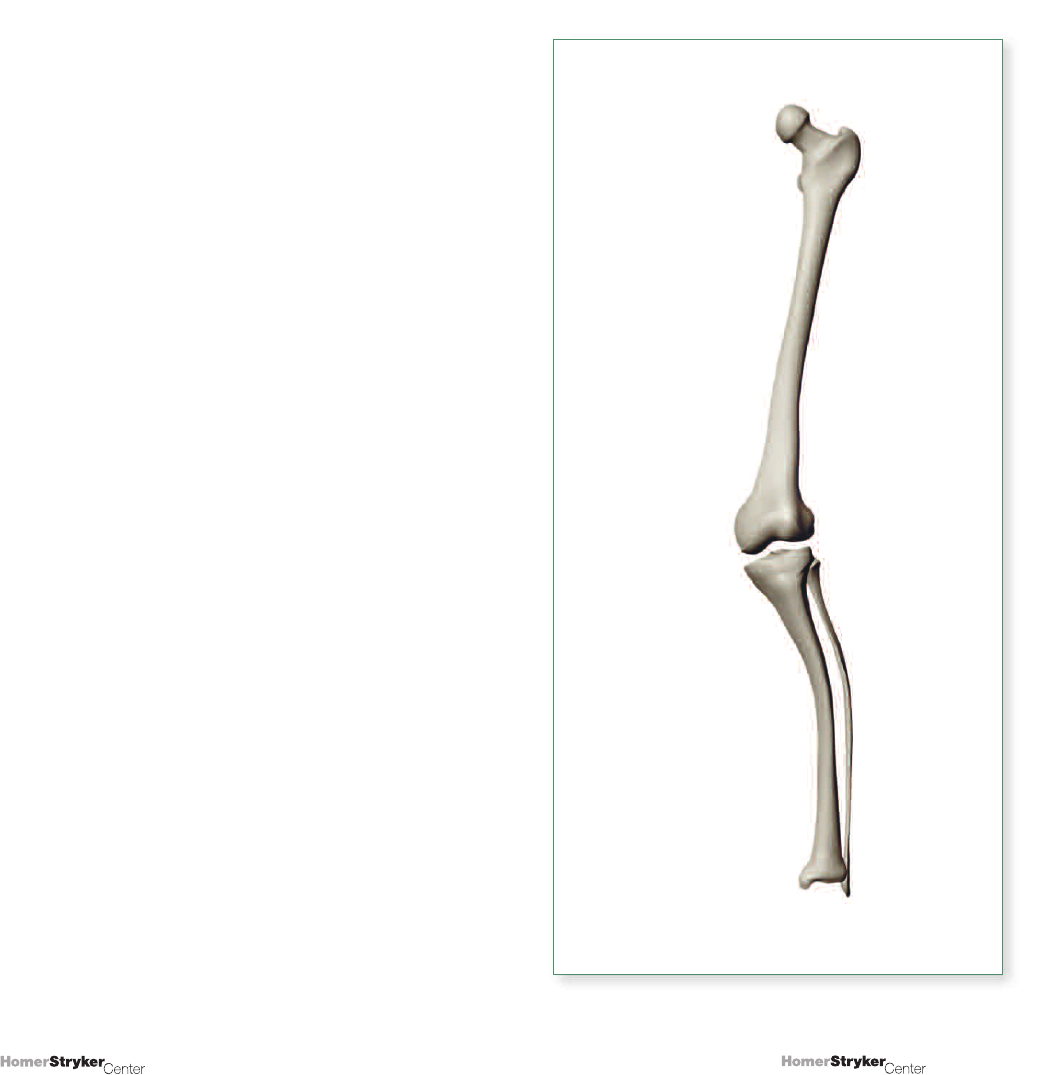
A standard method for determining normal alignment
of the knee is by drawing a line in the A/P plane that
begins at the center of the femoral head, passes through
the center of the knee, and continues to the center of
the ankle (Figure 1.1). This line is often referred to as the
mechanical axis of the lower extremity (MA-LE). If
the line passes medially to the knee center, a varus
deformity is present; if the line passes laterally to the
knee center or center of the distal femur, a valgus
deformity exists.
Distinctions can be made between the knee center and
center of the distal femur. In cases of medial or lateral
subluxation of the knee, for example, they may represent
2 different points. They may also be different from the
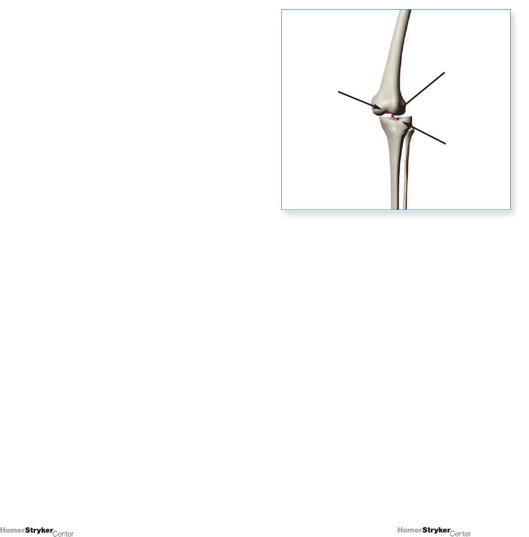
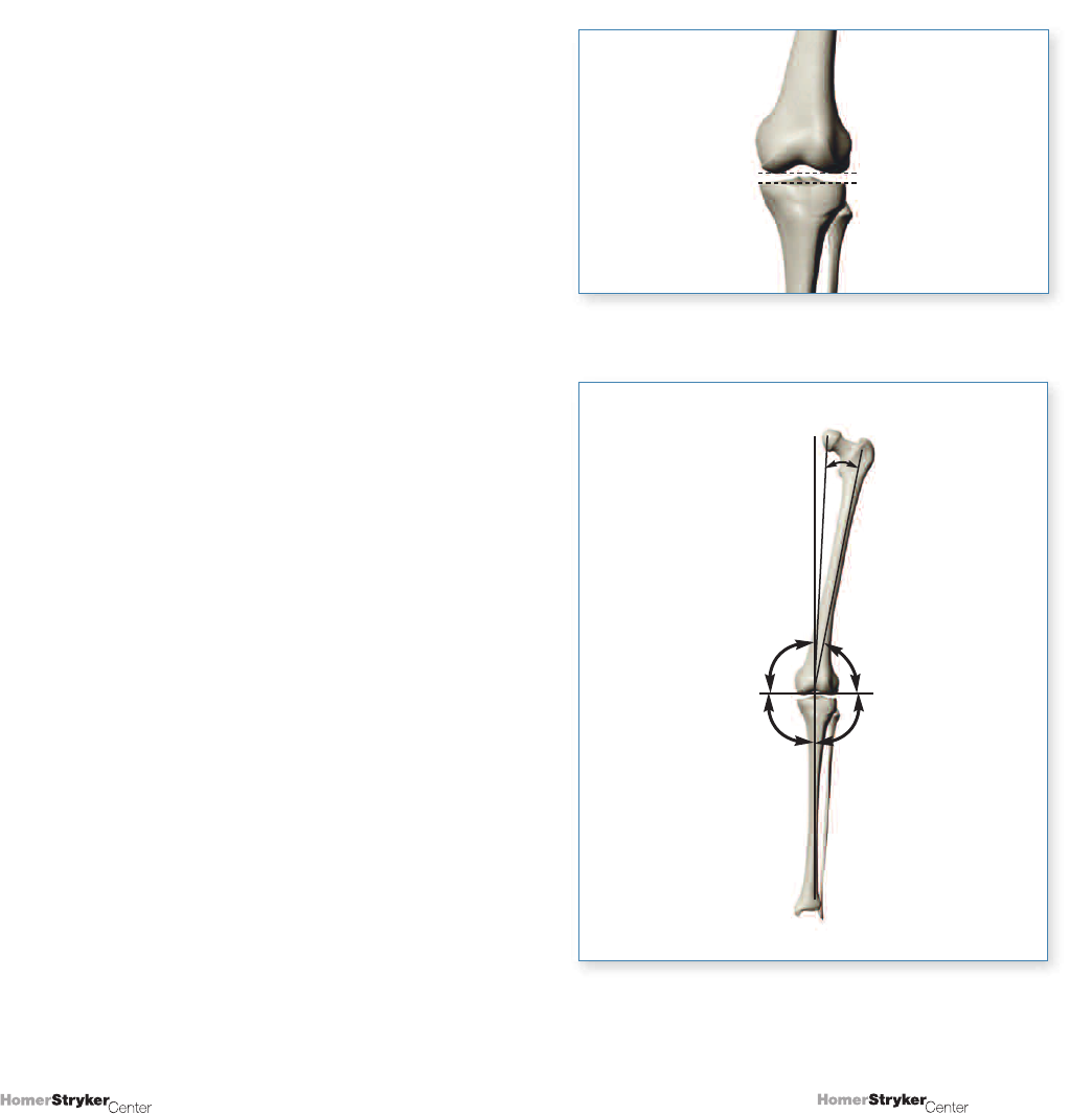
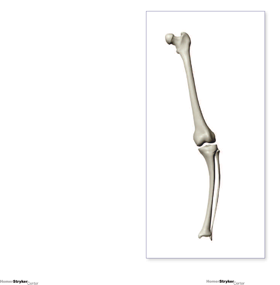
center of the proximal tibia. Figure 1.2 shows a lateral
tibial subluxation, where the center of the distal femur
and the center of the knee at different points.
Normal Alignment
ValgusVarus
MA-LE
Figure 1.1
Normal mechanical alignment and mechanical axis
of the lower extremity in common deformities.
Figure 1.2
Lateral tibial subluxation.
4
3
Copyright © 2008 Stryker.
Copyright © 2008 Stryker.
Several other lines (or axes) are used to describe lower
extremity alignment; all are drawn in the A/P plane.
These are not “axes” in a true sense, although the
nomenclature has found its way into general orthopaedic
terminology. These axes include:
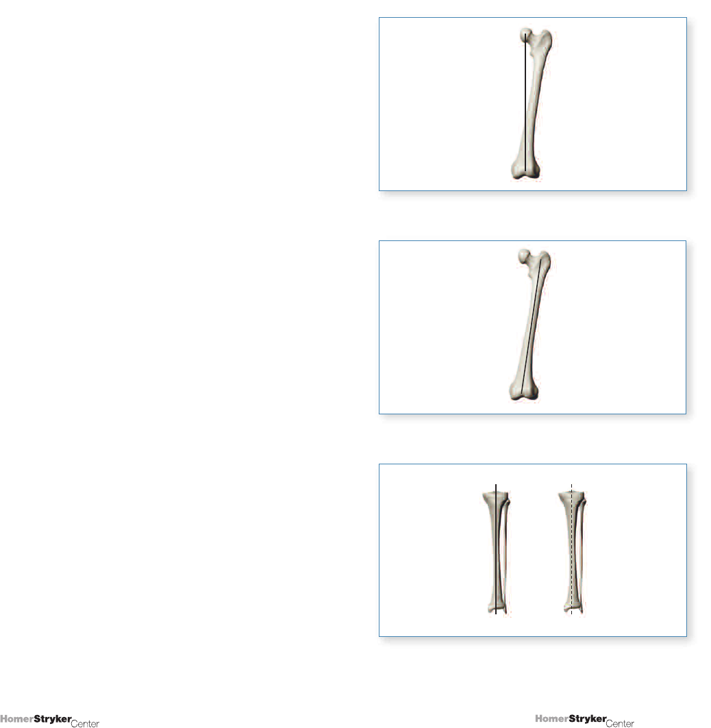
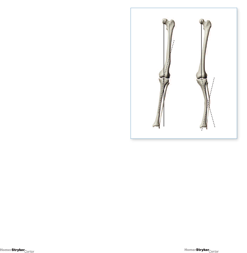
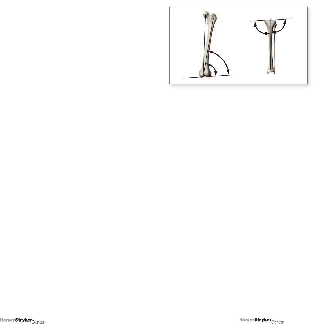
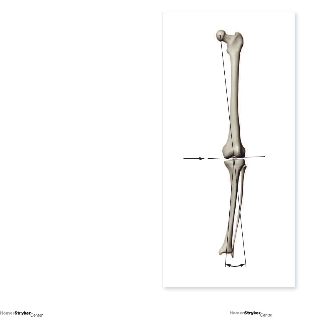
1. Mechanical axis of the femur (MAF):
A line from the center of the femoral head to the center
of the distal femur or center of the knee (Figure 1.3).
2. Femoral shaft axis (FShA):
A line drawn from the center of the proximal femur
to the center of the distal femur or center of the knee,
indicating the overall position of the femoral shaft
(Figure 1.4).
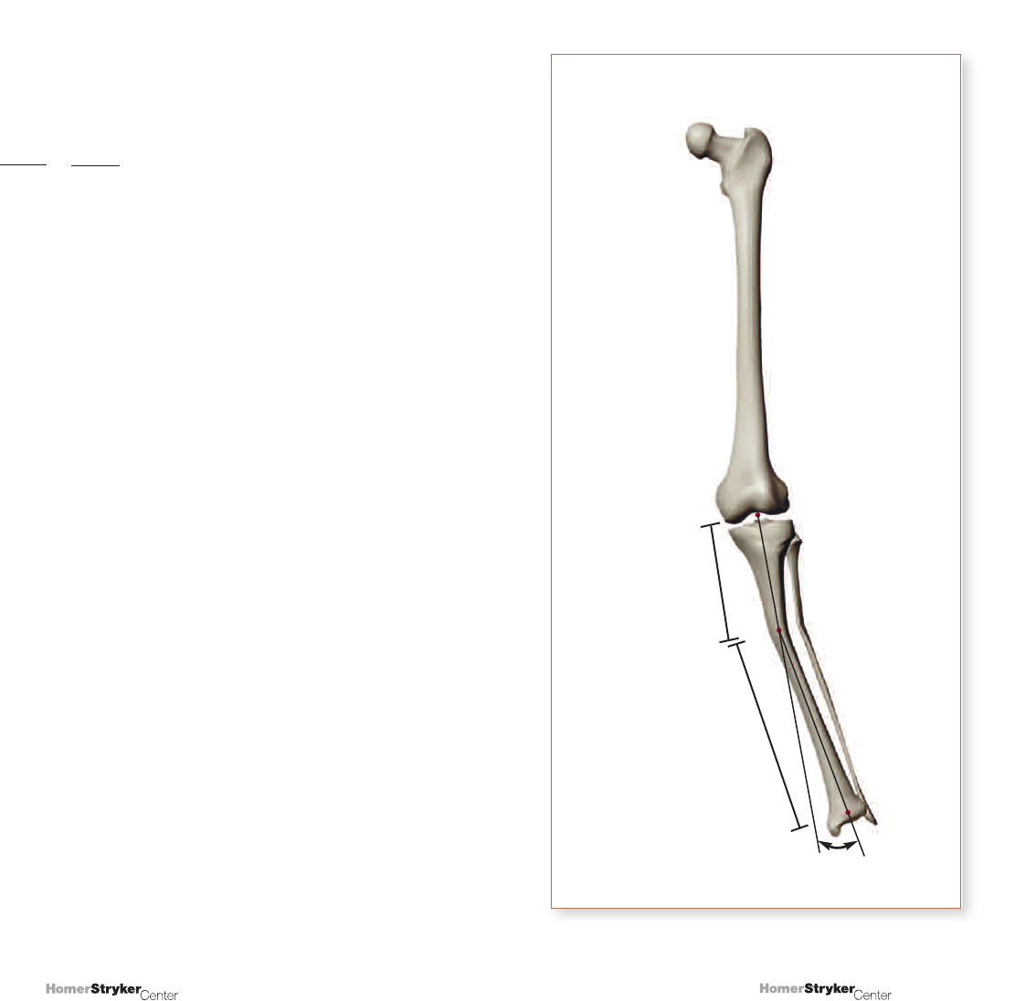
3. Tibial shaft axis (TShA) and Mechanical axis
of the tibia (MAT):
These 2 terms are often used interchangeably, and
both describe a line extending from the center of the
proximal tibia to the center of the ankle (Figure 1.5).
Figure 1.4
Figure 1.3
MAF
FShA
Figure 1.5
MAT
TShA
6
5
Copyright © 2008 Stryker.
Copyright © 2008 Stryker.
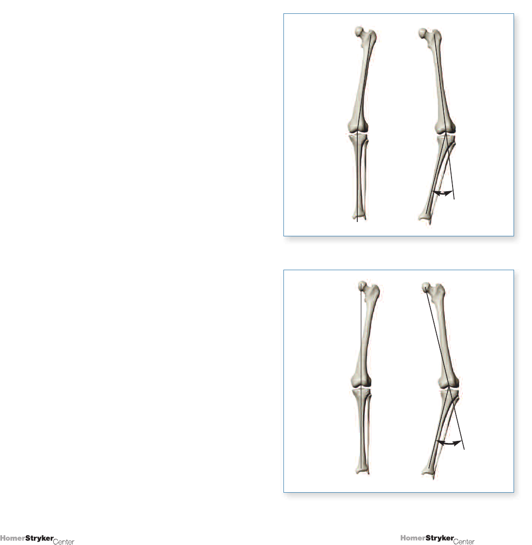
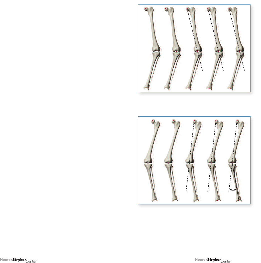
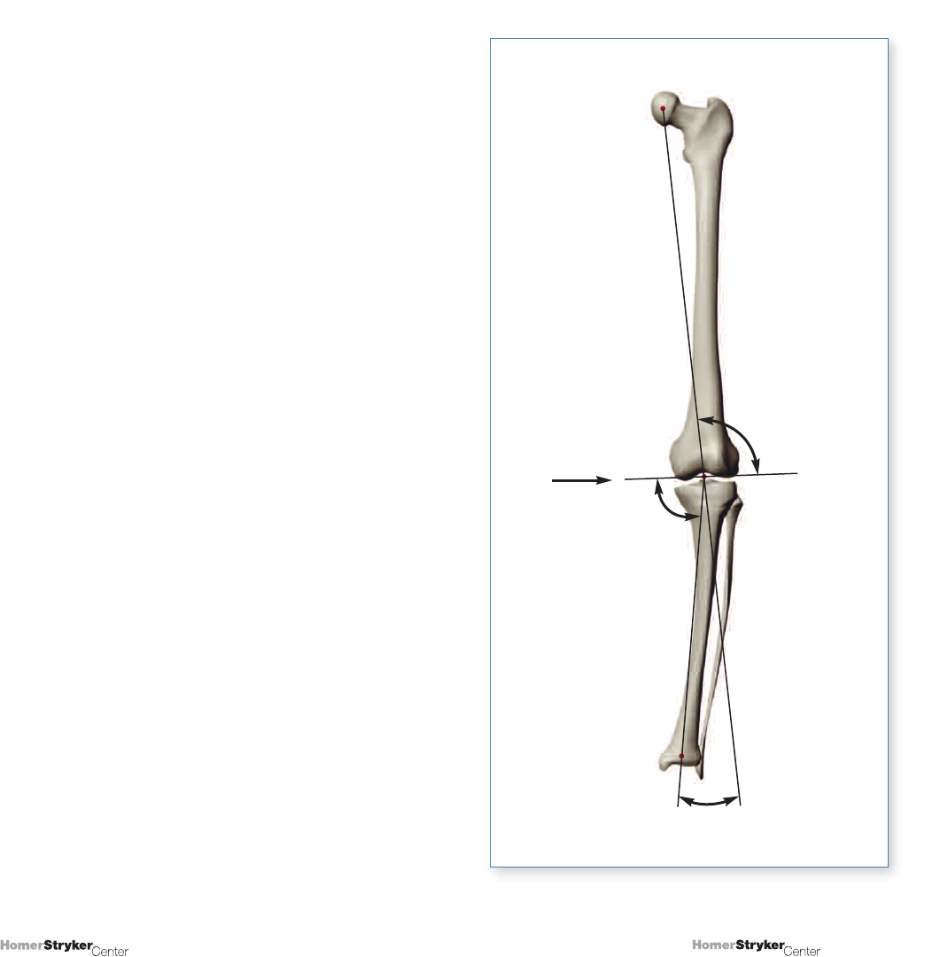
4. Anatomic tibiofemoral angle:
The angle formed when the line that forms the femoral
shaft axis is extended through the distal femur to
form an angle between the femoral shaft axis and the
tibial shaft axis (Figure 1.6). The angle is represented
by numbers that supplement the normal angle of
alignment (e.g., 3°, 6°, etc.) and indicates the extent
of anatomic misalignment or deformity.
5. Mechanical tibiofemoral angle (or mechanical
axis deviation):
The angle formed when the line that forms the
mechanical axis of the femur is extended through the
distal femur to form an angle between the mechanical
axis of the femur and the tibial shaft axis (Figure 1.7).
As with the anatomic tibiofemoral angle, this angle is
represented by numbers that supplement the normal
angle of alignment (e.g., 3°, 6°, etc.) and indicates the
extent of mechanical misalignment or deformity.
Comments
The descriptions that follow are based on some
assumptions that may not be completely precise in an
actual clinical setting including 1) that the knee is seen
in full extension, and 2) that the knee extremity is seen
in neutral rotation.
Additionally, our measurements are represented as being
made on a long standing radiograph, showing essentially
all of each tibia and femur. The femoral head and ankle
would ideally be shown, which may not be the case;
alternative management will be indicated.
Anatomic TF
⬔⬔
Figure 1.6
Figure 1.7
Mechanical TF
⬔⬔
8
7
Copyright © 2008 Stryker.
Copyright © 2008 Stryker.
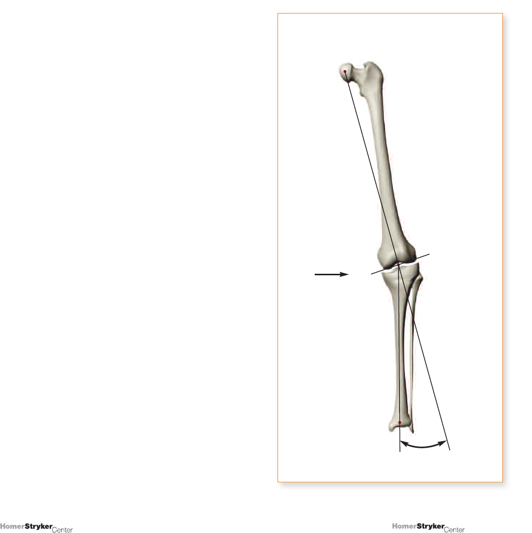
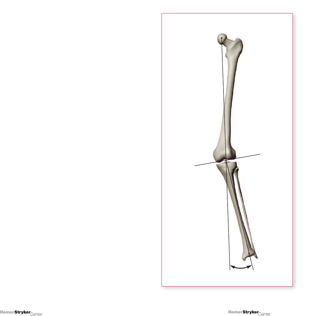
UNIT 2
Measurement of Overall Varus/Valgus
Deformity at the Knee
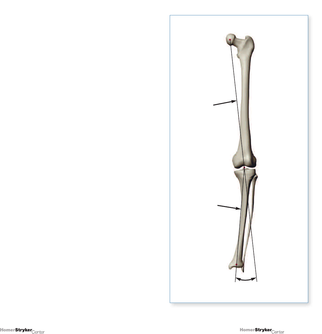
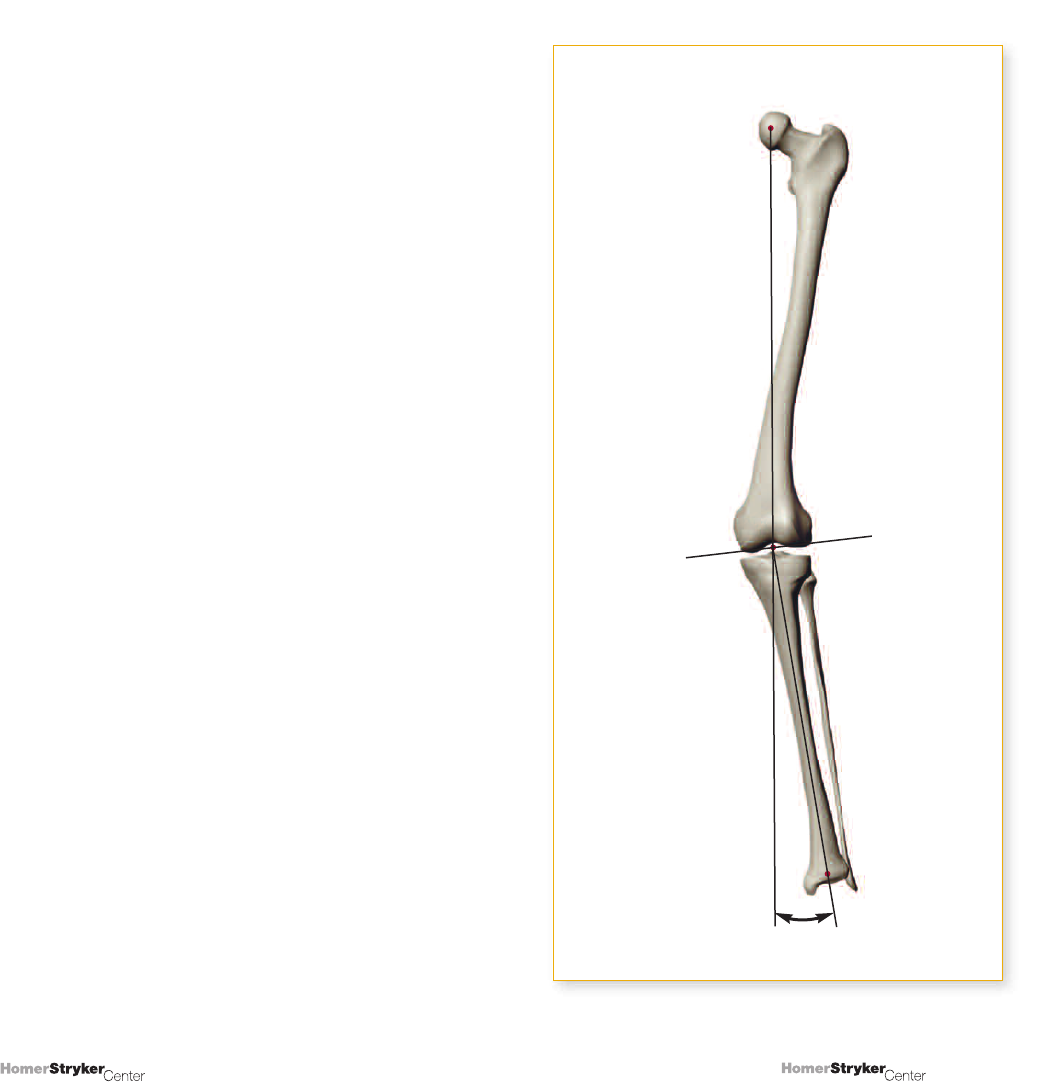
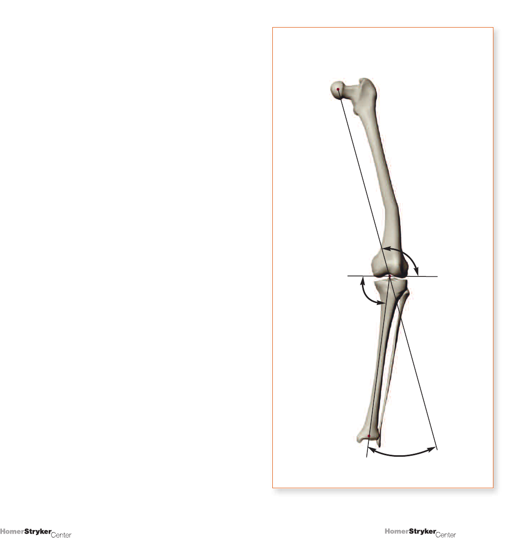
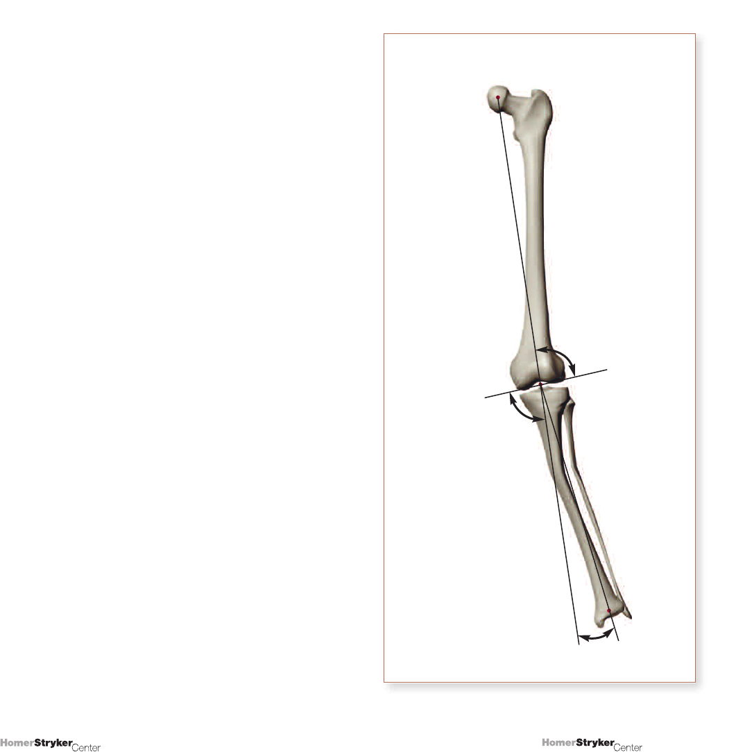
2.1 Measurement when the long standing
films indicate the center of the femoral head
and the center of the ankle
Our definition of normal alignment is when a line drawn
from the center of the hip to the center of the knee
continues toward and transverses the center of the
ankle. The question we want to answer is, “When this
is not the case, how much deformity exists?”. To answer,
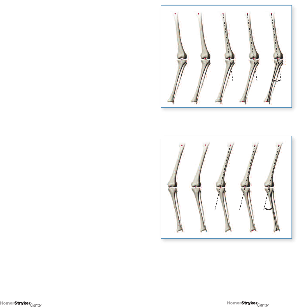
we draw a straight line from the center of the femoral
head to the center of the knee (the mechanical axis of
the femur) and project that line beyond the knee
downward, ideally until the level of the ankle.
The angle formed by the portion of the line projected
beyond the knee and the tibial shaft axis represents
the degree of deformity (a). Figure 2.1 shows how
this deformity is measured; 2.1(A) represents a varus
deformity, and 2.1(B), a valgus deformity.
Varus
= deformity
Valgus
Mechanical
Axis of
Femur
Femoral
Shaft Axis
Projection
of Mechanic
Axis of Femur
Tibial
Shaft Axis
ª
AB
Figure 2.1
Measurement of lower extremity deformity.
ª
ª
10
9
Copyright © 2008 Stryker.
Copyright © 2008 Stryker.
Note that deformity has been described and quantified
without using the mechanical axis of the lower extremity
(the line from the femoral head to the ankle). We have
observed that a majority of orthopaedic residents find the
presence of this line confusing, and it provides no useful
information for planning purposes.
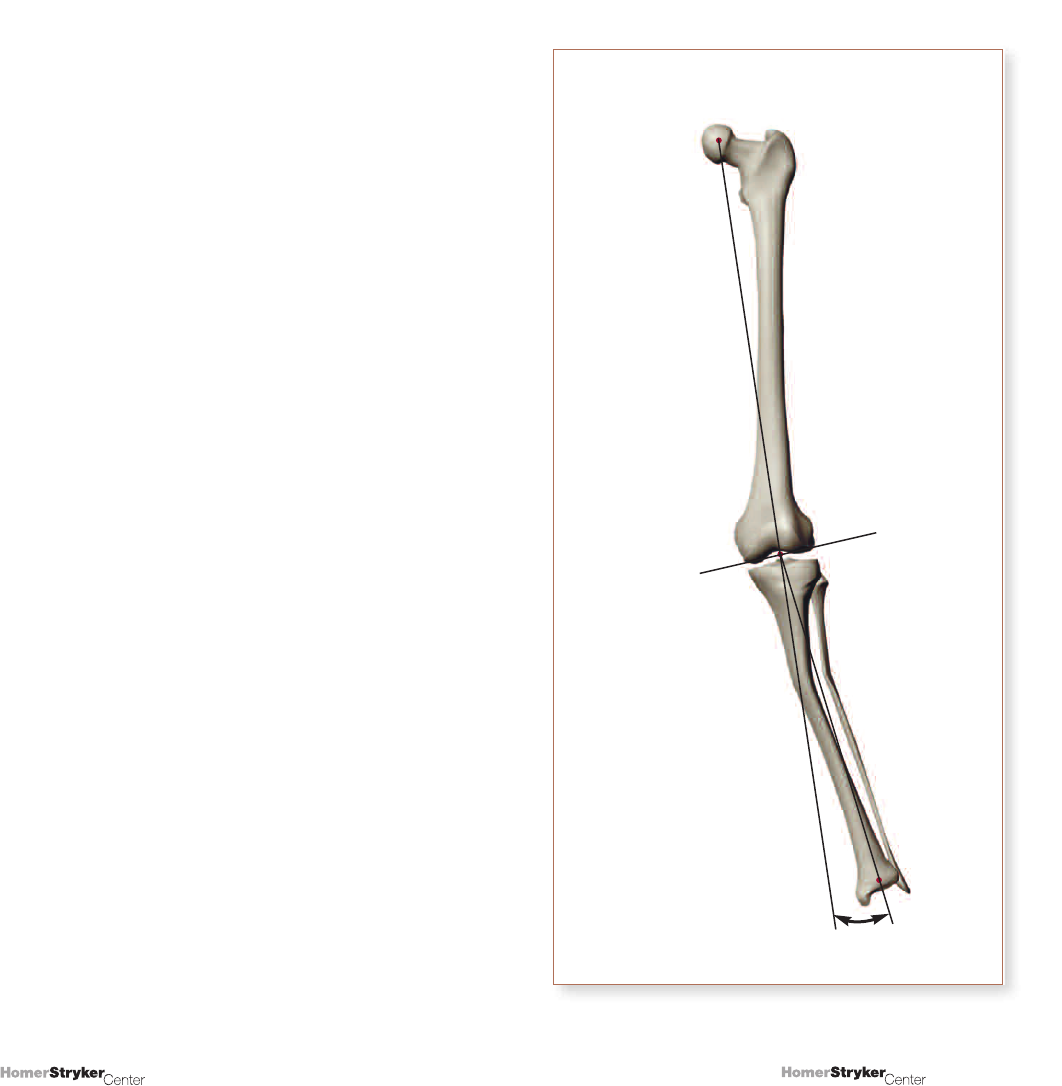
2.2 Measurement when the femoral
head is not visible on long standing lower
extremity radiographs
Our measurements thus far have been based on the
center of the femoral head; we have not used the “normal
tibiofemoral angle,” which includes the femoral shaft.
Certain characteristics, however, may impede visibility
of the femoral head, including height, obesity, and
radiograph quality. In these cases, the tibiofemoral angle
that is present is measured and compared to an assumed
value (such as 6° valgus), and the difference is taken as
the amount of deformity. This concept is illustrated in
Figure 2.2. The measured tibiofemoral angle is 20°
valgus and, when compared to the assumed 6° valgus,
leaves the estimated deformity at 14° valgus.
20°
The Anatomic TF Angle = 20°
valgus. If normal is assumed
to be 6° valgus, the deformity
is 14° valgus.
Figure 2.2
Measurement of lower extremity deformity when
femoral head is not visible - using the anatomic
tibio-femoral angle.
12
11
Copyright © 2008 Stryker.
Copyright © 2008 Stryker.
Why did we select 6° of valgus as the ‘normal’ or
‘average’ tibiofemoral angle? There is little disagreement
that the value should be between 5 and 7° of valgus.
The works of others (Krackow
1
, Moreland et. al
2
,
Yoshioka et. al
3
, Chao et. al
4
) suggest approximately
5.5 to 6°; therefore, for accuracy and simplicity, 6° is
recommended. There are certain instances with
arthroplasty patients in which normal valgus may be
different, such as 2 to 4°. Examples are the presence
of a total hip replacement, hip dysplasia with femoral
anteversion, etc.
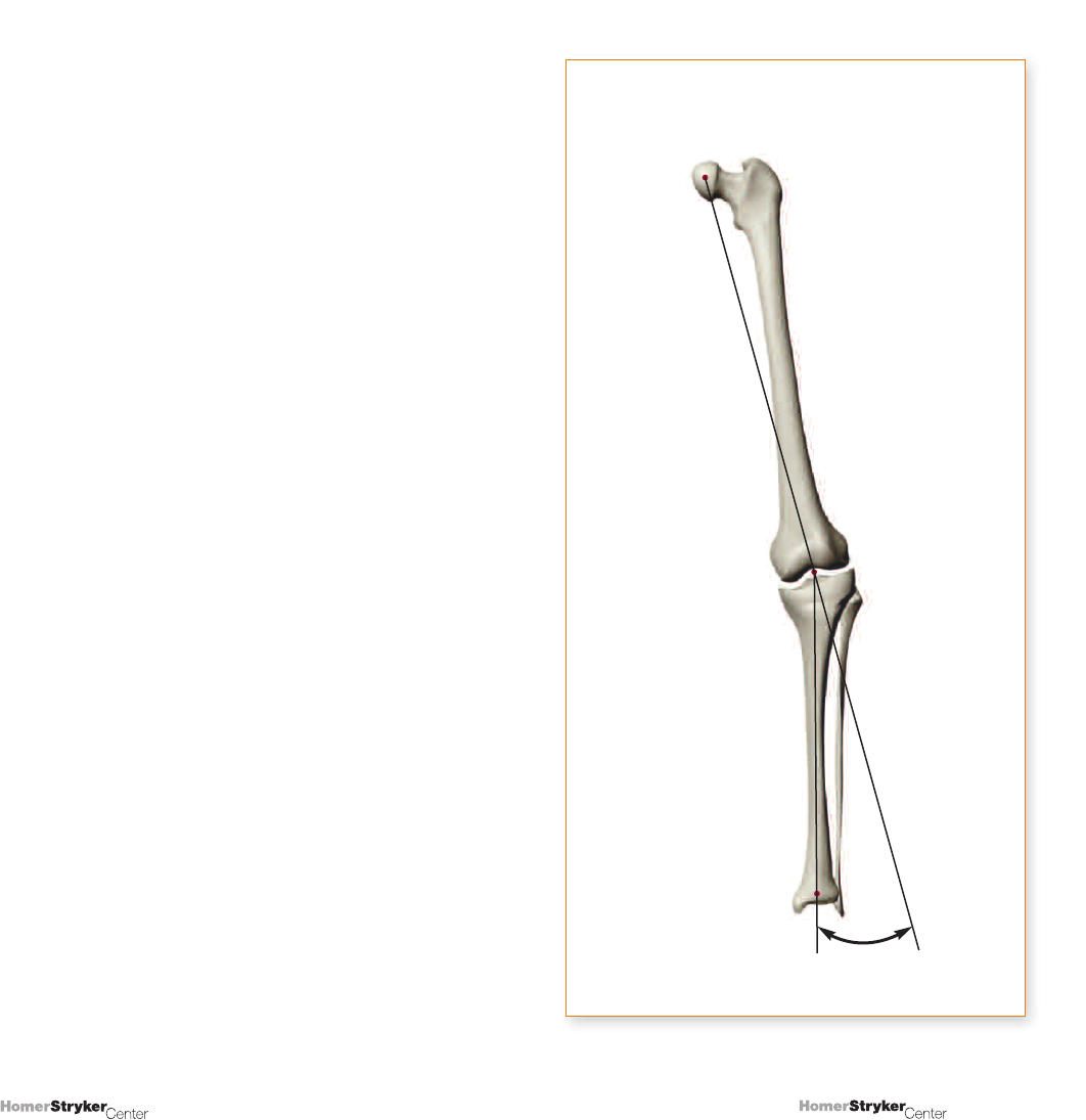
The distal and proximal points for the femoral shaft axis
are characterized somewhat differently. The distal point
can be clear if no uncertainty exists regarding the center
of the knee. One suggestion is to use the midpoint at
the superior aspect of the intercondylar sulcus. This point
can also be thought of as the functional center of the
distal femur and relates directly to patellar tracking, as it
is midway between the medial and lateral condyles.
The proximal point is not as clearly defined. One
suggestion is to use the midpoint of the proximal aspect
of the femur, in the region of the lesser trochanter
(Figure 2.3). Draw a transverse line just above or below
the lesser trochanter; its endosteal midpoint represents
the desired point. This makes it relatively easy to
approximate the overall course of the femoral shaft. In
the case of femoral bowing, place a mark at the proximal
femur and use the line defined by connecting the
proximal and distal marks.
Figure 2.3
Drawing femoral shaft axis when the femoral head
is not visible.
14
13
Copyright © 2008 Stryker.
Copyright © 2008 Stryker.
Important Consideration
When discussing normal knee alignment, it is
necessary to take into account that an individual’s
normal tibiofemoral angle is determined solely by
the femur, and equals the angle between the mechanical
axis of the femur (MAF) and femoral shaft axis (FShA)
(Figure 2.4). This angle is also the individual’s anatomic
tibiofemoral angle. The ability to see the tibia is not
necessary to obtain this angle; therefore, a neutrally
rotated A/P view of the entire femur can be used to
determine a patient’s ideal tibiofemoral angle.
A second important consideration is that of an indistinct
ankle joint. An unpublished study of long standing
lower extremity radiographs (LSLE) showed that a line
drawn from the center of the proximal tibia to the center
of the ankle crosses the tibial metaphysis approximately
50% of the way (medial-lateral) to the midpoint.
Therefore, marking the distal tibia to indicate the tibial
shaft axis at the midpoint across the visible end of the
tibia seems appropriate.
Figure 2.4
Location of the mechanical axis of the femur and the
femoral shaft axis. The angle between these two lines
is the ideal anatomic tibial femoral angle in this case.
ß
Mechanical Axis of
the Femur (MAF)
Femoral Shaft Axis
(FShA)
16
15
Copyright © 2008 Stryker.
Copyright © 2008 Stryker.
UNIT 3
Medial Lateral Tibiofemoral
Translation – Subluxation
Additional elements to knee pathology and deformity
are clearly present when viewing a radiograph with
medial-lateral tibiofemoral subluxation. Clinical
implications are dependent on how this translation is
quantified. In general, we are asking how this translation
affects various measurement conventions. Specifically,
we want to know if the various lines drawn that reference
the center of the knee are going to give similar, mildly
different, or significantly different determinations with
respect to tibiofemoral angle measurements and
deformity assessments.
This question is addressed in Figure 3.1. The choices
implied are to draw the femoral and tibial axes connected
to the middle point (K), the center or ‘midpoint’ of the
knee), or to the distal femoral (F) or proximal tibial (T) point.
Figure 3.1
Location of the center of the knee.
F
T
K
18
17
Copyright © 2008 Stryker.
Copyright © 2008 Stryker.
UNIT 4
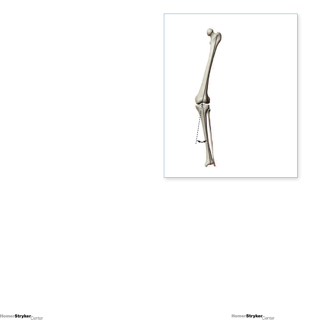
Extra-articular Deformity
It is sometimes necessary to analyze X-rays with
significant extra-articular deformity secondary to fracture
or developmental considerations (Figure 4.1). The
previous analyses largely ignored the intermediate shape
of the respective tibial and femoral shafts.
These cases can be analyzed using modern computer
programs, tracing paper, or basic trigonometry/geometry,
which is explained below.
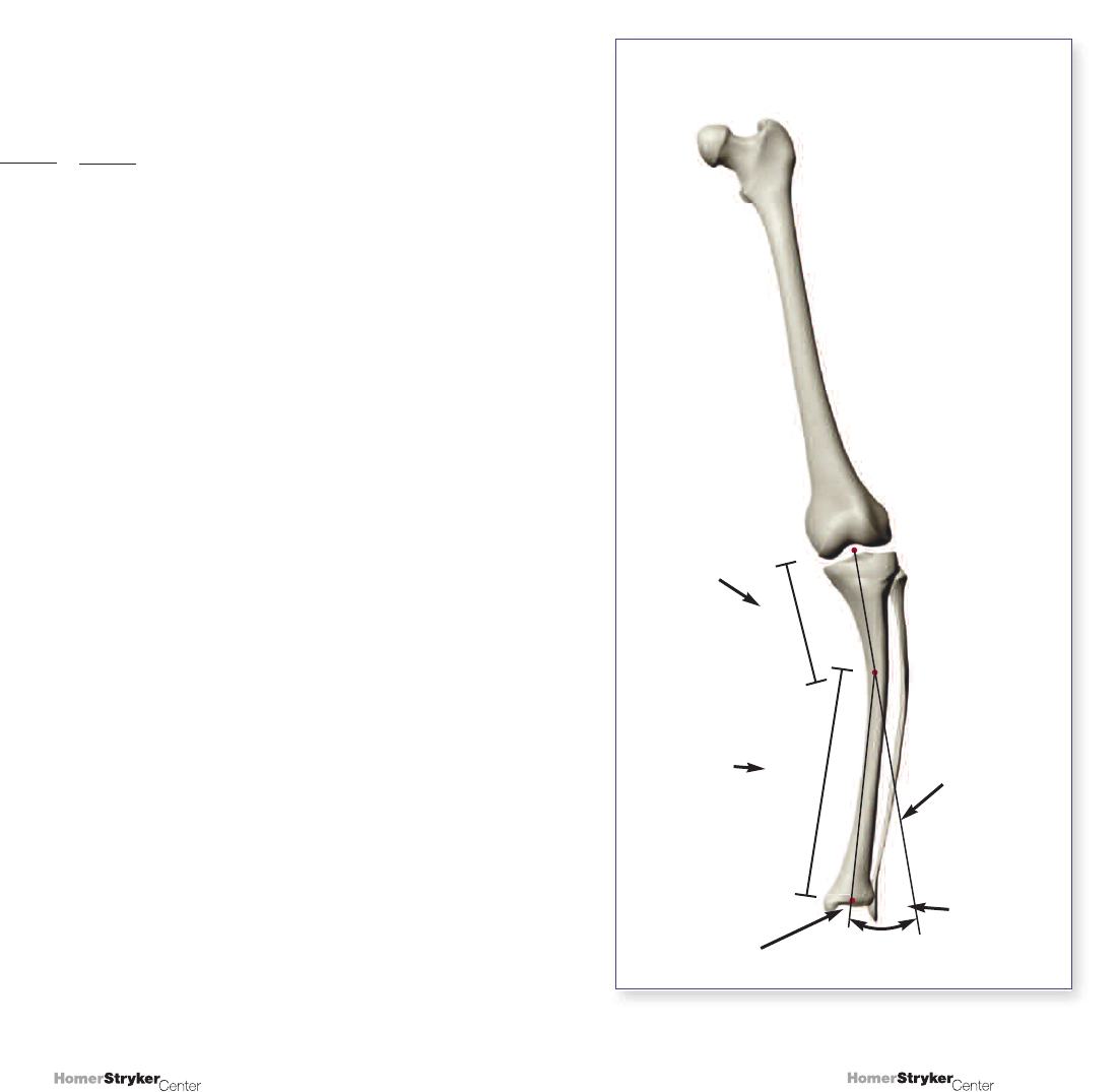
Extra-articular approximation theorem:
A tibial or femoral shaft extra-articular deformity of a
certain angular amount creates a corresponding
deformity at the knee in approximate proportion to the
percentage of the way that deformity is located toward
the knee.
Example 1 (Figure 4.1-A):
A 10° varus deformity 80% of the way from the hip to
the knee, or 20% of the way above the knee, would
impart an approximately 8° varus deformity at the knee,
which would be 100% on the femoral side.
Example 2 (Figure 4.1-B):
A 10° varus deformity 80% of the way from the ankle
to the knee, or 20% of the way below the knee, would
impart an approximately 8° varus deformity at the knee,
which would be 100% on the tibial side.
Figure 4.1
Measurement of extra-articular deformity.
B
A
20
19
Copyright © 2008 Stryker.
Copyright © 2008 Stryker.
The approximation relates to the angle and length
differences noted when drawing lines form the apex of
an isosceles triangle to its base. Drawing a line from the
vertex to the midpoint of the base creates a bisection
of both the base and the vertex angle. Drawing lines to
the points which define a trisection of the base length
does not. However, it does provide 3 equal angles at
the vertex of the triangle.
Summary: Measurement of
Varus/Valgus Deformity
If the femoral head is visible (Figure 4.2):
1. Locate the center of the knee and center of the
femoral head.
2. Draw a line connecting these two points.
3. Locate (or approximate) the center of the ankle.
4. Draw a line connecting the center of the knee to the
center of the ankle.
5. Measure the angle between the 2 lines. A
measurement of 0°/180° implies no deformity;
otherwise, the observed angle is the angle of varus
or valgus present (valgus if foot is lateral, varus if
foot is medial).
Figure 4.2 (Varus)
An uncomplicated varus deformity.
Figure 4.2 (Valgus)
An uncomplicated valgus deformity.
22
21
Copyright © 2008 Stryker.
Copyright © 2008 Stryker.
If the femoral head is not visible (Figure 4.3):
1. Locate the midpoint of the proximal femur in the
region of the lesser trochanter.
2. Locate the center of the knee.
3. Draw a line from the proximal femur to the center
of the knee.
4. Locate (or approximate) the center of the ankle.
5. Draw a line from the center of the knee to the center
of the ankle.
6. Measure the angle between the 2 lines and label as
varus or valgus, depending on position of tibia
(pointed inward or laterally).
7. Compare the measured angle to a normal value
(i.e., 6° valgus).
Figure 4.3 (Varus)
Varus deformity with the femoral head not visible.
Figure 4.3 (Valgus)
Valgus deformity with the femoral head not visible.
24
23
Copyright © 2008 Stryker.
Copyright © 2008 Stryker.
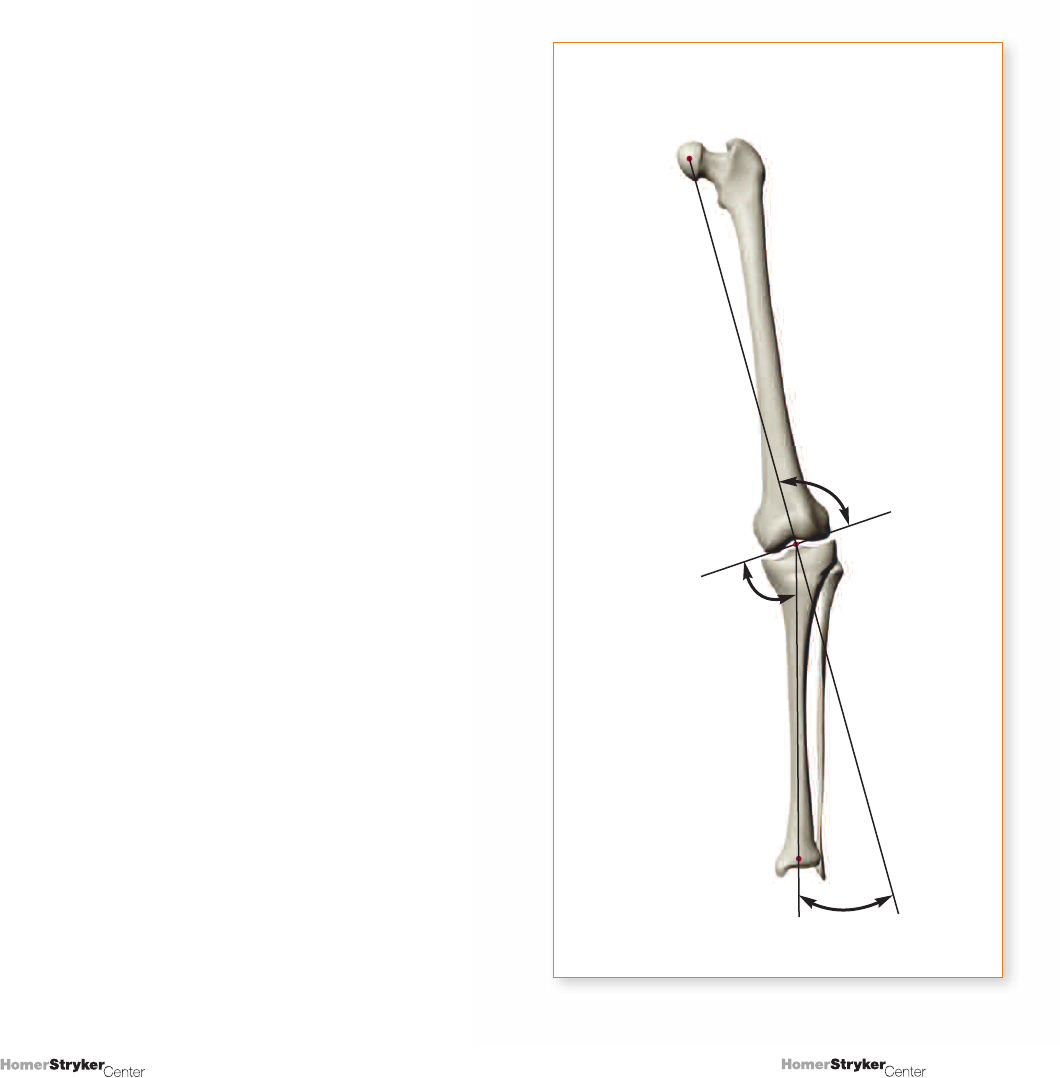
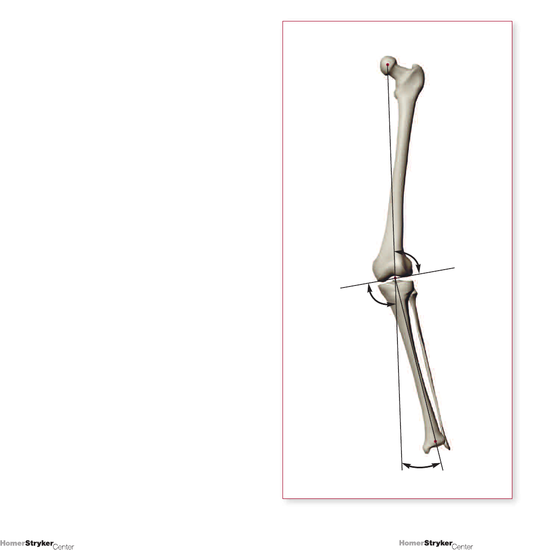
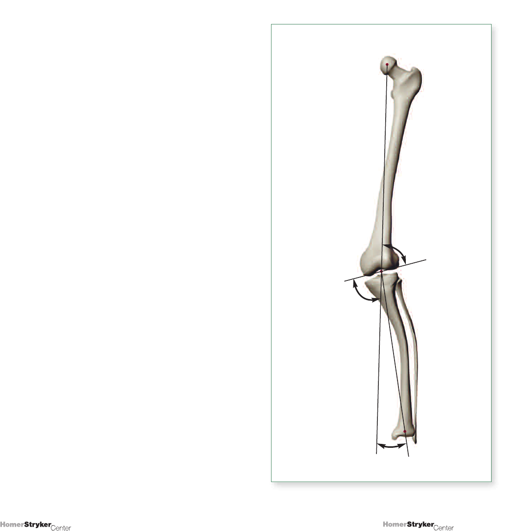
UNIT 5
Characterizing Deformity About the Knee
Determination of a varus/valgus deformity only tells us
that the deformity exists; details such as its location
are not revealed. Additional analyses, utilizing joint-line
orientation, allow prediction of outcomes of particular
osteotomy methods and anticipation of certain TKA
problems. Previous discussion has only considered
knee position as a center-point (i.e., centered on the
mechanical axis). Deformity can be characterized
according to 4 ‘origins’:
1. Deformity on the Femoral Side of the Joint
Due either to developmental abnormality or to attrition
of bone very close to the joint as a result of fracture,
degenerative wear, avascular necrosis, collapse, etc.
2. Deformity on the Tibial Side of the Joint
Due either to developmental abnormality,
degeneration, etc.
3. Deformity Within the Joint Itself
Due to asymmetric wear.
4. Deformity Due to Discreet
Extra-articular Angulation
Generally exemplified by new angulation after fracture
or osteotomy.
Considering these origins of deformity requires
establishing standards for normal (average) values
indicating joint line orientation, with any variation alluding
to the deformities just described. In Figure 5.1, the
normal articular cartilage space (medial vs. lateral) is
approximately equal – lines across the distal femoral
condyles and across the medial and lateral tibial
plateaus are essentially parallel.
In Figure 5.2, the overall joint line is typically slightly
different from perpendicular (2 to 3°, on average).
Figure 5.1
Joint lines added.
Figure 5.2
Representative angles for a non-deformed knee.
6°
90°
87-8°
81°
92-3°
26
25
Copyright © 2008 Stryker.
Copyright © 2008 Stryker.
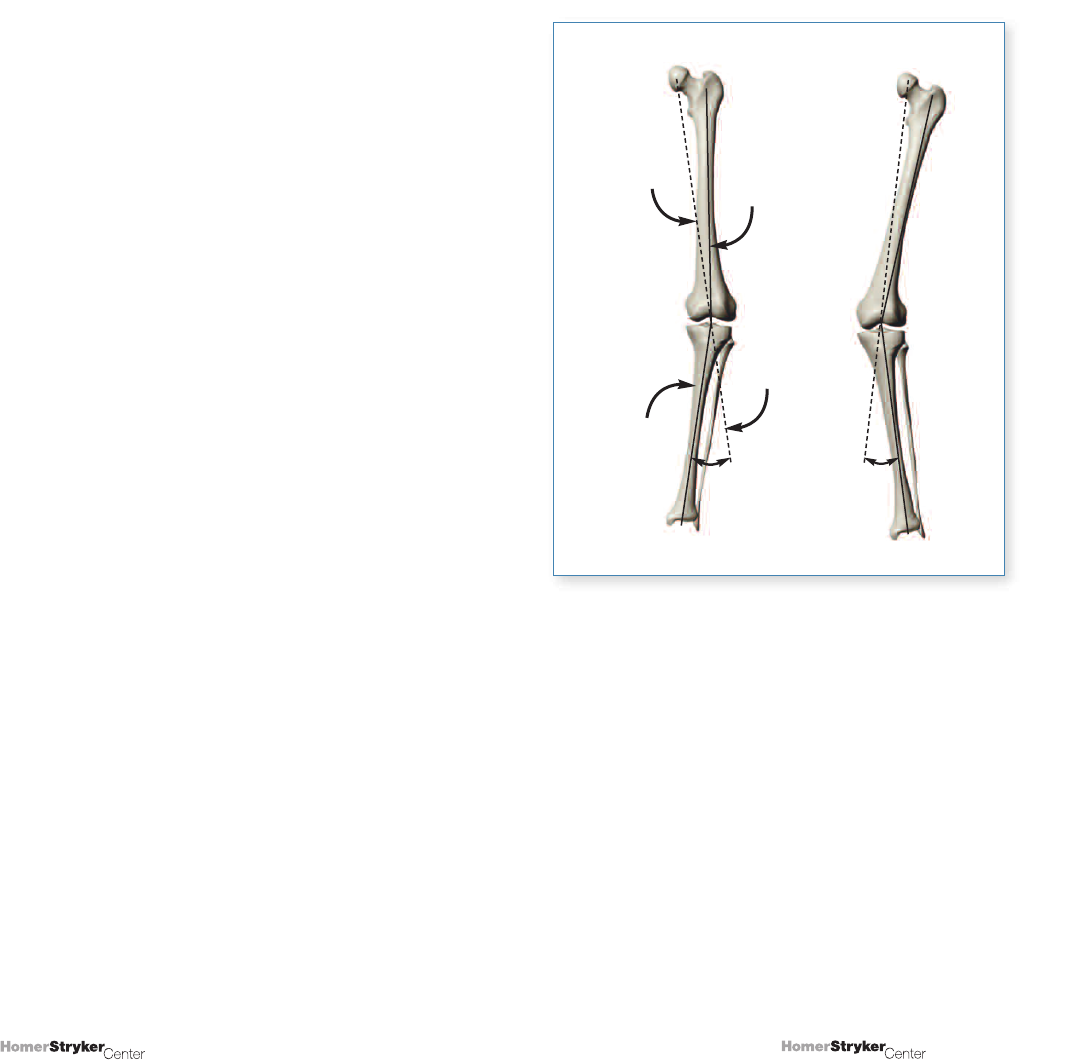
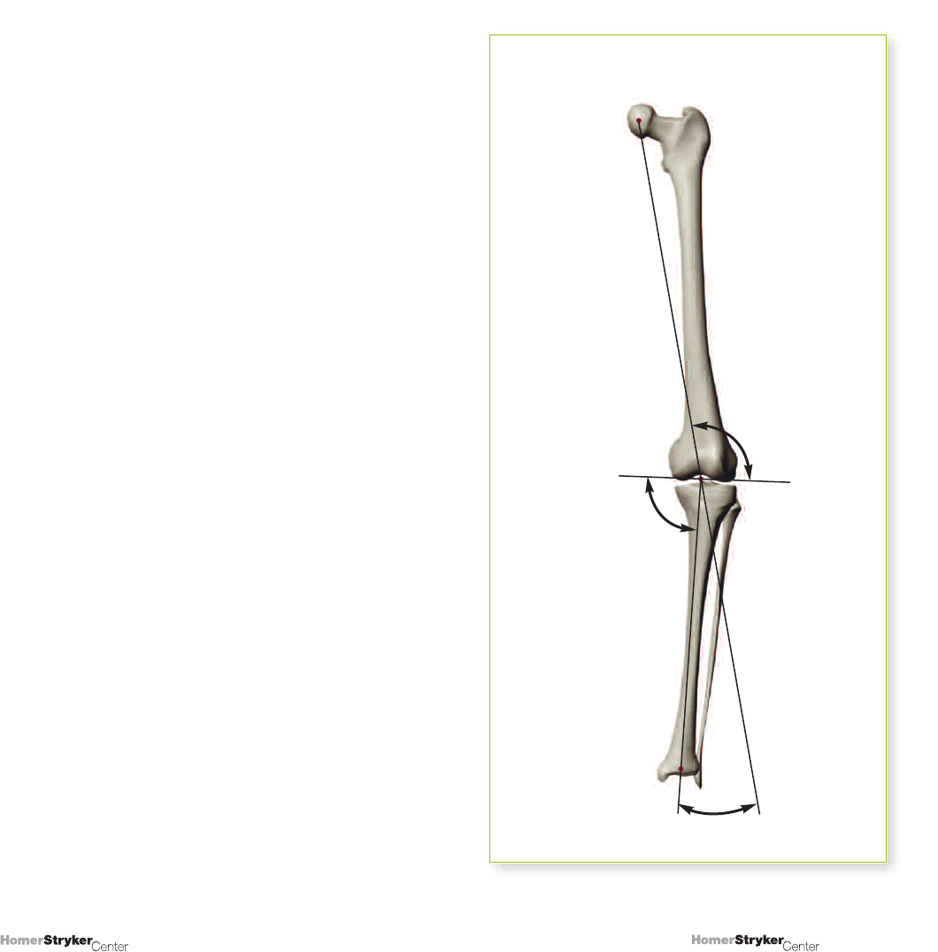
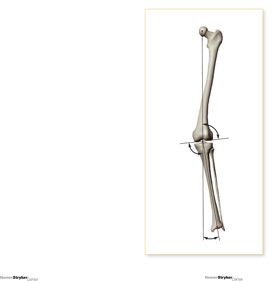
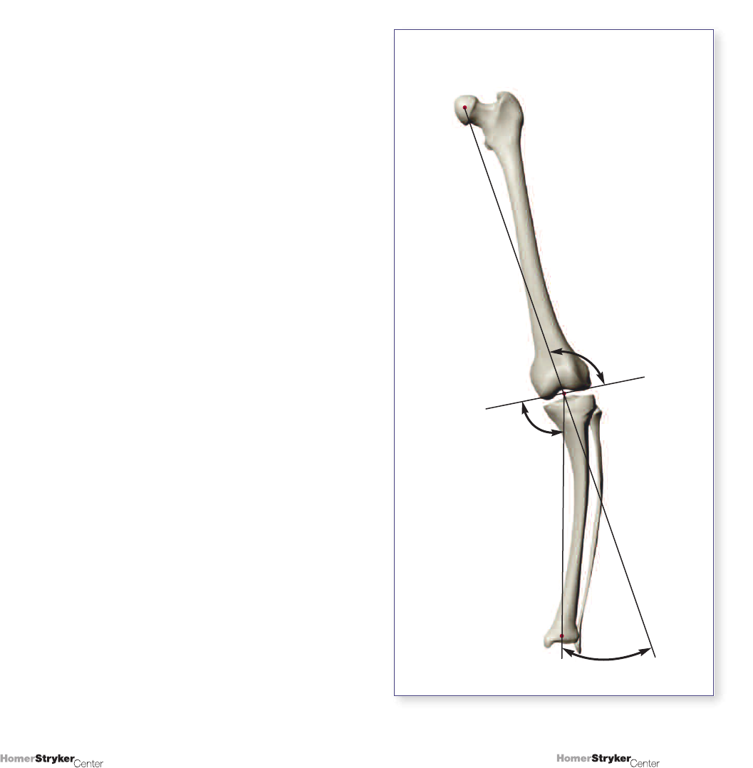
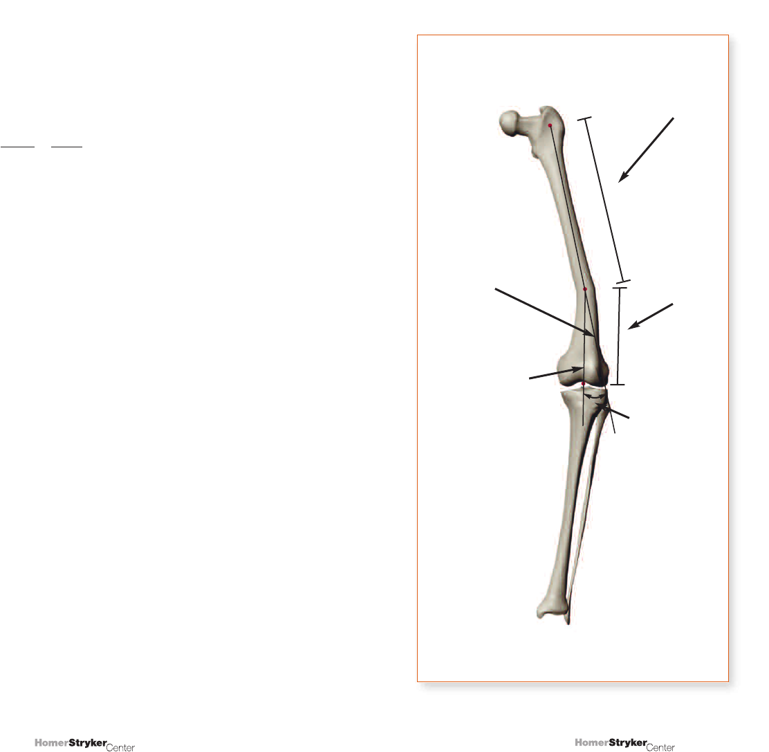
The normal femoral joint angle (FJA) is 2 to 3° valgus to
the mechanical axis of the femur, or 8 to 9° valgus
to the normal femoral shaft axis (Figure 5.3-A). The
normal tibial joint angle (TJA) is 2 to 3° varus to the
mechanical axis of the tibia (equivalent to the tibial shaft
axis (Figure 5.3-B). Smaller numbers are typically used
when describing these terms (e.g., a 3° varus TJA vs.
a medial TJA of 87°).
Here the joint line is being measured relative to the tibial
shaft axis and the mechanical axis of the femur. Recall
that the mechanical axis of the femur is a conceptual line
that does not exist on a radiograph (it must be drawn),
as opposed to the femoral shaft axis. Comparison of
the joint line to the femoral shaft is often the simplest
measure. Comparison with the mechanical axis of the
femur requires a visible femoral head center on the
radiograph. When not visible, the previous assumption
that the angle between the mechanical axis of the femur
and the femoral shaft axis is 6° must be used. Thus,
the joint line is actually compared to the femoral shaft
axis, and the 6° assumption is added. This value is
subsequently added to the normal 2 to 3° angulation
present between the mechanical axis and an otherwise
perpendicular joint.
Figure 5.3
Femoral (A) and tibial (B) joint angles.
87°
81°
87-8°
92-3°
(B)
(A)

28
27
Copyright © 2008 Stryker.
Copyright © 2008 Stryker.
Asymmetry within the joint line can be drawn
different ways:
1. A single line, with no distinction for any joint line
asymmetry (Figure 5.4).
2. Two lines, showing the intra-articular asymmetry (bony
distal femoral joint line and proximal tibial joint line,
(Figure 5.5).
3. A single line that bisects the bony joint lines shows
joint line asymmetry (Figure 5.6).
Figure 5.4
One line to indicate the joint line.
Figure 5.5
Two lines, the bony distal femoral joint line and
proximal tibial joint line, forming an intra-articular
angle due to joint wear and/or ligament instability.
Figure 5.6
The same physical situation as in Figure 5.5 only
with a single joint line to represent the mean overall
position of the joint line. This method is used in our
examples and problems.

30
29
Copyright © 2008 Stryker.
Copyright © 2008 Stryker.
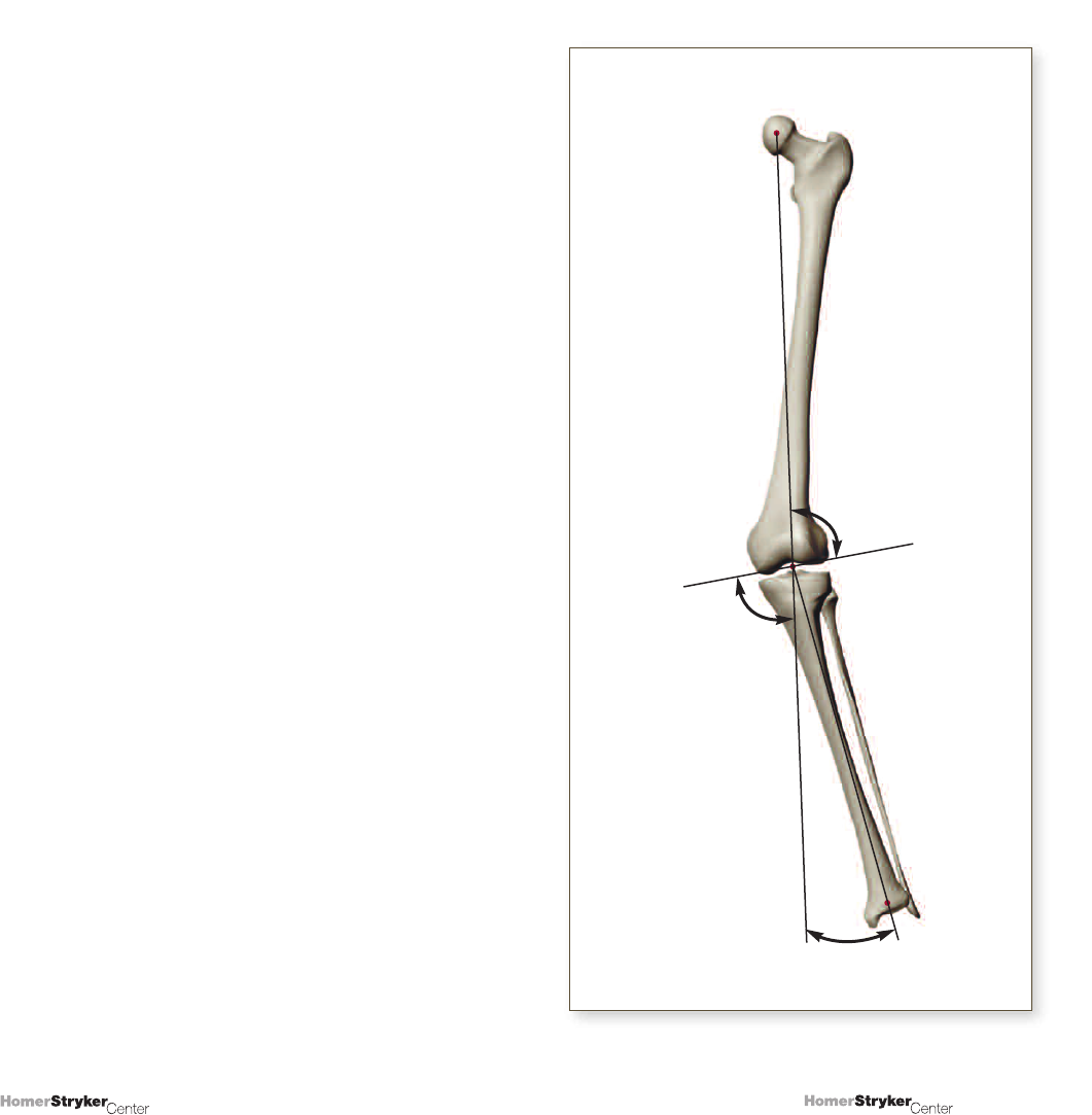
When dealing with cases of varus/valgus deformity with
extra-articular elements, the analysis can be relatively
straightforward by answering a sequence of questions:
1. How much varus/valgus deformity exists?
Answer:
Construct the mechanical axis of the femur and tibial
shaft axis; measure and label the angle between them.
2. How much of the deformity is:
a. In the distal femur (as it currently lies)?
b. In the proximal tibia (as it currently lies)?
c. Within the joint space (if asymmetric, and a
separate evaluation is desired)?
Answers:
a. Draw the femoral joint angle (FJA) and compare to
standard (2 to 3° valgus).
b. Draw the tibial joint angle (TJA) and compare to
standard (2 to 3° varus).
c. Draw the intra-articular angle (IAA) and compare to
standard (0°).
3. How much of the tibial or femoral deformity
is due to shaft angulation?
Answers:
a. Measure angulation in the shaft.
b. Determine its proportional distance away from the hip
or ankle.
c. Multiply the proportion with the shaft angulation and
compare to the deformity angles obtained during
FJA, TJA, and IAA comparisons (Question 2, above).
UNIT 6
INSTRUCTIONAL EXAMPLES
The alignment analyses are shown
step-by-step in the following 10 examples.
32
31
Copyright © 2008 Stryker.
Copyright © 2008 Stryker.
EXAMPLE 1
Varus Deformity of the Femur and Tibia

34
33
Copyright © 2008 Stryker.
Copyright © 2008 Stryker.
> Identify the center of the femoral head, knee,
and ankle.
Example 1A
Center of
Femoral Head
Center of Ankle
Center of Knee
36
35
Copyright © 2008 Stryker.
Copyright © 2008 Stryker.
> The axes are marked, and an overall varus deformity
of 11° is measured.
Example 1B
11˚
MAF – Mechanical
Axis of the Femur
MAT – Mechanical
Axis of the Tibia
38
37
Copyright © 2008 Stryker.
Copyright © 2008 Stryker.
> Add the joint line.
11˚
Example 1C
Joint Line
40
39
Copyright © 2008 Stryker.
Copyright © 2008 Stryker.
> The joint angles (FJA, TJA) are measured and
compared to normal joint angles.
> ∅ FJA = Observed FJA compared to 87° lateral.
> ∅ TJA = Observed TJA compared to 87° medial.
> The overall deformity shown here is 11° varus. The
femoral joint angle (FJA) is normally 87°. Since the
femoral joint angle shown here is 95°, there is an
“error”, or deviation, that we call the ∅ FJA. In this
case, the ∅ FJA is 8°. Therefore, we have an 8°
varus deformity at the femur.
> The tibial joint angle (TJA) is normally 87°. Since the
angle in this example is 84°, the ∅ TJA is equal to 3°.
This results in a deformity at the tibia of 3° varus.
> To analyze and check your work, make sure the sum
of the ∅ FJA and the ∅ TJA are equal to the overall
deformity; ∅ FJA and ∅ TJA = overall deformity; 8°
varus and 3° varus = 11° varus.
> We can also summarize saying this example shows an
11° overall varus deformity of which 8° is in the distal
femur and 3° is in the proximal tibia.
11˚
Joint Line
Example 1D
84˚
95˚
42
41
Copyright © 2008 Stryker.
Copyright © 2008 Stryker.
EXAMPLE 2
Varus Deformity of the Tibia
44
43
Copyright © 2008 Stryker.
Copyright © 2008 Stryker.
> The axes are marked and the overall deformity is
shown to be in 13°of varus.
Example 2A
13˚
46
45
Copyright © 2008 Stryker.
Copyright © 2008 Stryker.
> Add the joint line.
Example 2B
13˚
Joint Line
48
47
Copyright © 2008 Stryker.
Copyright © 2008 Stryker.
> Determine all the important angles.
Example 2C
74˚
87˚
13˚
50
49
Copyright © 2008 Stryker.
Copyright © 2008 Stryker.
EXAMPLE 3
Varus Deformity at the Femur with Minor
“Compensation” at the Tibia

52
51
Copyright © 2008 Stryker.
Copyright © 2008 Stryker.
> The axes are marked and the overall deformity is
indicated to be 14° varus.
Example 3A
14˚
54
53
Copyright © 2008 Stryker.
Copyright © 2008 Stryker.
> Determine all the important angles.
> The overall deformity is 14° varus.
> The FJA here is 102°. Because the normal FJA is
87°, and angle of 102° represents a ∅ FJA of 15°
varus, or a varus deformity of 15° in the femur.
> The TJA here is 88°. Because the normal TJA is 87°,
an angle of 88° represents a ∅ TJA of 1° valgus.
Therefore, the deformity in the tibia is 1of valgus
angulation.
> ∅ FJA + ∅ TJA = Overall Deformity.
> 15° varus and 1° valgus = 14° varus.
> Therefore, the overall varus deformity is 14° because
there is 15° of varus deformity from the femur and 1°
of valgus compensation at the tibial side.
14˚
Example 3B
88˚
102˚
56
55
Copyright © 2008 Stryker.
Copyright © 2008 Stryker.
EXAMPLE 4
Valgus Deformity at Both the Femur and Tibia
58
57
Copyright © 2008 Stryker.
Copyright © 2008 Stryker.
> The axes are marked and the overall deformity is
indicated to be 11° of valgus.
Example 4A
11˚
60
59
Copyright © 2008 Stryker.
Copyright © 2008 Stryker.
> Determine all the important angles.
> The overall deformity is 11º valgus.
> The FJA is 84º. Normally, the FJA is 87º. Therefore,
84º represents a ∅ FJA of 3º. This also equals a 3º
valgus deformity at the femur.
> The TJA is 95º. Since the normal TJA is 87º, an
angle of 95º represents a ∅ TJA of 8º. Thus, there
is an 8º valgus deformity at the tibia.
> ∅ FJA and ∅ TJA = overall deformity.
> 3º valgus and 8º valgus = 11º valgus.
> There is an overall valgus deformity of 11º, 3º from
the femur and 8º from the tibia.
11˚
Example 4B
84˚
95˚
62
61
Copyright © 2008 Stryker.
Copyright © 2008 Stryker.
EXAMPLE 5
Valgus Deformity at the Femur and Tibia
64
63
Copyright © 2008 Stryker.
Copyright © 2008 Stryker.
> The axes are marked and the overall deformity is
indicated to be 14° valgus.
Example 5A
14˚
66
65
Copyright © 2008 Stryker.
Copyright © 2008 Stryker.
> All important angles are determined.
> The overall deformity is 14° valgus.
> The FJA is 80°. Normally, the FJA is 87°. Therefore,
80° represents a ∅ FJA of 7°, indicating a 7° valgus
deformity at the femur.
> The TJA is 94°. Normally, the TJA is 87°. Therefore,
94° represents a ∅ TJA of 7°, indicating a 7° valgus
deformity at the tibia.
> ∅ FJA and ∅ TJA = overall deformity.
> 7° valgus and 7° valgus = 14° valgus.
> There is an overall deformity of 14° valgus, with half
from the femur and half from the tibia.
14˚
Example 5B
80˚
94˚
68
67
Copyright © 2008 Stryker.
Copyright © 2008 Stryker.
EXAMPLE 6
Valgus Deformity at the Femur and Tibia

70
69
Copyright © 2008 Stryker.
Copyright © 2008 Stryker.
> The axes are marked and the overall deformity is
indicated to be 15° valgus.
Example 6A
15˚
72
71
Copyright © 2008 Stryker.
Copyright © 2008 Stryker.
> All important angels are measured.
> The overall deformity is 15° valgus.
> The FJA is 81°. Normally, the FJA is 87°. Therefore,
81° represents a ∅ FJA of 6° and a valgus deformity
at the femur of 6°.
> The TJA is 96°. Normally, the TJA is 87°. Therefore,
96° represents a ∅ TJA of 9°. Thus, there is a 9°
valgus deformity at the tibia.
> ∅ FJA and ∅ TJA = overall deformity.
> 6° valgus and 9° valgus = 15° valgus.
> There is a 15° valgus overall deformity, with 6°from
the femur and 9°from the tibia.
15˚
Example 6B
81˚
96˚
74
73
Copyright © 2008 Stryker.
Copyright © 2008 Stryker.
EXAMPLE 7
Extra-articular Varus Angulation of the Tibia
76
75
Copyright © 2008 Stryker.
Copyright © 2008 Stryker.
> The axes are marked and the overall deformity is
indicated to be 22° varus.
Note:
The mechanical axis of the tibia is drawn from the center
of the proximal tibia to the center of the ankle, ignoring the
shaft angulation.
Example 7A
22˚
Tibial Shaft Axis
78
77
Copyright © 2008 Stryker.
Copyright © 2008 Stryker.
> The angulation at the extra-articular deformity is 16°.
> The proportional distance from the ankle to the knee
is calculated as follows:
48 = 48 = 62%;
48+29 77
therefore, 62% of 16°equals 9.92°which is about 10°.
> Thus, the contribution of extra-articular angulation to
the overall knee alignment is about 10°.
Note:
The length units used in the proportional distance
are meaningless here because they “cancel” due
to proportionality.
Example 7B
16˚
*These lengths need
not be meaningful as
absolute numbers, they
are just considered for
the X-ray or diagram
being measured.
Approximate*
Length of the
Proximal Tibial
Segment
Axis of the
Proximal Tibial
Segment
Axis of the Distal
Tibial Segment
Angulation
of the Extra-
articular
Deformity
29mm
48mm
Approximate*
Length of the
Distal Tibial
Segment
80
79
Copyright © 2008 Stryker.
Copyright © 2008 Stryker.
> All the important angels are determined.
> The overall deformity is 22° varus.
> The FJA is 99°. Normally the FJA is 87°. Therefore,
99° represents a ∅ FJA of 12° and indicates a 12°
varus deformity at the femur.
> The TJA is 77°. Normally, the TJA is 87°. Therefore, 77°
represents a ∅ TJA of 10° and indicates a 10° varus
deformity at the tibia.
> ∅ FJA and ∅ TJA = overall deformity.
> 12° varus and 10° varus = 22° varus.
> The tibial extra-articular angulation of 16° (derived from
the calculation on the previous page) contributed 10°
of varus to the knee alignment. Therefore, the 10°
overall tibial contribution is due essentially solely to the
extra-articular deformity.
Summary:
There is a 22° overall varus knee alignment. 12° of the
deformity is located at the femur, and 10° is found within
the tibia. The 10° at the proximal tibia is due to the
extra-articular tibial deformity, which is 16° at its apex
and contributes 10° at the joint level.
Example 7C
22˚
99˚
77˚
82
81
Copyright © 2008 Stryker.
Copyright © 2008 Stryker.
EXAMPLE 8
Valgus Deformity at the Femur and
Extra-articular Varus Tibial Angulation
84
83
Copyright © 2008 Stryker.
Copyright © 2008 Stryker.
> The axes are marked and the overall deformity is
indicated to be 8° valgus.
Example 8A
8˚

86
85
Copyright © 2008 Stryker.
Copyright © 2008 Stryker.
> The extra-articular tibial angulation.
> The angulation of the deformity is 18°.
> The proportional distance from the ankle to the knee
is calculated as follows:
36 = 36 = 50%;
36+37 73
therefore, 50% of 18° equals 9°.
> Thus, the contribution of extra-articular angulation to
the overall knee alignment is 9°.
Example 8B
18˚
37mm
36mm
88
87
Copyright © 2008 Stryker.
Copyright © 2008 Stryker.
> All important angles are determined.
> The overall deformity is 8° valgus.
> The FJA is 69°. Normally, the FJA is 87°. Therefore,
an angle of 69° represents a ∅ FJA of 18° and
indicates an 18° valgus deformity at the femur.
> The TJA is 77°. Normally, the TJA is 87°. Therefore,
an angle of 77° represents a ∅ TJA of 10° and
indicates a 10° varus deformity at the tibia.
> ∅ FJA and ∅ TJA = overall deformity.
> 18° valgus and 10° varus = 8° valgus.
> There is an 8° overall valgus knee deformity, an 18°
valgus deformity at the femur and a 10° proximal
tibial varus deformity. 9° of the 10° is due to tibial
shaft angulation.
Example 8C
8˚
69˚
77˚
90
89
Copyright © 2008 Stryker.
Copyright © 2008 Stryker.
EXAMPLE 9
Extra-articular Varus Angulation of the Femur

92
91
Copyright © 2008 Stryker.
Copyright © 2008 Stryker.
> The axes are marked and the overall deformity is
indicated to be 22° varus.
Example 9A
22˚
94
93
Copyright © 2008 Stryker.
Copyright © 2008 Stryker.
> Extra-articular femoral angulation of 15°.
> The angulation of the deformity is 15°.
> The proportional distance from the hip to the knee is
calculated as follows:
56 = 56 = 66.7% ≈ 67%;
56+28 84
therefore, 67% of 15° equals 10°.
> Thus, the contribution of extra-articular angulation
to the overall knee alignment is about 10°.
Example 9B
Approximate
Length of the
Distal Femoral
Segment
56mm
28mm
Extra-articular
Angulation of the
Femur is
approximately 15˚
Axis of the Distal
Femoral Segment
Axis of the Proximal
Femoral Segment
Approximate Length
of the Proximal
Femoral Segment
96
95
Copyright © 2008 Stryker.
Copyright © 2008 Stryker.
> All important angles are determined.
> The overall deformity is 22° varus.
> The FJA is 105°. Normally, the FJA is 87°. Therefore,
105° represents a ∅ FJA of 18° and indicates an
18° varus deformity at the femur.
> The TJA is 83°. Normally, the TJA is 87°. Therefore,
an angle of 83° represents a ∅ TJA of 4° and
indiates a 4° varus deformity at the tibia.
> ∅ FJA and ∅ TJA = overall deformity.
> 18° varus and 4° varus = 22° varus.
> The extra-articular contribution is 10° varus.
Summary:
There is 22° of varus angulation at the knee, 18° of
which is due to the deformity at the femur, with 10° of
this is due to the 15° extra-articular deformity at the
distal femur. There is 4° of varus at the tibia.
22˚
Example 9C
105˚
83˚
98
97
Copyright © 2008 Stryker.
Copyright © 2008 Stryker.
EXAMPLE 10
Valgus Deformity of the Femur with
Extra-articular Valgus Tibial Angulation
100
99
Copyright © 2008 Stryker.
Copyright © 2008 Stryker.
> The axes are marked and the overall deformity is
indicated to be 11° valgus.
Example 10A
11˚
102
101
Copyright © 2008 Stryker.
Copyright © 2008 Stryker.
> The angulation of the deformity is 10°.
> The proportional distance from the ankle to the knee
is calculated as follows:
49 = 49 = 70%;
49+21 70
therefore, 70% of 10° equals 7°.
> Thus, the contribution of extra-articular angulation to
the overall knee deformity is about 7°.
10˚
Example 10B
21mm
49mm
104
103
Copyright © 2008 Stryker.
Copyright © 2008 Stryker.
> All important angels are determined.
> The overall deformity is 11° valgus.
> The FJA is 84°. Normally, the FJA is 87°. Therefore,
84° represents a ∅ FJA of 3°. Thus, there is a 3°
valgus deformity at the femur.
> The TJA is 95°. Normally, the TJA is 87°. Therefore, 95°
represents a ∅ TJA of 8°. Thus, there is an 8° valgus
deformity at the tibia.
> ∅ FJA and ∅ TJA = overall deformity.
> 3° valgus and 8° valgus = 11° valgus.
> The 7° of valgus at the tibia is due to a 10° valgus
extra-articular tibial deformity.
Summary:
There is an overall valgus knee deformity of 11°, with 3°
of the 11° coming from the deformity at the femur, and
8° of the 11° coming from deformity at the proximal tibia;
7° of this 8° is from the tibial shaft angulation of 10°.
11˚
Example 10C
84˚
95˚

106
105
Copyright © 2008 Stryker.
Copyright © 2008 Stryker.
UNIT 7
ONLINE INTERACTIVE
PRACTICE
The interactive problems provided here are intended
to be used as learning tools. Improved accuracy in
measuring axial deformities of the knee comes from
constant practice only. Therefore, this interactive section
is designed in such a way as to encourage and reinforce
learning by repetition while taking different learning styles
into account. These problems do not comprise a test;
instead, they offer a dynamic way to use instructional
tools that are specifically designed to aid each user in
achieving mastery of this topic at a comfortable pace.
Completing these problems successfully will help
contribute toward addressing the challenge of measuring
axial deformities of the knee.
To access the Interactive Practice, please enter the URL
shown below into your internet browser.
http://www.homerstrykercenter.com/publications/
axialdeformity/
REFERENCES
1. Krackow KA. The Technique of Total Knee Arthroplasty.
St. Louis; C.V. Mosby Company; 1990.
2. Moreland JR, Bassett LW, Hanker GJ. Radiographic
analysis of the axial alignment of the lower extremity.
J Bone Joint Surg (Am) 1987;69-A:745-49.
3. Yoshioka Y, Siu D, Cooke DV. The anatomy and
functional axes of the femur. J Bone Joint Surg (Am)
1987;69-A:7873-80.
4. Chao EY, Neluheni EV, Hsu RW, Paley D. Biomechanics
of alignment. Orthop Clin N Am 1994;25:379-86.

Copyright © 2008 Stryker.
Copyright © 2008 Stryker.
